The University Grants Committee, Research Grants Council (RGC)
Hong Kong
Citation
Journal: Nat Commun / Year: 2023 Title: Cryo-EM structure of cyanophage P-SCSP1u offers insights into DNA gating and evolution of T7-like viruses. Authors: Lanlan Cai / Hang Liu / Wen Zhang / Shiwei Xiao / Qinglu Zeng / Shangyu Dang / Abstract: Cyanophages, together with their host cyanobacteria, play important roles in marine biogeochemical cycles and control of marine food webs. The recently identified MPP-C (Marine Picocyanobacteria ...Cyanophages, together with their host cyanobacteria, play important roles in marine biogeochemical cycles and control of marine food webs. The recently identified MPP-C (Marine Picocyanobacteria Podovirus clade C) cyanophages, belonging to the T7-like podoviruses, contain the smallest genomes among cyanopodoviruses and exhibit distinct infection kinetics. However, understanding of the MPP-C cyanophage infection process is hindered by the lack of high-resolution structural information. Here, we report the cryo-EM structure of the cyanophage P-SCSP1u, a representative member of the MPP-C phages, in its native form at near-atomic resolution, which reveals the assembly mechanism of the capsid and molecular interaction of the portal-tail complex. Structural comparison of the capsid proteins of P-SCSP1u and other podoviruses with known structures provides insights into the evolution of T7-like viruses. Furthermore, our study provides the near-atomic resolution structure of portal-tail complex for T7-like viruses. On the basis of previously reported structures of phage T7, we identify an additional valve and gate to explain the DNA gating mechanism for the T7-like viruses.
S: Portal protein(gp 16) of the cyanophage P-SCSP1u T: Portal protein(gp 16) of the cyanophage P-SCSP1u U: Portal protein(gp 16) of the cyanophage P-SCSP1u V: Portal protein(gp 16) of the cyanophage P-SCSP1u W: Portal protein(gp 16) of the cyanophage P-SCSP1u X: Portal protein(gp 16) of the cyanophage P-SCSP1u Y: Portal protein(gp 16) of the cyanophage P-SCSP1u Z: Portal protein(gp 16) of the cyanophage P-SCSP1u a: Portal protein(gp 16) of the cyanophage P-SCSP1u b: Portal protein(gp 16) of the cyanophage P-SCSP1u c: Portal protein(gp 16) of the cyanophage P-SCSP1u d: Portal protein(gp 16) of the cyanophage P-SCSP1u A: Nozzle protein(gp 23) of the cyanophage P-SCSP1u B: Nozzle protein(gp 23) of the cyanophage P-SCSP1u C: Nozzle protein(gp 23) of the cyanophage P-SCSP1u D: Nozzle protein(gp 23) of the cyanophage P-SCSP1u E: Nozzle protein(gp 23) of the cyanophage P-SCSP1u F: Nozzle protein(gp 23) of the cyanophage P-SCSP1u G: Adaptor protein(gp22) of the cyanophage P-SCSP1u H: Adaptor protein(gp22) of the cyanophage P-SCSP1u I: Adaptor protein(gp22) of the cyanophage P-SCSP1u J: Adaptor protein(gp22) of the cyanophage P-SCSP1u K: Adaptor protein(gp22) of the cyanophage P-SCSP1u L: Adaptor protein(gp22) of the cyanophage P-SCSP1u q: Adaptor protein(gp22) of the cyanophage P-SCSP1u r: Adaptor protein(gp22) of the cyanophage P-SCSP1u s: Adaptor protein(gp22) of the cyanophage P-SCSP1u t: Adaptor protein(gp22) of the cyanophage P-SCSP1u u: Adaptor protein(gp22) of the cyanophage P-SCSP1u v: Adaptor protein(gp22) of the cyanophage P-SCSP1u e: Fiber protein(gp 28) of the cyanophage P-SCSP1u f: Fiber protein(gp 28) of the cyanophage P-SCSP1u g: Fiber protein(gp 28) of the cyanophage P-SCSP1u h: Fiber protein(gp 28) of the cyanophage P-SCSP1u j: Fiber protein(gp 28) of the cyanophage P-SCSP1u l: Fiber protein(gp 28) of the cyanophage P-SCSP1u i: Fiber protein(gp 28) of the cyanophage P-SCSP1u k: Fiber protein(gp 28) of the cyanophage P-SCSP1u m: Fiber protein(gp 28) of the cyanophage P-SCSP1u n: Fiber protein(gp 28) of the cyanophage P-SCSP1u o: Fiber protein(gp 28) of the cyanophage P-SCSP1u p: Fiber protein(gp 28) of the cyanophage P-SCSP1u M: Fiber protein(gp 28) of the cyanophage P-SCSP1u N: Fiber protein(gp 28) of the cyanophage P-SCSP1u O: Fiber protein(gp 28) of the cyanophage P-SCSP1u P: Fiber protein(gp 28) of the cyanophage P-SCSP1u Q: Fiber protein(gp 28) of the cyanophage P-SCSP1u R: Fiber protein(gp 28) of the cyanophage P-SCSP1u
In the structure databanks used in Yorodumi, some data are registered as the other names, "COVID-19 virus" and "2019-nCoV". Here are the details of the virus and the list of structure data.
Jan 31, 2019. EMDB accession codes are about to change! (news from PDBe EMDB page)
EMDB accession codes are about to change! (news from PDBe EMDB page)
The allocation of 4 digits for EMDB accession codes will soon come to an end. Whilst these codes will remain in use, new EMDB accession codes will include an additional digit and will expand incrementally as the available range of codes is exhausted. The current 4-digit format prefixed with “EMD-” (i.e. EMD-XXXX) will advance to a 5-digit format (i.e. EMD-XXXXX), and so on. It is currently estimated that the 4-digit codes will be depleted around Spring 2019, at which point the 5-digit format will come into force.
The EM Navigator/Yorodumi systems omit the EMD- prefix.
Related info.:Q: What is EMD? / ID/Accession-code notation in Yorodumi/EM Navigator
Yorodumi is a browser for structure data from EMDB, PDB, SASBDB, etc.
This page is also the successor to EM Navigator detail page, and also detail information page/front-end page for Omokage search.
The word "yorodu" (or yorozu) is an old Japanese word meaning "ten thousand". "mi" (miru) is to see.
Related info.:EMDB / PDB / SASBDB / Comparison of 3 databanks / Yorodumi Search / Aug 31, 2016. New EM Navigator & Yorodumi / Yorodumi Papers / Jmol/JSmol / Function and homology information / Changes in new EM Navigator and Yorodumi
 Movie
Movie Controller


 Open data
 Basic information
 Components
 Keywords
 VIRUS / Whole virus /
  Prochlorococcus phage P-SCSP1u (virus)
 Authors
 Hong Kong, 1items
  Citation
 Structure visualization
 Molmil
 Downloads & links
 Other downloads
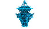


 PDBj
PDBj Assembly
 Sample preparation
 Electron microscopy imaging
 Processing