 Movie
Movie Controller


+ Open data

- Basic information
| Entry | Database: PDB / ID: 8gs3 | ||||||
|---|---|---|---|---|---|---|---|
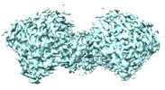
| Title | Cryo-EM structure of human Neuroligin 3 | ||||||
 Components Components | Neuroligin-3 NLGN3 NLGN3 | ||||||
 Keywords Keywords | MEMBRANE PROTEIN / Neuroligin / synapse protein / plasma membrane | ||||||
| Function / homology |  Function and homology information Function and homology informationasymmetric, glutamatergic, excitatory synapse / symmetric, GABA-ergic, inhibitory synapse / rhythmic synaptic transmission / postsynaptic membrane assembly / presynaptic membrane assembly / regulation of respiratory gaseous exchange by nervous system process / neuron cell-cell adhesion / presynapse assembly / neurexin family protein binding / vocalization behavior ...asymmetric, glutamatergic, excitatory synapse / symmetric, GABA-ergic, inhibitory synapse / rhythmic synaptic transmission / postsynaptic membrane assembly / presynaptic membrane assembly / regulation of respiratory gaseous exchange by nervous system process / neuron cell-cell adhesion / presynapse assembly / neurexin family protein binding / vocalization behavior / inhibitory postsynaptic potential / axon extension / Neurexins and neuroligins / positive regulation of synapse assembly / positive regulation of AMPA receptor activity / adult behavior / social behavior / positive regulation of excitatory postsynaptic potential / synaptic vesicle endocytosis / excitatory synapse / endocytic vesicle / cell adhesion molecule binding / synapse assembly / receptor-mediated endocytosis / positive regulation of synaptic transmission, glutamatergic / learning / synapse organization / modulation of chemical synaptic transmission / presynapse / signaling receptor activity / scaffold protein binding / chemical synaptic transmission / postsynaptic membrane / molecular adaptor activity / synapse / cell surface / membrane / plasma membraneSimilarity search - Function | ||||||
| Biological species |  Homo sapiens (human) Homo sapiens (human) | ||||||
| Method | ELECTRON MICROSCOPY / single particle reconstruction / cryo EM / Resolution: 3.9 Å | ||||||
 Authors Authors | Zhang, H. / Zhang, Z. / Hou, M. | ||||||
| Funding support |  China, 1items China, 1items
| ||||||
 Citation Citation | Journal: Front Endocrinol (Lausanne) / Year: 2022 Title: Expression and structural analysis of human neuroligin 2 and neuroligin 3 implicated in autism spectrum disorders. Authors: Zhenzhen Zhang / Mengzhuo Hou / Huaxing Ou / Daping Wang / Zhifang Li / Huawei Zhang / Jianping Lu / Abstract: The development of autism spectrum disorders (ASDs) involves both environmental factors such as maternal diabetes and genetic factors such as neuroligins (NLGNs). NLGN2 and NLGN3 are two members of ...The development of autism spectrum disorders (ASDs) involves both environmental factors such as maternal diabetes and genetic factors such as neuroligins (NLGNs). NLGN2 and NLGN3 are two members of NLGNs with distinct distributions and functions in synapse development and plasticity. The relationship between maternal diabetes and NLGNs, and the distinct working mechanisms of different NLGNs currently remain unclear. Here, we first analyzed the expression levels of NLGN2 and NLGN3 in a streptozotocin-induced ASD mouse model and different brain regions to reveal their differences and similarities. Then, cryogenic electron microscopy (cryo-EM) structures of human NLGN2 and NLGN3 were determined. The overall structures are similar to their homologs in previous reports. However, structural comparisons revealed the relative rotations of two protomers in the homodimers of NLGN2 and NLGN3. Taken together with the previously reported NLGN2-MDGA1 complex, we speculate that the distinct assembly adopted by NLGN2 and NLGN3 may affect their interactions with MDGAs. Our results provide structural insights into the potential distinct mechanisms of NLGN2 and NLGN3 implicated in the development of ASD. | ||||||
| History |
|
- Structure visualization
| Structure viewer | Molecule: MolmilJmol/JSmol |
|---|
- Downloads & links
-Download
| PDBx/mmCIF format | 8gs3.cif.gz | 271.5 KB | Display | PDBx/mmCIF format |
|---|---|---|---|---|
| PDB format | pdb8gs3.ent.gz | 200.6 KB | Display | PDB format |
| PDBx/mmJSON format | 8gs3.json.gz | Tree view | PDBx/mmJSON format | |
| Others |  Other downloads Other downloads |
-Validation report
| Arichive directory | https://data.pdbj.org/pub/pdb/validation_reports/gs/8gs3ftp://data.pdbj.org/pub/pdb/validation_reports/gs/8gs3 | HTTPS FTP |
|---|
-Related structure data
| Related structure data |  34219MC 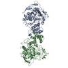 8gs4C M: map data used to model this data C: citing same article ( |
|---|---|
| Similar structure data |
-Links
 PDBj
PDBj

- Assembly
| Deposited unit | 
|
|---|---|
| 1 |
|
-Components
| #1: Protein | NLGN3 / Gliotactin homolog Mass: 93999.062 Da / Num. of mol.: 2 Source method: isolated from a genetically manipulated source Source: (gene. exp.) Homo sapiens (human) / Gene: NLGN3, NL3 / Cell line (production host): HEK293 / Production host: Homo sapiens (human) / References: UniProt: Q9NZ94 |
|---|
-Experimental details
-Experiment
| Experiment | Method: ELECTRON MICROSCOPY |
|---|---|
| EM experiment | Aggregation state: PARTICLE / 3D reconstruction method: single particle reconstruction |
- Sample preparation
| Component | Name: homodimer of Neuroligin 3 / Type: COMPLEX / Entity ID: all / Source: RECOMBINANT |
|---|---|
| Source (natural) | Organism: Homo sapiens (human) |
| Source (recombinant) | Organism: Homo sapiens (human) / Cell: HEK293 |
| Buffer solution | pH: 7.4 |
| Specimen | Embedding applied: NO / Shadowing applied: NO / Staining applied: NO / Vitrification applied: YES |
| Vitrification | Cryogen name: ETHANE |
- Electron microscopy imaging
| Experimental equipment |  Model: Titan Krios / Image courtesy: FEI Company |
|---|---|
| Microscopy | Model: FEI TITAN KRIOS |
| Electron gun | Electron source: FIELD EMISSION GUN / Accelerating voltage: 300 kV / Illumination mode: FLOOD BEAM |
| Electron lens | Mode: BRIGHT FIELDBright-field microscopy / Nominal defocus max: 2500 nm / Nominal defocus min: 1500 nm / Calibrated defocus min: 1500 nm |
| Image recording | Electron dose: 50 e/Å2 / Film or detector model: GATAN K2 SUMMIT (4k x 4k) |
- Processing
| CTF correction | Type: PHASE FLIPPING AND AMPLITUDE CORRECTION | ||||||||||||||||||||||||
|---|---|---|---|---|---|---|---|---|---|---|---|---|---|---|---|---|---|---|---|---|---|---|---|---|---|
| 3D reconstruction | Resolution: 3.9 Å / Resolution method: FSC 0.143 CUT-OFF / Num. of particles: 255939 / Symmetry type: POINT | ||||||||||||||||||||||||
| Refine LS restraints |
|
