 Movie
Movie Controller


+ Open data

- Basic information
| Entry | Database: PDB / ID: 8gf6 | ||||||||||||||||||||||||
|---|---|---|---|---|---|---|---|---|---|---|---|---|---|---|---|---|---|---|---|---|---|---|---|---|---|
| Title | Apo-apo MCR assembly intermediate | ||||||||||||||||||||||||
 Components Components |
| ||||||||||||||||||||||||
 Keywords Keywords |  TRANSFERASE / methanogenesis / MCR complex TRANSFERASE / methanogenesis / MCR complex | ||||||||||||||||||||||||
| Function / homology |  Function and homology informationcoenzyme-B sulfoethylthiotransferase / coenzyme-B sulfoethylthiotransferase activity / methanogenesis / metal ion binding / cytoplasm Function and homology informationcoenzyme-B sulfoethylthiotransferase / coenzyme-B sulfoethylthiotransferase activity / methanogenesis / metal ion binding / cytoplasmSimilarity search - Function | ||||||||||||||||||||||||
| Biological species |  Methanosarcina acetivorans C2A (archaea) Methanosarcina acetivorans C2A (archaea) | ||||||||||||||||||||||||
| Method | ELECTRON MICROSCOPY / single particle reconstruction / cryo EM / Resolution: 3.1 Å | ||||||||||||||||||||||||
 Authors Authors | Joiner, A.M.N. / Chadwick, G.L. / Nayak, D.D. | ||||||||||||||||||||||||
| Funding support |  United States, 7items United States, 7items
| ||||||||||||||||||||||||
 Citation Citation | Journal: Proc Natl Acad Sci U S A / Year: 2023 Title: McrD binds asymmetrically to methyl-coenzyme M reductase improving active-site accessibility during assembly. Authors: Grayson L Chadwick / Aaron M N Joiner / Sangeetha Ramesh / Douglas A Mitchell / Dipti D Nayak / Abstract: Methyl-coenzyme M reductase (MCR) catalyzes the formation of methane, and its activity accounts for nearly all biologically produced methane released into the atmosphere. The assembly of MCR is an ...Methyl-coenzyme M reductase (MCR) catalyzes the formation of methane, and its activity accounts for nearly all biologically produced methane released into the atmosphere. The assembly of MCR is an intricate process involving the installation of a complex set of posttranslational modifications and the unique Ni-containing tetrapyrrole called coenzyme F. Despite decades of research, details of MCR assembly remain largely unresolved. Here, we report the structural characterization of MCR in two intermediate states of assembly. These intermediate states lack one or both F cofactors and form complexes with the previously uncharacterized McrD protein. McrD is found to bind asymmetrically to MCR, displacing large regions of the alpha subunit and increasing active-site accessibility for the installation of F-shedding light on the assembly of MCR and the role of McrD therein. This work offers crucial information for the expression of MCR in a heterologous host and provides targets for the design of MCR inhibitors. | ||||||||||||||||||||||||
| History |
|
- Structure visualization
| Structure viewer | Molecule: MolmilJmol/JSmol |
|---|
- Downloads & links
-Download
| PDBx/mmCIF format | 8gf6.cif.gz | 401.6 KB | Display | PDBx/mmCIF format |
|---|---|---|---|---|
| PDB format | pdb8gf6.ent.gz | 331.3 KB | Display | PDB format |
| PDBx/mmJSON format | 8gf6.json.gz | Tree view | PDBx/mmJSON format | |
| Others |  Other downloads Other downloads |
-Validation report
| Arichive directory | https://data.pdbj.org/pub/pdb/validation_reports/gf/8gf6ftp://data.pdbj.org/pub/pdb/validation_reports/gf/8gf6 | HTTPS FTP |
|---|
-Related structure data
| Related structure data |  29979MC  8gf5C M: map data used to model this data C: citing same article ( |
|---|---|
| Similar structure data |

-Links
 PDBj
PDBj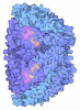
- Assembly
| Deposited unit | 
|
|---|---|
| 1 |
|
-Components
| #1: Protein | Coenzyme-B sulfoethylthiotransferase / Coenzyme-B sulfoethylthiotransferase alpha Mass: 62180.078 Da / Num. of mol.: 2 / Source method: isolated from a natural source / Source: (natural) Methanosarcina acetivorans C2A (archaea)References: UniProt: Q8THH1, coenzyme-B sulfoethylthiotransferase#2: Protein | Coenzyme-B sulfoethylthiotransferaseMass: 45174.070 Da / Num. of mol.: 2 / Source method: isolated from a natural source / Source: (natural) Methanosarcina acetivorans C2A (archaea) / References: UniProt: Q8THG7#3: Protein | Coenzyme-B sulfoethylthiotransferaseMass: 27630.184 Da / Num. of mol.: 2 / Source method: isolated from a natural source / Source: (natural) Methanosarcina acetivorans C2A (archaea) / References: UniProt: Q8THH0#4: Protein | | Coenzyme-B sulfoethylthiotransferaseMass: 21842.742 Da / Num. of mol.: 1 Source method: isolated from a genetically manipulated source Source: (gene. exp.) Methanosarcina acetivorans C2A (archaea)Gene: mcrD / Production host: Methanosarcina acetivorans C2A (archaea) / References: UniProt: Q8THG8Has ligand of interest | N | |
|---|
-Experimental details
-Experiment
| Experiment | Method: ELECTRON MICROSCOPY |
|---|---|
| EM experiment | Aggregation state: PARTICLE / 3D reconstruction method: single particle reconstruction |
- Sample preparation
| Component | Name: Assembly intermediate of the MCR complex bound to mcrD Type: COMPLEX / Entity ID: all / Source: NATURAL | |||||||||||||||
|---|---|---|---|---|---|---|---|---|---|---|---|---|---|---|---|---|
| Source (natural) | Organism: Methanosarcina acetivorans C2A (archaea) | |||||||||||||||
| Buffer solution | pH: 8 | |||||||||||||||
| Buffer component |
| |||||||||||||||
| Specimen | Conc.: 0.55 mg/ml / Embedding applied: NO / Shadowing applied: NO / Staining applied: NO / Vitrification applied: YES | |||||||||||||||
| Specimen support | Details: 25mA / Grid material: COPPER / Grid mesh size: 300 divisions/in. / Grid type: Quantifoil R2/1 | |||||||||||||||
| Vitrification | Instrument: FEI VITROBOT MARK IV / Cryogen name: ETHANE / Humidity: 100 % / Chamber temperature: 277 K / Details: Vitrification occurred under aerobic conditions |
- Electron microscopy imaging
| Experimental equipment |  Model: Talos Arctica / Image courtesy: FEI Company |
|---|---|
| Microscopy | Model: FEI TALOS ARCTICA |
| Electron gun | Electron source: FIELD EMISSION GUN / Accelerating voltage: 200 kV / Illumination mode: FLOOD BEAM |
| Electron lens | Mode: BRIGHT FIELDBright-field microscopy / Nominal magnification: 36000 X / Nominal defocus max: 2000 nm / Nominal defocus min: 800 nm |
| Specimen holder | Cryogen: NITROGEN / Specimen holder model: FEI TITAN KRIOS AUTOGRID HOLDER |
| Image recording | Electron dose: 50 e/Å2 / Film or detector model: GATAN K3 (6k x 4k) |
- Processing
| EM software | Name: SerialEM / Category: image acquisition | ||||||||||||||||||||||||
|---|---|---|---|---|---|---|---|---|---|---|---|---|---|---|---|---|---|---|---|---|---|---|---|---|---|
| CTF correction | Type: PHASE FLIPPING AND AMPLITUDE CORRECTION | ||||||||||||||||||||||||
| 3D reconstruction | Resolution: 3.1 Å / Resolution method: FSC 0.143 CUT-OFF / Num. of particles: 94000 / Symmetry type: POINT | ||||||||||||||||||||||||
| Refine LS restraints |
|
