 Movie
Movie Controller


+ Open data

- Basic information
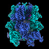
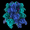
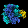
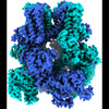
| Entry | Database: PDB / ID: 8ddr | ||||||
|---|---|---|---|---|---|---|---|
| Title | cryo-EM structure of TRPM3 ion channel in the absence of PIP2 | ||||||
 Components Components |
| ||||||
 Keywords Keywords |  MEMBRANE PROTEIN / TRPM3 / ion channel / PIP2 MEMBRANE PROTEIN / TRPM3 / ion channel / PIP2 | ||||||
| Function / homology |  Function and homology information Function and homology informationmonoatomic cation transmembrane transport / monoatomic cation transport / monoatomic cation channel activity / protein tetramerization / calcium ion transmembrane transport / calcium channel activity / membraneSimilarity search - Function | ||||||
| Biological species |  Mus musculus (house mouse) Mus musculus (house mouse) | ||||||
| Method | ELECTRON MICROSCOPY / single particle reconstruction / cryo EM / Resolution: 3.2 Å | ||||||
 Authors Authors | Zhao, C. / MacKinnon, R. | ||||||
| Funding support |  United States, 1items United States, 1items
| ||||||
 Citation Citation | Journal: Neuron / Year: 2023 Title: Structural and functional analyses of a GPCR-inhibited ion channel TRPM3. Authors: Chen Zhao / Roderick MacKinnon / Abstract: G-protein coupled receptors (GPCRs) govern the physiological response to stimuli by modulating the activity of downstream effectors, including ion channels. TRPM3 is an ion channel inhibited by GPCRs ...G-protein coupled receptors (GPCRs) govern the physiological response to stimuli by modulating the activity of downstream effectors, including ion channels. TRPM3 is an ion channel inhibited by GPCRs through direct interaction with G protein (Gβγ) released upon their activation. This GPCR-TRPM3 signaling pathway contributes to the analgesic effect of morphine. Here, we characterized Gβγ inhibition of TRPM3 using electrophysiology and single particle cryo-electron microscopy (cryo-EM). From electrophysiology, we obtained a half inhibition constant (IC50) of ∼240 nM. Using cryo-EM, we determined structures of mouse TRPM3 expressed in human cells with and without Gβγ and with and without PIP, a lipid required for TRPM3 activity, at resolutions of 2.7-4.7 Å. Gβγ-TRPM3 interfaces vary depending on PIP occupancy; however, in all cases, Gβγ appears loosely attached to TRPM3. The IC50 in electrophysiology experiments raises the possibility that additional unknown factors may stabilize the TRPM3-Gβγ complex. | ||||||
| History |
|
- Structure visualization
| Structure viewer | Molecule: MolmilJmol/JSmol |
|---|
- Downloads & links
-Download
| PDBx/mmCIF format | 8ddr.cif.gz | 709.5 KB | Display | PDBx/mmCIF format |
|---|---|---|---|---|
| PDB format | pdb8ddr.ent.gz | 577.5 KB | Display | PDB format |
| PDBx/mmJSON format | 8ddr.json.gz | Tree view | PDBx/mmJSON format | |
| Others |  Other downloads Other downloads |
-Validation report
| Arichive directory | https://data.pdbj.org/pub/pdb/validation_reports/dd/8ddrftp://data.pdbj.org/pub/pdb/validation_reports/dd/8ddr | HTTPS FTP |
|---|
-Related structure data
| Related structure data | 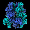 27339MC  8ddqC  8ddsC  8ddtC 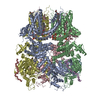 8dduC 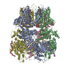 8ddvC  8ddwC  8ddxC 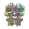 8ed7C  8ed8C 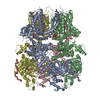 8ed9C C: citing same article ( M: map data used to model this data |
|---|---|
| Similar structure data |


-Links
 PDBj
PDBj- Assembly
| Deposited unit | 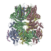
|
|---|---|
| 1 |
|
-Components
| #1: Protein | Mass: 154649.328 Da / Num. of mol.: 4 Source method: isolated from a genetically manipulated source Source: (gene. exp.) Mus musculus (house mouse) / Gene: Trpm3 / Production host:  Homo sapiens (human) / References: UniProt: Q5F4S7 Homo sapiens (human) / References: UniProt: Q5F4S7#2: Protein/peptide | Mass: 1464.797 Da / Num. of mol.: 4 Source method: isolated from a genetically manipulated source Details: a segment at the N-terminus of TRPM3 whose sequence cannot be identified from the cryo-EM density Source: (gene. exp.) Mus musculus (house mouse) / Production host: Homo sapiens (human)#3: Chemical | ChemComp-3PH / Phosphatidic acid  Mass: 704.998 Da / Num. of mol.: 4 / Source method: obtained synthetically / Formula: C39H77O8P Mass: 704.998 Da / Num. of mol.: 4 / Source method: obtained synthetically / Formula: C39H77O8P#4: Chemical | ChemComp-9Z9 / ( 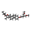  Mass: 544.805 Da / Num. of mol.: 4 / Source method: obtained synthetically / Formula: C34H56O5 / Comment: detergent*YM Mass: 544.805 Da / Num. of mol.: 4 / Source method: obtained synthetically / Formula: C34H56O5 / Comment: detergent*YM#5: Chemical |   Mass: 22.990 Da / Num. of mol.: 2 / Source method: obtained synthetically / Formula: Na Mass: 22.990 Da / Num. of mol.: 2 / Source method: obtained synthetically / Formula: NaHas ligand of interest | N | |
|---|
-Experimental details
-Experiment
| Experiment | Method: ELECTRON MICROSCOPY |
|---|---|
| EM experiment | Aggregation state: PARTICLE / 3D reconstruction method: single particle reconstruction |
- Sample preparation
| Component | Name: TRPM3 / Type: COMPLEX / Entity ID: #1-#2 / Source: RECOMBINANT |
|---|---|
| Molecular weight | Experimental value: NO |
| Source (natural) | Organism: Mus musculus (house mouse) |
| Source (recombinant) | Organism: Homo sapiens (human) |
| Buffer solution | pH: 8.5 |
| Specimen | Embedding applied: NO / Shadowing applied: NO / Staining applied: NO / Vitrification applied: YES |
| Vitrification | Cryogen name: ETHANE |
- Electron microscopy imaging
| Experimental equipment |  Model: Titan Krios / Image courtesy: FEI Company |
|---|---|
| Microscopy | Model: FEI TITAN KRIOS |
| Electron gun | Electron source: FIELD EMISSION GUN / Accelerating voltage: 300 kV / Illumination mode: FLOOD BEAM |
| Electron lens | Mode: BRIGHT FIELDBright-field microscopy / Nominal defocus max: 2500 nm / Nominal defocus min: 1000 nm |
| Image recording | Electron dose: 73 e/Å2 / Film or detector model: GATAN K2 IS (4k x 4k) |
- Processing
| Software | Name: PHENIX / Version: 1.20.1_4487: / Classification: refinement | ||||||||||||||||||||||||
|---|---|---|---|---|---|---|---|---|---|---|---|---|---|---|---|---|---|---|---|---|---|---|---|---|---|
| CTF correction | Type: PHASE FLIPPING AND AMPLITUDE CORRECTION | ||||||||||||||||||||||||
| Symmetry | Point symmetry: C4 (4 fold cyclic) | ||||||||||||||||||||||||
| 3D reconstruction | Resolution: 3.2 Å / Resolution method: FSC 0.143 CUT-OFF / Num. of particles: 32631 / Symmetry type: POINT | ||||||||||||||||||||||||
| Refine LS restraints |
|
