 ムービー
ムービー コントローラー


+ データを開く

- 基本情報
基本情報
| 登録情報 | データベース: PDB / ID: 8bka | ||||||||||||
|---|---|---|---|---|---|---|---|---|---|---|---|---|---|
| タイトル | Cryo-EM structure of mouse heavy-chain apoferritin at 2.7 A plunged 35ms after mixing with b-galactosidase | ||||||||||||
 要素 要素 | Ferritin heavy chain フェリチン フェリチン | ||||||||||||
 キーワード キーワード | METAL BINDING PROTEIN / iron storage / ferritin (フェリチン) / octahedral | ||||||||||||
| 機能・相同性 |  機能・相同性情報 機能・相同性情報Iron uptake and transport / Golgi Associated Vesicle Biogenesis / iron ion sequestering activity / オートファゴソーム / ferroxidase / ferroxidase activity / intracellular sequestering of iron ion / negative regulation of fibroblast proliferation / endocytic vesicle lumen / Neutrophil degranulation ...Iron uptake and transport / Golgi Associated Vesicle Biogenesis / iron ion sequestering activity / オートファゴソーム / ferroxidase / ferroxidase activity / intracellular sequestering of iron ion / negative regulation of fibroblast proliferation / endocytic vesicle lumen / Neutrophil degranulation / ferric iron binding / ferrous iron binding / iron ion transport / 免疫応答 / iron ion binding / negative regulation of cell population proliferation / ミトコンドリア / extracellular region / identical protein binding / 細胞質類似検索 - 分子機能 | ||||||||||||
| 生物種 |  Mus musculus (ハツカネズミ) Mus musculus (ハツカネズミ) | ||||||||||||
| 手法 | 電子顕微鏡法 / 単粒子再構成法 / クライオ電子顕微鏡法 / 解像度: 2.7 Å | ||||||||||||
 データ登録者 データ登録者 | Torino, S. / Dhurandhar, M. / Efremov, R. | ||||||||||||
| 資金援助 | European Union,  ベルギー, 3件 ベルギー, 3件
| ||||||||||||
 引用 引用 | ジャーナル: Nat Methods / 年: 2023 タイトル: Time-resolved cryo-EM using a combination of droplet microfluidics with on-demand jetting. 著者: Stefania Torino / Mugdha Dhurandhar / Annelore Stroobants / Raf Claessens / Rouslan G Efremov / 要旨: Single-particle cryogenic electron microscopy (cryo-EM) allows reconstruction of high-resolution structures of proteins in different conformations. Protein function often involves transient ...Single-particle cryogenic electron microscopy (cryo-EM) allows reconstruction of high-resolution structures of proteins in different conformations. Protein function often involves transient functional conformations, which can be resolved using time-resolved cryo-EM (trEM). In trEM, reactions are arrested after a defined delay time by rapid vitrification of protein solution on the EM grid. Despite the increasing interest in trEM among the cryo-EM community, making trEM samples with a time resolution below 100 ms remains challenging. Here we report the design and the realization of a time-resolved cryo-plunger that combines a droplet-based microfluidic mixer with a laser-induced generator of microjets that allows rapid reaction initiation and plunge-freezing of cryo-EM grids. Using this approach, a time resolution of 5 ms was achieved and the protein density map was reconstructed to a resolution of 2.1 Å. trEM experiments on GroEL:GroES chaperonin complex resolved the kinetics of the complex formation and visualized putative short-lived conformations of GroEL-ATP complex. | ||||||||||||
| 履歴 |
|
- 構造の表示
構造の表示
| 構造ビューア | 分子: MolmilJmol/JSmol |
|---|
- ダウンロードとリンク
-ダウンロード
| PDBx/mmCIF形式 | 8bka.cif.gz | 845.6 KB | 表示 | PDBx/mmCIF形式 |
|---|---|---|---|---|
| PDB形式 | pdb8bka.ent.gz | 702.5 KB | 表示 | PDB形式 |
| PDBx/mmJSON形式 | 8bka.json.gz | ツリー表示 | PDBx/mmJSON形式 | |
| その他 |  その他のダウンロード その他のダウンロード |
-検証レポート
| アーカイブディレクトリ | https://data.pdbj.org/pub/pdb/validation_reports/bk/8bkaftp://data.pdbj.org/pub/pdb/validation_reports/bk/8bka | HTTPS FTP |
|---|
-関連構造データ
| 関連構造データ |  16094MC  8bk7C  8bk8C  8bk9C  8bkbC  8bkgC  8bkzC  8bl2C  8bl7C  8blcC  8bldC  8bleC  8blfC  8blyC  8bm0C  8bm1C  8bmdC  8bmoC  8bmtC M: このデータのモデリングに利用したマップデータ C: 同じ文献を引用 ( |
|---|---|
| 類似構造データ |


-リンク
 PDBj
PDBj


- 集合体
集合体
| 登録構造単位 | 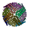
|
|---|---|
| 1 |
|
-要素
| #1: タンパク質 | フェリチン / Ferritin H subunit 分子量: 21097.631 Da / 分子数: 24 / 由来タイプ: 組換発現 / 由来: (組換発現) Mus musculus (ハツカネズミ) / 遺伝子: Fth1, Fth / 発現宿主:  Escherichia coli (大腸菌) / 参照: UniProt: P09528, ferroxidase Escherichia coli (大腸菌) / 参照: UniProt: P09528, ferroxidase#2: 化合物 | ChemComp-FE / 鉄  分子量: 55.845 Da / 分子数: 6 / 由来タイプ: 合成 / 式: Fe 分子量: 55.845 Da / 分子数: 6 / 由来タイプ: 合成 / 式: Fe#3: 水 | ChemComp-HOH / | 水 分子量: 18.015 Da / 分子数: 2271 / 由来タイプ: 天然 / 式: H2O 分子量: 18.015 Da / 分子数: 2271 / 由来タイプ: 天然 / 式: H2O研究の焦点であるリガンドがあるか | N | |
|---|
-実験情報
-実験
| 実験 | 手法: 電子顕微鏡法 |
|---|---|
| EM実験 | 試料の集合状態: PARTICLE / 3次元再構成法: 単粒子再構成法 |
- 試料調製
試料調製
| 構成要素 | 名称: Mouse heavy chain apoferritin from E.coli / タイプ: COMPLEX / Entity ID: #1 / 由来: RECOMBINANT | ||||||||||||||||||||
|---|---|---|---|---|---|---|---|---|---|---|---|---|---|---|---|---|---|---|---|---|---|
| 由来(天然) | 生物種: Mus musculus (ハツカネズミ) | ||||||||||||||||||||
| 由来(組換発現) | 生物種: Escherichia coli (大腸菌) | ||||||||||||||||||||
| 緩衝液 | pH: 7.5 / 詳細: contains Amaranth dye (acid red 27) 32 mM | ||||||||||||||||||||
| 緩衝液成分 |
| ||||||||||||||||||||
| 試料 | 濃度: 3 mg/ml / 包埋: NO / シャドウイング: NO / 染色: NO / 凍結: YES 詳細: Apoferritin in buffer of Amaranth dye (acid red 27 concentration 32 mM) | ||||||||||||||||||||
| 試料支持 | グリッドの材料: COPPER / グリッドのサイズ: 300 divisions/in. / グリッドのタイプ: Quantifoil R2/1 | ||||||||||||||||||||
| 急速凍結 | 凍結剤: ETHANE |
- 電子顕微鏡撮影
| 顕微鏡 | モデル: JEOL CRYO ARM 300 |
|---|---|
| 電子銃 | 電子線源: FIELD EMISSION GUN / 加速電圧: 300 kV / 照射モード: FLOOD BEAM |
| 電子レンズ | モード: BRIGHT FIELDBright-field microscopy / 倍率(公称値): 60000 X / 最大 デフォーカス(公称値): 3500 nm / 最小 デフォーカス(公称値): 500 nm / Cs: 2.55 mm / アライメント法: COMA FREE |
| 試料ホルダ | 凍結剤: NITROGEN / 試料ホルダーモデル: JEOL CRYOSPECPORTER |
| 撮影 | 平均露光時間: 2.796 sec. / 電子線照射量: 59 e/Å2 / フィルム・検出器のモデル: GATAN K3 (6k x 4k) / 撮影したグリッド数: 2 / 実像数: 722 |
| 電子光学装置 | エネルギーフィルター名称: In-column Omega Filter エネルギーフィルタースリット幅: 20 eV |
- 解析
解析
| ソフトウェア | 名称: PHENIX / バージョン: 1.20.1_4487: / 分類: 精密化 | ||||||||||||||||||||||||
|---|---|---|---|---|---|---|---|---|---|---|---|---|---|---|---|---|---|---|---|---|---|---|---|---|---|
| CTF補正 | タイプ: PHASE FLIPPING AND AMPLITUDE CORRECTION | ||||||||||||||||||||||||
| 3次元再構成 | 解像度: 2.7 Å / 解像度の算出法: FSC 0.143 CUT-OFF / 粒子像の数: 7432 / 対称性のタイプ: POINT | ||||||||||||||||||||||||
| 拘束条件 |
|
