 Movie
Movie Controller


+ Open data

- Basic information
| Entry | Database: PDB / ID: 8au1 | ||||||
|---|---|---|---|---|---|---|---|
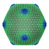
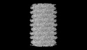
| Title | Jumbo Phage phi-kp24 tail outer sheath | ||||||
 Components Components | Putative tail sheath protein | ||||||
 Keywords Keywords |  STRUCTURAL PROTEIN / Jumbo Phage / Klebsiella pneumoniae / tail / sheath STRUCTURAL PROTEIN / Jumbo Phage / Klebsiella pneumoniae / tail / sheath | ||||||
| Function / homology | Putative tail sheath protein Function and homology information Function and homology information | ||||||
| Biological species |  Klebsiella phage vB_KpM_FBKp24 (virus) Klebsiella phage vB_KpM_FBKp24 (virus) | ||||||
| Method | ELECTRON MICROSCOPY / helical reconstruction / cryo EM / Resolution: 3 Å | ||||||
 Authors Authors | Ouyang, R. / Briegel, A. | ||||||
| Funding support |  Netherlands, 1items Netherlands, 1items
| ||||||
 Citation Citation | Journal: Nat Commun / Year: 2022 Title: High-resolution reconstruction of a Jumbo-bacteriophage infecting capsulated bacteria using hyperbranched tail fibers. Authors: Ruochen Ouyang / Ana Rita Costa / C Keith Cassidy / Aleksandra Otwinowska / Vera C J Williams / Agnieszka Latka / Phill J Stansfeld / Zuzanna Drulis-Kawa / Yves Briers / Daniël M Pelt / ...Authors: Ruochen Ouyang / Ana Rita Costa / C Keith Cassidy / Aleksandra Otwinowska / Vera C J Williams / Agnieszka Latka / Phill J Stansfeld / Zuzanna Drulis-Kawa / Yves Briers / Daniël M Pelt / Stan J J Brouns / Ariane Briegel /     Abstract: The Klebsiella jumbo myophage ϕKp24 displays an unusually complex arrangement of tail fibers interacting with a host cell. In this study, we combine cryo-electron microscopy methods, protein ...The Klebsiella jumbo myophage ϕKp24 displays an unusually complex arrangement of tail fibers interacting with a host cell. In this study, we combine cryo-electron microscopy methods, protein structure prediction methods, molecular simulations, microbiological and machine learning approaches to explore the capsid, tail, and tail fibers of ϕKp24. We determine the structure of the capsid and tail at 4.1 Å and 3.0 Å resolution. We observe the tail fibers are branched and rearranged dramatically upon cell surface attachment. This complex configuration involves fourteen putative tail fibers with depolymerase activity that provide ϕKp24 with the ability to infect a broad panel of capsular polysaccharide (CPS) types of Klebsiella pneumoniae. Our study provides structural and functional insight into how ϕKp24 adapts to the variable surfaces of capsulated bacterial pathogens, which is useful for the development of phage therapy approaches against pan-drug resistant K. pneumoniae strains. | ||||||
| History |
|
- Structure visualization
| Structure viewer | Molecule: MolmilJmol/JSmol |
|---|
- Downloads & links
-Download
| PDBx/mmCIF format | 8au1.cif.gz | 3.7 MB | Display | PDBx/mmCIF format |
|---|---|---|---|---|
| PDB format | pdb8au1.ent.gz | Display | PDB format | |
| PDBx/mmJSON format | 8au1.json.gz | Tree view | PDBx/mmJSON format | |
| Others |  Other downloads Other downloads |
-Validation report
| Arichive directory | https://data.pdbj.org/pub/pdb/validation_reports/au/8au1ftp://data.pdbj.org/pub/pdb/validation_reports/au/8au1 | HTTPS FTP |
|---|
-Related structure data
| Related structure data | 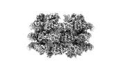 15669MC 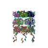 8bfkC 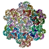 8bflC 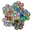 8bfpC  15776 8b09 8b33 8b36 M: map data used to model this data C: citing same article ( |
|---|---|
| Similar structure data |

-Links
 PDBj
PDBj- Assembly
| Deposited unit | 
|
|---|---|
| 1 |
|
-Components
| #1: Protein | Mass: 76402.500 Da / Num. of mol.: 18 / Source method: isolated from a natural source / Source: (natural) Klebsiella phage vB_KpM_FBKp24 (virus) / References: UniProt: A0A7U0GB71 |
|---|
-Experimental details
-Experiment
| Experiment | Method: ELECTRON MICROSCOPY |
|---|---|
| EM experiment | Aggregation state: PARTICLE / 3D reconstruction method: helical reconstruction |
- Sample preparation
| Component | Name: Jumbo Phage phi-kp24 tail outer sheath / Type: COMPLEX Details: The outer sheath in extension of Klebsiella Phage phi-kp24 Entity ID: all / Source: NATURAL |
|---|---|
| Molecular weight | Experimental value: NO |
| Source (natural) | Organism: Klebsiella phage vB_KpM_FBKp24 (virus) |
| Details of virus | Empty: NO / Enveloped: YES / Isolate: OTHER |
| Buffer solution | pH: 7 |
| Specimen | Embedding applied: NO / Shadowing applied: NO / Staining applied: NO / Vitrification applied: YES |
| Specimen support | Grid type: Quantifoil R2/2 |
| Vitrification | Cryogen name: ETHANE |
- Electron microscopy imaging
| Experimental equipment |  Model: Titan Krios / Image courtesy: FEI Company |
|---|---|
| Microscopy | Model: FEI TITAN KRIOS |
| Electron gun | Electron source: OTHER / Accelerating voltage: 300 kV / Illumination mode: OTHER |
| Electron lens | Mode: OTHER / Nominal defocus max: 5000 nm / Nominal defocus min: 1000 nm |
| Image recording | Electron dose: 30 e/Å2 / Film or detector model: GATAN K3 BIOQUANTUM (6k x 4k) |
- Processing
| Software |
| ||||||||||||||||||||||||
|---|---|---|---|---|---|---|---|---|---|---|---|---|---|---|---|---|---|---|---|---|---|---|---|---|---|
| EM software |
| ||||||||||||||||||||||||
| CTF correction | Type: PHASE FLIPPING AND AMPLITUDE CORRECTION | ||||||||||||||||||||||||
| Helical symmerty | Angular rotation/subunit: 20.89 ° / Axial rise/subunit: 39.03 Å / Axial symmetry: C6 | ||||||||||||||||||||||||
| 3D reconstruction | Resolution: 3 Å / Resolution method: FSC 0.143 CUT-OFF / Num. of particles: 81869 / Symmetry type: HELICAL | ||||||||||||||||||||||||
| Atomic model building | Protocol: FLEXIBLE FIT / Space: REAL | ||||||||||||||||||||||||
| Refinement | Cross valid method: NONE Stereochemistry target values: GeoStd + Monomer Library + CDL v1.2 | ||||||||||||||||||||||||
| Displacement parameters | Biso mean: 67.82 Å2 | ||||||||||||||||||||||||
| Refine LS restraints |
|
