Movie
Movie Controller


+ Open data

- Basic information
| Entry | Database: PDB / ID: 8adn | |||||||||
|---|---|---|---|---|---|---|---|---|---|---|
| Title | Vairimorpha necatrix 20S proteasome from spores | |||||||||
 Components Components |
| |||||||||
 Keywords Keywords |  HYDROLASE / Peptidase Activity / Reductive Evolution / Bound Peptide / Microsporidia HYDROLASE / Peptidase Activity / Reductive Evolution / Bound Peptide / Microsporidia | |||||||||
| Biological species |  Vairimorpha necatrix (fungus) Vairimorpha necatrix (fungus) | |||||||||
| Method | ELECTRON MICROSCOPY / single particle reconstruction / cryo EM / Resolution: 2.77 Å | |||||||||
 Authors Authors | Jespersen, N. / Ehrenbolger, K. / Winiger, R. / Svedberg, D. / Vossbrinck, C.R. / Barandun, J. | |||||||||
| Funding support |  Sweden, European Union, 2items Sweden, European Union, 2items
| |||||||||
 Citation Citation | Journal: Nat Commun / Year: 2022 Title: Structure of the reduced microsporidian proteasome bound by PI31-like peptides in dormant spores. Authors: Nathan Jespersen / Kai Ehrenbolger / Rahel R Winiger / Dennis Svedberg / Charles R Vossbrinck / Jonas Barandun /  Abstract: Proteasomes play an essential role in the life cycle of intracellular pathogens with extracellular stages by ensuring proteostasis in environments with limited resources. In microsporidia, divergent ...Proteasomes play an essential role in the life cycle of intracellular pathogens with extracellular stages by ensuring proteostasis in environments with limited resources. In microsporidia, divergent parasites with extraordinarily streamlined genomes, the proteasome complexity and structure are unknown, which limits our understanding of how these unique pathogens adapt and compact essential eukaryotic complexes. We present cryo-electron microscopy structures of the microsporidian 20S and 26S proteasome isolated from dormant or germinated Vairimorpha necatrix spores. The discovery of PI31-like peptides, known to inhibit proteasome activity, bound simultaneously to all six active sites within the central cavity of the dormant spore proteasome, suggests reduced activity in the environmental stage. In contrast, the absence of the PI31-like peptides and the existence of 26S particles post-germination in the presence of ATP indicates that proteasomes are reactivated in nutrient-rich conditions. Structural and phylogenetic analyses reveal that microsporidian proteasomes have undergone extensive reductive evolution, lost at least two regulatory proteins, and compacted nearly every subunit. The highly derived structure of the microsporidian proteasome, and the minimized version of PI31 presented here, reinforce the feasibility of the development of specific inhibitors and provide insight into the unique evolution and biology of these medically and economically important pathogens. | |||||||||
| History |
|
- Structure visualization
| Structure viewer | Molecule:  MolmilJmol/JSmol MolmilJmol/JSmol |
|---|
- Downloads & links
-Download
| PDBx/mmCIF format | 8adn.cif.gz | 1.1 MB | Display | PDBx/mmCIF format |
|---|---|---|---|---|
| PDB format | pdb8adn.ent.gz | 960.5 KB | Display | PDB format |
| PDBx/mmJSON format | 8adn.json.gz | Tree view | PDBx/mmJSON format | |
| Others |  Other downloads Other downloads |
-Validation report
| Arichive directory | https://data.pdbj.org/pub/pdb/validation_reports/ad/8adnftp://data.pdbj.org/pub/pdb/validation_reports/ad/8adn | HTTPS FTP |
|---|
-Related structure data
-Links
 PDBj
PDBj- Assembly
| Deposited unit | 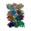
|
|---|---|
| 1 |
|
-Components
-Protein , 1 types, 2 molecules 34
| #1: Protein | Mass: 17070.094 Da / Num. of mol.: 2 Source method: isolated from a genetically manipulated source Source: (gene. exp.) Vairimorpha necatrix (fungus) / Production host: Vairimorpha necatrix (fungus) |
|---|
-Proteasome subunit alpha type- ... , 7 types, 14 molecules AOBPCQDRESFTGU
| #2: Protein | Mass: 25849.268 Da / Num. of mol.: 2 Source method: isolated from a genetically manipulated source Source: (gene. exp.) Vairimorpha necatrix (fungus) / Production host: Vairimorpha necatrix (fungus)#3: Protein | Mass: 26848.580 Da / Num. of mol.: 2 Source method: isolated from a genetically manipulated source Source: (gene. exp.) Vairimorpha necatrix (fungus) / Production host: Vairimorpha necatrix (fungus)#4: Protein | Mass: 25596.154 Da / Num. of mol.: 2 Source method: isolated from a genetically manipulated source Source: (gene. exp.) Vairimorpha necatrix (fungus) / Production host: Vairimorpha necatrix (fungus)#5: Protein | Mass: 26520.125 Da / Num. of mol.: 2 Source method: isolated from a genetically manipulated source Source: (gene. exp.) Vairimorpha necatrix (fungus) / Production host: Vairimorpha necatrix (fungus)#6: Protein | Mass: 26297.920 Da / Num. of mol.: 2 Source method: isolated from a genetically manipulated source Source: (gene. exp.) Vairimorpha necatrix (fungus) / Production host: Vairimorpha necatrix (fungus)#7: Protein | Mass: 27835.818 Da / Num. of mol.: 2 Source method: isolated from a genetically manipulated source Source: (gene. exp.) Vairimorpha necatrix (fungus) / Production host: Vairimorpha necatrix (fungus)#8: Protein | Mass: 26308.912 Da / Num. of mol.: 2 Source method: isolated from a genetically manipulated source Source: (gene. exp.) Vairimorpha necatrix (fungus) / Production host: Vairimorpha necatrix (fungus) |
|---|
-Proteasome subunit beta type- ... , 7 types, 14 molecules HVIWJXKYLZMaNb
| #9: Protein | PSMB2 Mass: 24776.287 Da / Num. of mol.: 2 Source method: isolated from a genetically manipulated source Source: (gene. exp.) Vairimorpha necatrix (fungus) / Production host: Vairimorpha necatrix (fungus)#10: Protein | PSMB3Mass: 23016.334 Da / Num. of mol.: 2 Source method: isolated from a genetically manipulated source Source: (gene. exp.) Vairimorpha necatrix (fungus) / Production host: Vairimorpha necatrix (fungus)#11: Protein | PSMB4Mass: 21945.895 Da / Num. of mol.: 2 Source method: isolated from a genetically manipulated source Source: (gene. exp.) Vairimorpha necatrix (fungus) / Production host: Vairimorpha necatrix (fungus)#12: Protein | PSMB5Mass: 25290.760 Da / Num. of mol.: 2 Source method: isolated from a genetically manipulated source Source: (gene. exp.) Vairimorpha necatrix (fungus) / Production host: Vairimorpha necatrix (fungus)#13: Protein | Mass: 34081.102 Da / Num. of mol.: 2 Source method: isolated from a genetically manipulated source Source: (gene. exp.) Vairimorpha necatrix (fungus) / Production host: Vairimorpha necatrix (fungus)#14: Protein | Mass: 23806.217 Da / Num. of mol.: 2 Source method: isolated from a genetically manipulated source Source: (gene. exp.) Vairimorpha necatrix (fungus) / Production host: Vairimorpha necatrix (fungus)#15: Protein | PSMB1Mass: 23654.027 Da / Num. of mol.: 2 Source method: isolated from a genetically manipulated source Source: (gene. exp.) Vairimorpha necatrix (fungus) / Production host: Vairimorpha necatrix (fungus) |
|---|
-Experimental details
-Experiment
| Experiment | Method: ELECTRON MICROSCOPY |
|---|---|
| EM experiment | Aggregation state: PARTICLE / 3D reconstruction method: single particle reconstruction |
- Sample preparation
| Component | Name: 20S proteasomeProteasome / Type: COMPLEX / Details: Purified from spores / Entity ID: all / Source: NATURAL | ||||||||||||||||||||
|---|---|---|---|---|---|---|---|---|---|---|---|---|---|---|---|---|---|---|---|---|---|
| Source (natural) | Organism: Vairimorpha necatrix (fungus) | ||||||||||||||||||||
| Buffer solution | pH: 7.4 | ||||||||||||||||||||
| Buffer component |
| ||||||||||||||||||||
| Specimen | Embedding applied: NO / Shadowing applied: NO / Staining applied: NO / Vitrification applied: YES | ||||||||||||||||||||
| Specimen support | Details: 15 mA / Grid material: COPPER / Grid mesh size: 200 divisions/in. / Grid type: Quantifoil R2/1 | ||||||||||||||||||||
| Vitrification | Instrument: FEI VITROBOT MARK IV / Cryogen name: ETHANE / Humidity: 100 % / Chamber temperature: 277 K |
- Electron microscopy imaging
| Experimental equipment |  Model: Titan Krios / Image courtesy: FEI Company | |||||||||||||||||||||
|---|---|---|---|---|---|---|---|---|---|---|---|---|---|---|---|---|---|---|---|---|---|---|
| Microscopy | Model: TFS KRIOS | |||||||||||||||||||||
| Electron gun | Electron source: FIELD EMISSION GUN / Accelerating voltage: 300 kV / Illumination mode: FLOOD BEAM | |||||||||||||||||||||
| Electron lens | Mode: BRIGHT FIELDBright-field microscopy / Nominal magnification: 130000 X / Nominal defocus max: 2500 nm / Nominal defocus min: 700 nm / Cs: 2.7 mm | |||||||||||||||||||||
| Specimen holder | Cryogen: NITROGEN / Specimen holder model: FEI TITAN KRIOS AUTOGRID HOLDER | |||||||||||||||||||||
| Image recording |
|
- Processing
| Software | Name: UCSF ChimeraX / Version: 1.4/v9 / Classification: model building / URL: https://www.rbvi.ucsf.edu/chimerax/ / Os: macOS / Type: package | ||||||||||||||||||||||||||||||||||||
|---|---|---|---|---|---|---|---|---|---|---|---|---|---|---|---|---|---|---|---|---|---|---|---|---|---|---|---|---|---|---|---|---|---|---|---|---|---|
| EM software |
| ||||||||||||||||||||||||||||||||||||
| CTF correction | Type: PHASE FLIPPING AND AMPLITUDE CORRECTION | ||||||||||||||||||||||||||||||||||||
| Symmetry | Point symmetry: C2 (2 fold cyclic) | ||||||||||||||||||||||||||||||||||||
| 3D reconstruction | Resolution: 2.77 Å / Resolution method: FSC 0.143 CUT-OFF / Num. of particles: 52679 / Num. of class averages: 1 / Symmetry type: POINT | ||||||||||||||||||||||||||||||||||||
| Atomic model building | Protocol: OTHER | ||||||||||||||||||||||||||||||||||||
| Atomic model building | PDB-ID: 5CZ4 Accession code: 5CZ4 / Source name: PDB / Type: experimental model |