 Movie
Movie Controller

[English] 日本語
 Yorodumi
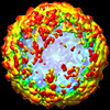
- PDB-7zuf: Saccharomyces cerevisiae L-BC virus, open particle, C5 reconstruction -
+ Open data

- Basic information
| Entry | Database: PDB / ID: 7zuf | ||||||
|---|---|---|---|---|---|---|---|
| Title | Saccharomyces cerevisiae L-BC virus, open particle, C5 reconstruction | ||||||
 Components Components | Major capsid protein | ||||||
 Keywords Keywords |  VIRUS / open particle / totivirus / C5 asymmetric unit VIRUS / open particle / totivirus / C5 asymmetric unit | ||||||
| Function / homology | Major coat protein, L-A virus / L-A virus major coat protein superfamily / L-A virus, major coat protein / viral capsid / RNA binding / Major capsid protein Function and homology information Function and homology information | ||||||
| Biological species |  Saccharomyces cerevisiae (brewer's yeast) Saccharomyces cerevisiae (brewer's yeast) | ||||||
| Method | ELECTRON MICROSCOPY / single particle reconstruction / cryo EM / Resolution: 10 Å | ||||||
 Authors Authors | Grybchuk, D. / Prochazkova, M. / Fuzik, T. / Konovalovas, A. / Serva, S. / Yurchenko, V. / Plevka, P. | ||||||
| Funding support |  Czech Republic, 1items Czech Republic, 1items
| ||||||
 Citation Citation | Journal: Commun Biol / Year: 2022 Title: Structures of L-BC virus and its open particle provide insight into Totivirus capsid assembly. Authors: Danyil Grybchuk / Michaela Procházková / Tibor Füzik / Aleksandras Konovalovas / Saulius Serva / Vyacheslav Yurchenko / Pavel Plevka / Abstract: L-BC virus persists in the budding yeast Saccharomyces cerevisiae, whereas other viruses from the family Totiviridae infect a diverse group of organisms including protists, fungi, arthropods, and ...L-BC virus persists in the budding yeast Saccharomyces cerevisiae, whereas other viruses from the family Totiviridae infect a diverse group of organisms including protists, fungi, arthropods, and vertebrates. The presence of totiviruses alters the fitness of the host organisms, for example, by maintaining the killer system in yeast or increasing the virulence of Leishmania guyanensis. Despite the importance of totiviruses for their host survival, there is limited information about Totivirus structure and assembly. Here we used cryo-electron microscopy to determine the structure of L-BC virus to a resolution of 2.9 Å. The L-BC capsid is organized with icosahedral symmetry, with each asymmetric unit composed of two copies of the capsid protein. Decamers of capsid proteins are stabilized by domain swapping of the C-termini of subunits located around icosahedral fivefold axes. We show that capsids of 9% of particles in a purified L-BC sample were open and lacked one decamer of capsid proteins. The existence of the open particles together with domain swapping within a decamer provides evidence that Totiviridae capsids assemble from the decamers of capsid proteins. Furthermore, the open particles may be assembly intermediates that are prepared for the incorporation of the virus (+) strand RNA. | ||||||
| History |
|
- Structure visualization
| Structure viewer | Molecule: MolmilJmol/JSmol |
|---|
- Downloads & links
-Download
| PDBx/mmCIF format | 7zuf.cif.gz | 1.6 MB | Display | PDBx/mmCIF format |
|---|---|---|---|---|
| PDB format | pdb7zuf.ent.gz | Display | PDB format | |
| PDBx/mmJSON format | 7zuf.json.gz | Tree view | PDBx/mmJSON format | |
| Others |  Other downloads Other downloads |
-Validation report
| Arichive directory | https://data.pdbj.org/pub/pdb/validation_reports/zu/7zufftp://data.pdbj.org/pub/pdb/validation_reports/zu/7zuf | HTTPS FTP |
|---|
-Related structure data
| Related structure data | 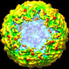 14975MC  7qwxC  7qwzC  7ztsC M: map data used to model this data C: citing same article ( |
|---|---|
| Similar structure data |

-Links
 PDBj
PDBj- Assembly
| Deposited unit | 
|
|---|---|
| 1 | x 5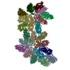
|
-Components
| #1: Protein | Mass: 78393.812 Da / Num. of mol.: 22 / Source method: isolated from a natural source / Source: (natural) Saccharomyces cerevisiae (brewer's yeast) / References: UniProt: Q87026 |
|---|
-Experimental details
-Experiment
| Experiment | Method: ELECTRON MICROSCOPY |
|---|---|
| EM experiment | Aggregation state: PARTICLE / 3D reconstruction method: single particle reconstruction |
- Sample preparation
| Component | Name: Saccharomyces cerevisiae virus L-BC (La) / Type: VIRUS Details: Virus was isolated by shearing with glass beads and overnight precipitation in 5% PEG-4000 and 500 mM NaCl Entity ID: all / Source: NATURAL |
|---|---|
| Molecular weight | Experimental value: NO |
| Source (natural) | Organism:  Saccharomyces cerevisiae virus L-BC (La) Saccharomyces cerevisiae virus L-BC (La) |
| Details of virus | Empty: YES / Enveloped: NO / Isolate: OTHER / Type: VIRUS-LIKE PARTICLE |
| Natural host | Organism: Saccharomyces cerevisiae / Strain: BY4741 |
| Virus shell | Diameter: 400 nm / Triangulation number (T number): 2 |
| Buffer solution | pH: 7.5 / Details: 20 mM Tris-HCl pH 7.5, 50 mM KCl, 10 mM MgCl2 |
| Specimen | Conc.: 1.5 mg/ml / Embedding applied: NO / Shadowing applied: NO / Staining applied: NO / Vitrification applied: YES Details: Virus was isolated by shearing with glass beads and overnight precipitation in 5% PEG-4000 and 500 mM NaCl |
| Specimen support | Details: Glow discharge current 7 mA / Grid material: COPPER / Grid mesh size: 200 divisions/in. / Grid type: Quantifoil R2/2 |
| Vitrification | Instrument: FEI VITROBOT MARK IV / Cryogen name: ETHANE-PROPANE / Humidity: 100 % / Chamber temperature: 277 K / Details: blot force 0, blot time 3 s, 4 C, 100% humidity |
- Electron microscopy imaging
| Experimental equipment |  Model: Titan Krios / Image courtesy: FEI Company |
|---|---|
| Microscopy | Model: FEI TITAN KRIOS |
| Electron gun | Electron source: FIELD EMISSION GUN / Accelerating voltage: 300 kV / Illumination mode: FLOOD BEAM |
| Electron lens | Mode: BRIGHT FIELDBright-field microscopy / Nominal magnification: 130000 X / Nominal defocus max: 1700 nm / Nominal defocus min: 700 nm / Cs: 2.7 mm / C2 aperture diameter: 30 µm / Alignment procedure: ZEMLIN TABLEAU |
| Specimen holder | Cryogen: NITROGEN / Specimen holder model: FEI TITAN KRIOS AUTOGRID HOLDER / Temperature (max): 80 K / Temperature (min): 70 K |
| Image recording | Average exposure time: 6 sec. / Electron dose: 36 e/Å2 / Detector mode: COUNTING / Film or detector model: GATAN K2 SUMMIT (4k x 4k) / Num. of grids imaged: 1 / Num. of real images: 11977 |
| EM imaging optics | Energyfilter name: GIF Bioquantum / Energyfilter slit width: 10 eV |
| Image scans | Sampling size: 5 µm / Width: 3600 / Height: 3600 / Movie frames/image: 30 / Used frames/image: 1-30 |
- Processing
| EM software |
| ||||||||||||||||||||||||||||||||||||||||||||||||||
|---|---|---|---|---|---|---|---|---|---|---|---|---|---|---|---|---|---|---|---|---|---|---|---|---|---|---|---|---|---|---|---|---|---|---|---|---|---|---|---|---|---|---|---|---|---|---|---|---|---|---|---|
| CTF correction | Type: PHASE FLIPPING AND AMPLITUDE CORRECTION | ||||||||||||||||||||||||||||||||||||||||||||||||||
| Particle selection | Num. of particles selected: 72705 | ||||||||||||||||||||||||||||||||||||||||||||||||||
| 3D reconstruction | Resolution: 10 Å / Resolution method: FSC 0.143 CUT-OFF / Num. of particles: 1120 / Algorithm: FOURIER SPACE / Num. of class averages: 1 / Symmetry type: POINT | ||||||||||||||||||||||||||||||||||||||||||||||||||
| Atomic model building | Protocol: FLEXIBLE FIT / Space: REAL / Details: Initial model generated by RaptorX server |
