 Movie
Movie Controller

[English] 日本語
 Yorodumi
- PDB-7zlg: Cryo-EM structure of C-mannosyltransferase CeDPY19, in complex wi... -
+ Open data

- Basic information
| Entry | Database: PDB / ID: 7zlg | ||||||
|---|---|---|---|---|---|---|---|
| Title | Cryo-EM structure of C-mannosyltransferase CeDPY19, in complex with acceptor peptide and bound to CMT2-Fab and anti-Fab nanobody | ||||||
 Components Components |
| ||||||
 Keywords Keywords |  MEMBRANE PROTEIN / C-mannosyltransferase MEMBRANE PROTEIN / C-mannosyltransferase | ||||||
| Function / homology |  Function and homology information Function and homology informationprotein C-linked glycosylation via 2'-alpha-mannosyl-L-tryptophan / mannosyltransferase activity / nuclear inner membrane / Transferases; Glycosyltransferases; Hexosyltransferases / nervous system development / cell differentiation / carbohydrate metabolic process / endoplasmic reticulum membrane / perinuclear region of cytoplasm / extracellular region / cytoplasmSimilarity search - Function | ||||||
| Biological species | synthetic construct (others) Caenorhabditis elegans (invertebrata) Caenorhabditis elegans (invertebrata) | ||||||
| Method | ELECTRON MICROSCOPY / single particle reconstruction / cryo EM / Resolution: 2.72 Å | ||||||
 Authors Authors | Bloch, J.S. / Mukherjee, S. / Mao, R. / Irobalieva, R. / Kossiakoff, A.A. / Goddard-Borger, E.D. / Locher, K.P. | ||||||
| Funding support |  Switzerland, 1items Switzerland, 1items
| ||||||
 Citation Citation | Journal: Nat Chem Biol / Year: 2023 Title: Structure, sequon recognition and mechanism of tryptophan C-mannosyltransferase. Authors: Joël S Bloch / Alan John / Runyu Mao / Somnath Mukherjee / Jérémy Boilevin / Rossitza N Irobalieva / Tamis Darbre / Nichollas E Scott / Jean-Louis Reymond / Anthony A Kossiakoff / Ethan D ...Authors: Joël S Bloch / Alan John / Runyu Mao / Somnath Mukherjee / Jérémy Boilevin / Rossitza N Irobalieva / Tamis Darbre / Nichollas E Scott / Jean-Louis Reymond / Anthony A Kossiakoff / Ethan D Goddard-Borger / Kaspar P Locher /   Abstract: C-linked glycosylation is essential for the trafficking, folding and function of secretory and transmembrane proteins involved in cellular communication processes. The tryptophan C- ...C-linked glycosylation is essential for the trafficking, folding and function of secretory and transmembrane proteins involved in cellular communication processes. The tryptophan C-mannosyltransferase (CMT) enzymes that install the modification attach a mannose to the first tryptophan of WxxW/C sequons in nascent polypeptide chains by an unknown mechanism. Here, we report cryogenic-electron microscopy structures of Caenorhabditis elegans CMT in four key states: apo, acceptor peptide-bound, donor-substrate analog-bound and as a trapped ternary complex with both peptide and a donor-substrate mimic bound. The structures indicate how the C-mannosylation sequon is recognized by this CMT and its paralogs, and how sequon binding triggers conformational activation of the donor substrate: a process relevant to all glycosyltransferase C superfamily enzymes. Our structural data further indicate that the CMTs adopt an unprecedented electrophilic aromatic substitution mechanism to enable the C-glycosylation of proteins. These results afford opportunities for understanding human disease and therapeutic targeting of specific CMT paralogs. | ||||||
| History |
|
- Structure visualization
| Structure viewer | Molecule: MolmilJmol/JSmol |
|---|
- Downloads & links
-Download
| PDBx/mmCIF format | 7zlg.cif.gz | 220.9 KB | Display | PDBx/mmCIF format |
|---|---|---|---|---|
| PDB format | pdb7zlg.ent.gz | 177.4 KB | Display | PDB format |
| PDBx/mmJSON format | 7zlg.json.gz | Tree view | PDBx/mmJSON format | |
| Others |  Other downloads Other downloads |
-Validation report
| Arichive directory | https://data.pdbj.org/pub/pdb/validation_reports/zl/7zlgftp://data.pdbj.org/pub/pdb/validation_reports/zl/7zlg | HTTPS FTP |
|---|
-Related structure data
| Related structure data |  14779MC  7zlhC  7zliC  7zljC M: map data used to model this data C: citing same article ( |
|---|---|
| Similar structure data |


-Links
 PDBj
PDBj


- Assembly
| Deposited unit | 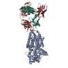
|
|---|---|
| 1 |
|
-Components
-Antibody , 3 types, 3 molecules HKL
| #1: Antibody | Mass: 25000.723 Da / Num. of mol.: 1 Source method: isolated from a genetically manipulated source Source: (gene. exp.) synthetic construct (others) / Production host:  Escherichia coli (E. coli) Escherichia coli (E. coli) |
|---|---|
| #2: Antibody | Mass: 13390.644 Da / Num. of mol.: 1 Source method: isolated from a genetically manipulated source Source: (gene. exp.) synthetic construct (others) / Production host: Escherichia coli (E. coli) |
| #3: Antibody | Mass: 23238.750 Da / Num. of mol.: 1 Source method: isolated from a genetically manipulated source Source: (gene. exp.) synthetic construct (others) / Production host: Escherichia coli (E. coli) / References: UniProt: Q7Z3Y4 |
-Protein / Protein/peptide / Non-polymers , 3 types, 12 molecules AP
| #4: Protein | Mass: 80892.602 Da / Num. of mol.: 1 Source method: isolated from a genetically manipulated source Source: (gene. exp.) Caenorhabditis elegans (invertebrata) / Gene: dpy-19, F22B7.10 / Production host:   Spodoptera frugiperda (fall armyworm) Spodoptera frugiperda (fall armyworm)References: UniProt: P34413, Transferases; Glycosyltransferases; Hexosyltransferases |
|---|---|
| #5: Protein/peptide | Mass: 821.900 Da / Num. of mol.: 1 Source method: isolated from a genetically manipulated source Details: GSWAKWS / Source: (gene. exp.) synthetic construct (others) / Production host: synthetic construct (others) |
| #6: Water | ChemComp-HOH / WaterMass: 18.015 Da / Num. of mol.: 10 / Source method: isolated from a natural source / Formula: H2O |
-Experimental details
-Experiment
| Experiment | Method: ELECTRON MICROSCOPY |
|---|---|
| EM experiment | Aggregation state: 3D ARRAY / 3D reconstruction method: single particle reconstruction |
- Sample preparation
| Component | Name: C-mannosyltransferase CeDPY19, in complex with acceptor peptide and bound to CMT2-Fab and anti-Fab nanobody Type: COMPLEX / Entity ID: #1-#5 / Source: RECOMBINANT |
|---|---|
| Source (natural) | Organism: Caenorhabditis elegans (invertebrata) |
| Source (recombinant) | Organism: Spodoptera frugiperda (fall armyworm) |
| Buffer solution | pH: 7.4 |
| Specimen | Embedding applied: NO / Shadowing applied: NO / Staining applied: NO / Vitrification applied: YES |
| Vitrification | Cryogen name: ETHANE-PROPANE |
- Electron microscopy imaging
| Experimental equipment |  Model: Titan Krios / Image courtesy: FEI Company |
|---|---|
| Microscopy | Model: FEI TITAN KRIOS |
| Electron gun | Electron source: FIELD EMISSION GUN / Accelerating voltage: 300 kV / Illumination mode: SPOT SCAN |
| Electron lens | Mode: BRIGHT FIELDBright-field microscopy / Nominal defocus max: 800 nm / Nominal defocus min: 600 nm |
| Image recording | Electron dose: 48 e/Å2 / Film or detector model: GATAN K3 BIOQUANTUM (6k x 4k) |
- Processing
| Software | Name: PHENIX / Version: 1.19.2_4158: / Classification: refinement | ||||||||||||||||||||||||
|---|---|---|---|---|---|---|---|---|---|---|---|---|---|---|---|---|---|---|---|---|---|---|---|---|---|
| CTF correction | Type: PHASE FLIPPING AND AMPLITUDE CORRECTION | ||||||||||||||||||||||||
| 3D reconstruction | Resolution: 2.72 Å / Resolution method: FSC 0.143 CUT-OFF / Num. of particles: 324852 / Symmetry type: POINT | ||||||||||||||||||||||||
| Refine LS restraints |
|
