Movie
Movie Controller


+ Open data

- Basic information
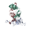
| Entry | Database: PDB / ID: 7z90 | ||||||
|---|---|---|---|---|---|---|---|
| Title | Leishmania RNA virus 1 virion | ||||||
 Components Components | Capsid protein,Major capsid protein | ||||||
 Keywords Keywords | VIRUS / virion / full particle | ||||||
| Function / homology | Totivirus coat / Totivirus coat protein / Capsid protein Function and homology information Function and homology information | ||||||
| Biological species |  Leishmania RNA virus 1 - 4 Leishmania RNA virus 1 - 4 | ||||||
| Method | ELECTRON MICROSCOPY / single particle reconstruction / cryo EM / Resolution: 3.88 Å | ||||||
 Authors Authors | Prochazkova, M. / Plevka, P. | ||||||
| Funding support |  Czech Republic, 1items Czech Republic, 1items
| ||||||
 Citation Citation | Journal: Virology / Year: 2022 Title: Virion structure of Leishmania RNA virus 1. Authors: Michaela Procházková / Tibor Füzik / Danyil Grybchuk / Vyacheslav Yurchenko / Pavel Plevka / Abstract: The presence of Leishmania RNA virus 1 (LRV1) enables Leishmania protozoan parasites to cause more severe disease than the virus-free strains. The structure of LRV1 virus-like particles has been ...The presence of Leishmania RNA virus 1 (LRV1) enables Leishmania protozoan parasites to cause more severe disease than the virus-free strains. The structure of LRV1 virus-like particles has been determined previously, however, the structure of the LRV1 virion has not been characterized. Here we used cryo-electron microscopy and single-particle reconstruction to determine the structures of the LRV1 virion and empty particle isolated from Leishmania guyanensis to resolutions of 4.0 Å and 3.6 Å, respectively. The capsid of LRV1 is built from sixty dimers of capsid proteins organized with icosahedral symmetry. RNA genomes of totiviruses are replicated inside the virions by RNA polymerases expressed as C-terminal extensions of a sub-population of capsid proteins. Most of the virions probably contain one or two copies of the RNA polymerase, however, the location of the polymerase domains in LRV1 capsid could not be identified, indicating that it varies among particles. Importance. Every year over 200 000 people contract leishmaniasis and more than five hundred people die of the disease. The mucocutaneous form of leishmaniasis produces lesions that can destroy the mucous membranes of the nose, mouth, and throat. Leishmania parasites carrying Leishmania RNA virus 1 (LRV1) are predisposed to cause aggravated symptoms in the mucocutaneous form of leishmaniasis. Here, we present the structure of the LRV1 virion determined using cryo-electron microscopy. #1: Journal: J Virol / Year: 2021Title: Capsid Structure of Leishmania RNA virus 1 Authors: Prochazkova, M. / Fuzik, T. / Grybchuk, D. / Falginella, F.L. / Podesvova, L. / Yurchenko, V. / Vacha, R. / Plevka, P. | ||||||
| History |
|
- Structure visualization
| Structure viewer | Molecule: MolmilJmol/JSmol |
|---|
- Downloads & links
-Download
| PDBx/mmCIF format | 7z90.cif.gz | 218.9 KB | Display | PDBx/mmCIF format |
|---|---|---|---|---|
| PDB format | pdb7z90.ent.gz | 177.7 KB | Display | PDB format |
| PDBx/mmJSON format | 7z90.json.gz | Tree view | PDBx/mmJSON format | |
| Others |  Other downloads Other downloads |
-Validation report
| Arichive directory | https://data.pdbj.org/pub/pdb/validation_reports/z9/7z90ftp://data.pdbj.org/pub/pdb/validation_reports/z9/7z90 | HTTPS FTP |
|---|
-Related structure data
| Related structure data | 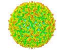 14566MC 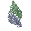 7ns2C C: citing same article ( M: map data used to model this data |
|---|---|
| Similar structure data |

-Links
 PDBj
PDBj- Assembly
| Deposited unit | 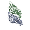
|
|---|---|
| 1 | x 60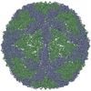
|
| 2 |
|
| 3 | x 5
|
-Components
| #1: Protein | Mass: 86431.742 Da / Num. of mol.: 2 / Source method: isolated from a natural source / Source: (natural) Leishmania RNA virus 1 - 4 / References: UniProt: L7XUU7 |
|---|
-Experimental details
-Experiment
| Experiment | Method: ELECTRON MICROSCOPY |
|---|---|
| EM experiment | Aggregation state: PARTICLE / 3D reconstruction method: single particle reconstruction |
- Sample preparation
| Component | Name: Leishmania RNA virus 1 - 4 / Type: VIRUS / Entity ID: all / Source: NATURAL |
|---|---|
| Molecular weight | Value: 9.72 MDa / Experimental value: NO |
| Source (natural) | Organism: Leishmania RNA virus 1 - 4 |
| Details of virus | Empty: NO / Enveloped: NO / Isolate: STRAIN / Type: VIRION |
| Natural host | Organism: Leishmania guyanensis / Strain: MHOM/BR/75/M4147 |
| Virus shell | Name: capsid / Diameter: 420 nm / Triangulation number (T number): 2 |
| Buffer solution | pH: 7.4 |
| Specimen | Embedding applied: NO / Shadowing applied: NO / Staining applied: NO / Vitrification applied: YES |
| Vitrification | Cryogen name: ETHANE-PROPANE |
- Electron microscopy imaging
| Experimental equipment |  Model: Titan Krios / Image courtesy: FEI Company |
|---|---|
| Microscopy | Model: FEI TITAN KRIOS |
| Electron gun | Electron source: FIELD EMISSION GUN / Accelerating voltage: 300 kV / Illumination mode: FLOOD BEAM |
| Electron lens | Mode: BRIGHT FIELDBright-field microscopy / Nominal defocus max: 2500 nm / Nominal defocus min: 1200 nm |
| Image recording | Electron dose: 1.98 e/Å2 / Detector mode: COUNTING / Film or detector model: FEI FALCON III (4k x 4k) / Num. of grids imaged: 1 / Num. of real images: 4542 |
- Processing
| Software | Name: PHENIX / Version: 1.18.2_3874: / Classification: refinement | ||||||||||||||||||||||||
|---|---|---|---|---|---|---|---|---|---|---|---|---|---|---|---|---|---|---|---|---|---|---|---|---|---|
| EM software |
| ||||||||||||||||||||||||
| CTF correction | Type: PHASE FLIPPING AND AMPLITUDE CORRECTION | ||||||||||||||||||||||||
| Particle selection | Num. of particles selected: 7284 | ||||||||||||||||||||||||
| Symmetry | Point symmetry: C1 (asymmetric) | ||||||||||||||||||||||||
| 3D reconstruction | Resolution: 3.88 Å / Resolution method: FSC 0.143 CUT-OFF / Num. of particles: 1156 / Num. of class averages: 1 / Symmetry type: POINT | ||||||||||||||||||||||||
| Atomic model building | B value: 75 / Protocol: RIGID BODY FIT / Space: REAL / Target criteria: correlation coefficient | ||||||||||||||||||||||||
| Atomic model building | PDB-ID: 7SN2 Pdb chain-ID: A | ||||||||||||||||||||||||
| Refinement | Highest resolution: 3.88 Å | ||||||||||||||||||||||||
| Refine LS restraints |
|