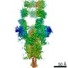
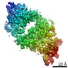
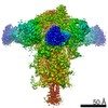
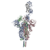
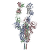
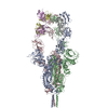
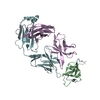
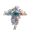
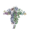
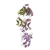
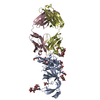
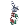
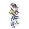
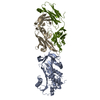
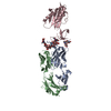
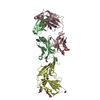
[日本語] English
- PDB-7wcr: RBD-1 of SARS-CoV-2 Beta spike in complex with S5D2 Fab -

+
データを開く


IDまたはキーワード:

読み込み中...

-
基本情報

登録情報
データベース: PDB / ID: 7wcr
タイトルRBD-1 of SARS-CoV-2 Beta spike in complex with S5D2 Fab
要素
  • Heavy chain of S5D2 Fab
  • Light chain of S5D2 Fab
  • Spike glycoproteinスパイクタンパク質
キーワードVIRAL PROTEIN (ウイルスタンパク質) / SARS-CoV-2 (SARSコロナウイルス2) / coronavirus (オルトコロナウイルス亜科) / Beta variant (SARSコロナウイルス2-ベータ株) / B.1.351 lineage (SARSコロナウイルス2-ベータ株) / spike protein (スパイクタンパク質)
機能・相同性
機能・相同性情報


Maturation of spike protein / viral translation / Translation of Structural Proteins / Virion Assembly and Release / host cell surface / host extracellular space / suppression by virus of host tetherin activity / Induction of Cell-Cell Fusion / structural constituent of virion / host cell endoplasmic reticulum-Golgi intermediate compartment membrane ...Maturation of spike protein / viral translation / Translation of Structural Proteins / Virion Assembly and Release / host cell surface / host extracellular space / suppression by virus of host tetherin activity / Induction of Cell-Cell Fusion / structural constituent of virion / host cell endoplasmic reticulum-Golgi intermediate compartment membrane / entry receptor-mediated virion attachment to host cell / receptor-mediated endocytosis of virus by host cell / Attachment and Entry / membrane fusion / positive regulation of viral entry into host cell / receptor-mediated virion attachment to host cell / receptor ligand activity / host cell surface receptor binding / fusion of virus membrane with host plasma membrane / fusion of virus membrane with host endosome membrane / エンベロープ (ウイルス) / virion attachment to host cell / SARS-CoV-2 activates/modulates innate and adaptive immune responses / host cell plasma membrane / virion membrane / 生体膜 / identical protein binding / 細胞膜
類似検索 - 分子機能
Spike (S) protein S1 subunit, receptor-binding domain, SARS-CoV-2 / Spike (S) protein S1 subunit, N-terminal domain, SARS-CoV-like / Betacoronavirus spike (S) glycoprotein S1 subunit N-terminal (NTD) domain profile. / Spike glycoprotein, N-terminal domain superfamily / Betacoronavirus spike (S) glycoprotein S1 subunit C-terminal (CTD) domain profile. / Spike glycoprotein, betacoronavirus / Spike (S) protein S1 subunit, receptor-binding domain, betacoronavirus / Spike S1 subunit, receptor binding domain superfamily, betacoronavirus / Betacoronavirus spike glycoprotein S1, receptor binding / Spike glycoprotein S1, N-terminal domain, betacoronavirus-like ...Spike (S) protein S1 subunit, receptor-binding domain, SARS-CoV-2 / Spike (S) protein S1 subunit, N-terminal domain, SARS-CoV-like / Betacoronavirus spike (S) glycoprotein S1 subunit N-terminal (NTD) domain profile. / Spike glycoprotein, N-terminal domain superfamily / Betacoronavirus spike (S) glycoprotein S1 subunit C-terminal (CTD) domain profile. / Spike glycoprotein, betacoronavirus / Spike (S) protein S1 subunit, receptor-binding domain, betacoronavirus / Spike S1 subunit, receptor binding domain superfamily, betacoronavirus / Betacoronavirus spike glycoprotein S1, receptor binding / Spike glycoprotein S1, N-terminal domain, betacoronavirus-like / Betacoronavirus-like spike glycoprotein S1, N-terminal / Spike glycoprotein S2, coronavirus, heptad repeat 1 / Spike glycoprotein S2, coronavirus, heptad repeat 2 / Coronavirus spike (S) glycoprotein S2 subunit heptad repeat 2 (HR2) region profile. / Coronavirus spike (S) glycoprotein S2 subunit heptad repeat 1 (HR1) region profile. / Spike glycoprotein S2 superfamily, coronavirus / Spike glycoprotein S2, coronavirus / Coronavirus spike glycoprotein S2 / Coronavirus spike glycoprotein S1, C-terminal / Coronavirus spike glycoprotein S1, C-terminal
類似検索 - ドメイン・相同性
スパイクタンパク質
類似検索 - 構成要素
生物種Severe acute respiratory syndrome coronavirus 2 (SARSコロナウイルス2)
Mus musculus (ハツカネズミ)
手法電子顕微鏡法 / 単粒子再構成法 / クライオ電子顕微鏡法 / 解像度: 3.5 Å
データ登録者Wang, Y.F. / Cong, Y.
資金援助 中国, 1件
組織認可番号
Chinese Academy of Sciences 中国
引用ジャーナル: Emerg Microbes Infect / : 2022
タイトル: Mapping cross-variant neutralizing sites on the SARS-CoV-2 spike protein.
著者: Shiqi Xu / Yifan Wang / Yanxing Wang / Chao Zhang / Qin Hong / Chenjian Gu / Rong Xu / Tingfeng Wang / Yong Yang / Jinkai Zang / Yu Zhou / Zuyang Li / Qixing Liu / Bingjie Zhou / Lulu Bai / ...著者: Shiqi Xu / Yifan Wang / Yanxing Wang / Chao Zhang / Qin Hong / Chenjian Gu / Rong Xu / Tingfeng Wang / Yong Yang / Jinkai Zang / Yu Zhou / Zuyang Li / Qixing Liu / Bingjie Zhou / Lulu Bai / Yuanfei Zhu / Qiang Deng / Haikun Wang / Dimitri Lavillette / Gary Wong / Youhua Xie / Yao Cong / Zhong Huang /
要旨: The emergence of multiple severe acute respiratory syndrome coronavirus 2 (SARS-CoV-2) variants of concern threatens the efficacy of currently approved vaccines and authorized therapeutic monoclonal ...The emergence of multiple severe acute respiratory syndrome coronavirus 2 (SARS-CoV-2) variants of concern threatens the efficacy of currently approved vaccines and authorized therapeutic monoclonal antibodies (MAbs). It is hence important to continue searching for SARS-CoV-2 broadly neutralizing MAbs and defining their epitopes. Here, we isolate 9 neutralizing mouse MAbs raised against the spike protein of a SARS-CoV-2 prototype strain and evaluate their neutralizing potency towards a panel of variants, including B.1.1.7, B.1.351, B.1.617.1, and B.1.617.2. By using a combination of biochemical, virological, and cryo-EM structural analyses, we identify three types of cross-variant neutralizing MAbs, represented by S5D2, S5G2, and S3H3, respectively, and further define their epitopes. S5D2 binds the top lateral edge of the receptor-binding motif within the receptor-binding domain (RBD) with a binding footprint centred around the loop, and efficiently neutralizes all variant pseudoviruses, but the potency against B.1.617.2 was observed to decrease significantly. S5G2 targets the highly conserved RBD core region and exhibits comparable neutralization towards the variant panel. S3H3 binds a previously unreported epitope located within the evolutionarily stable SD1 region and is able to near equally neutralize all of the variants tested. Our work thus defines three distinct cross-variant neutralizing sites on the SARS-CoV-2 spike protein, providing guidance for design and development of broadly effective vaccines and MAb-based therapies.
履歴
登録2021年12月20日登録サイト: PDBJ / 処理サイト: PDBJ
改定 1.02022年2月2日Provider: repository / タイプ: Initial release

-
構造の表示

ムービー
  • 登録構造単位
  • Jmolによる作画
  • ダウンロード
  • EMマップとの重ね合わせ
  • マップデータ: EMDB-32428
  • UCSF Chimeraによる作画
  • ダウンロード
ムービービューア
構造ビューア分子:
MolmilJmol/JSmol

ダウンロードとリンク

-
集合体

登録構造単位
A: Spike glycoprotein
a: Heavy chain of S5D2 Fab
b: Light chain of S5D2 Fab


分子量 (理論値)分子数
合計 (水以外)186,8373
ポリマ-186,8373
非ポリマー00
0
1


  • 登録構造と同一
  • 登録者が定義した集合体
タイプ名称対称操作
identity operation1_5551
Buried area4790 Å2
ΔGint-28 kcal/mol
Surface area31670 Å2

-
要素

#1: タンパク質 Spike glycoprotein / スパイクタンパク質 / S glycoprotein / E2 / Peplomer protein


分子量: 139650.516 Da / 分子数: 1 / 由来タイプ: 組換発現
由来: (組換発現) Severe acute respiratory syndrome coronavirus 2 (SARSコロナウイルス2)
遺伝子: S, 2 / 細胞株 (発現宿主): HEK293 / 発現宿主: Homo sapiens (ヒト) / 参照: UniProt: P0DTC2
#2: 抗体 Heavy chain of S5D2 Fab


分子量: 23121.744 Da / 分子数: 1 / 由来タイプ: 天然 / 由来: (天然) Mus musculus (ハツカネズミ)
#3: 抗体 Light chain of S5D2 Fab


分子量: 24064.529 Da / 分子数: 1 / 由来タイプ: 天然 / 由来: (天然) Mus musculus (ハツカネズミ)

-
実験情報

-
実験

実験手法: 電子顕微鏡法
EM実験試料の集合状態: PARTICLE / 3次元再構成法: 単粒子再構成法

-
試料調製

構成要素
ID名称タイプEntity IDParent-ID由来
1RBD-1 of SARS-CoV-2 Beta spike in complex with S5D2 FabCOMPLEXall0MULTIPLE SOURCES
2SARS-CoV-2 spike proteinCoronavirus spike proteinCOMPLEX#11RECOMBINANT
3Heavy chain of S5D2 FabCOMPLEX#21NATURAL
4Light chain of S5D2 FabCOMPLEX#31NATURAL
由来(天然)
IDEntity assembly-ID生物種Ncbi tax-ID
21Severe acute respiratory syndrome coronavirus 2 (SARSコロナウイルス2)2697049
32Mus musculus (ハツカネズミ)10090
43Mus musculus (ハツカネズミ)10090
由来(組換発現)生物種: Homo sapiens (ヒト) / 細胞: HEK293
緩衝液pH: 7.5
試料包埋: NO / シャドウイング: NO / 染色: NO / 凍結: YES
急速凍結凍結剤: ETHANE

-
電子顕微鏡撮影

実験機器
モデル: Titan Krios / 画像提供: FEI Company
顕微鏡モデル: FEI TITAN KRIOS
電子銃電子線源: FIELD EMISSION GUN / 加速電圧: 300 kV / 照射モード: FLOOD BEAM
電子レンズモード: BRIGHT FIELDBright-field microscopy / 最大 デフォーカス(公称値): 2500 nm / 最小 デフォーカス(公称値): 800 nm
撮影電子線照射量: 50 e/Å2 / フィルム・検出器のモデル: GATAN K3 (6k x 4k)

-
解析

CTF補正タイプ: PHASE FLIPPING AND AMPLITUDE CORRECTION
3次元再構成解像度: 3.5 Å / 解像度の算出法: FSC 0.143 CUT-OFF / 粒子像の数: 100064 / 対称性のタイプ: POINT

+
万見について

-
お知らせ

-
2022年2月9日: EMDBエントリの付随情報ファイルのフォーマットが新しくなりました

EMDBエントリの付随情報ファイルのフォーマットが新しくなりました

  • EMDBのヘッダファイルのバージョン3が、公式のフォーマットとなりました。
  • これまでは公式だったバージョン1.9は、アーカイブから削除されます。

関連情報:EMDBヘッダ

外部リンク:wwPDBはEMDBデータモデルのバージョン3へ移行します

-
2020年8月12日: 新型コロナ情報

新型コロナ情報

URL: https://pdbj.org/emnavi/covid19.php

新ページ: EM Navigatorに新型コロナウイルスの特設ページを開設しました。

関連情報:Covid-19情報 / 2020年3月5日: 新型コロナウイルスの構造データ

+
2020年3月5日: 新型コロナウイルスの構造データ

新型コロナウイルスの構造データ

関連情報:万見生物種 / 2020年8月12日: 新型コロナ情報

外部リンク:COVID-19特集ページ - PDBj / 今月の分子2020年2月:コロナウイルスプロテーアーゼ

+
2019年1月31日: EMDBのIDの桁数の変更

EMDBのIDの桁数の変更

  • EMDBエントリに付与されているアクセスコード(EMDB-ID)は4桁の数字(例、EMD-1234)でしたが、間もなく枯渇します。これまでの4桁のID番号は4桁のまま変更されませんが、4桁の数字を使い切った後に発行されるIDは5桁以上の数字(例、EMD-12345)になります。5桁のIDは2019年の春頃から発行される見通しです。
  • EM Navigator/万見では、接頭語「EMD-」は省略されています。

関連情報:Q: 「EMD」とは何ですか? / 万見/EM NavigatorにおけるID/アクセスコードの表記

外部リンク:EMDB Accession Codes are Changing Soon! / PDBjへお問い合わせ

+
2017年7月12日: PDB大規模アップデート

PDB大規模アップデート

  • 新バージョンのPDBx/mmCIF辞書形式に基づくデータがリリースされました。
  • 今回の更新はバージョン番号が4から5になる大規模なもので、全エントリデータの書き換えが行われる「Remediation」というアップデートに該当します。
  • このバージョンアップで、電子顕微鏡の実験手法に関する多くの項目の書式が改定されました(例:em_softwareなど)。
  • EM NavigatorとYorodumiでも、この改定に基づいた表示内容になります。

外部リンク:wwPDB Remediation / OneDepデータ基準に準拠した、より強化された内容のモデル構造ファイルが、PDBアーカイブで公開されました。

-
万見 (Yorodumi)

幾万の構造データを、幾万の視点から

  • 万見(Yorodumi)は、EMDB/PDB/SASBDBなどの構造データを閲覧するためのページです。
  • EM Navigatorの詳細ページの後継、Omokage検索のフロントエンドも兼ねています。

関連情報:EMDB / PDB / SASBDB / 3つのデータバンクの比較 / 万見検索 / 2016年8月31日: 新しいEM Navigatorと万見 / 万見文献 / Jmol/JSmol / 機能・相同性情報 / 新しいEM Navigatorと万見の変更点

他の情報も見る