 Movie
Movie Controller

[English] 日本語
 Yorodumi
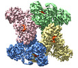
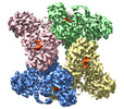
- PDB-7wad: Trichodesmium erythraeum cyanophycin synthetase 1 (TeCphA1) with ... -
+ Open data

- Basic information
| Entry | Database: PDB / ID: 7wad | |||||||||
|---|---|---|---|---|---|---|---|---|---|---|
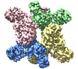
| Title | Trichodesmium erythraeum cyanophycin synthetase 1 (TeCphA1) with ATPgammaS | |||||||||
 Components Components | Cyanophycin synthase | |||||||||
 Keywords Keywords | LIGASE / Cyanophycin / Non-ribosomal peptide synthesis / ATP / Aspartate / Arginine | |||||||||
| Function / homology |  Function and homology informationcyanophycin synthase (L-aspartate-adding) / cyanophycin synthase (L-arginine-adding) / cyanophycin synthetase activity (L-aspartate-adding) / cyanophycin synthetase activity (L-arginine-adding) / tetrahydrofolylpolyglutamate synthase activity / macromolecule biosynthetic process / ATP binding / metal ion binding Function and homology informationcyanophycin synthase (L-aspartate-adding) / cyanophycin synthase (L-arginine-adding) / cyanophycin synthetase activity (L-aspartate-adding) / cyanophycin synthetase activity (L-arginine-adding) / tetrahydrofolylpolyglutamate synthase activity / macromolecule biosynthetic process / ATP binding / metal ion bindingSimilarity search - Function | |||||||||
| Biological species |  Trichodesmium erythraeum IMS101 (bacteria) Trichodesmium erythraeum IMS101 (bacteria) | |||||||||
| Method | ELECTRON MICROSCOPY / single particle reconstruction / cryo EM / Resolution: 2.96 Å | |||||||||
 Authors Authors | Kawasaki, M. / Miyakawa, T. / Yang, J. / Adachi, N. / Fujii, A. / Miyauchi, Y. / Muramatsu, T. / Moriya, T. / Senda, T. / Tanokura, M. | |||||||||
| Funding support |  Japan, 2items Japan, 2items
| |||||||||
 Citation Citation | Journal: Nat Commun / Year: 2022 Title: Structural bases for aspartate recognition and polymerization efficiency of cyanobacterial cyanophycin synthetase. Authors: Takuya Miyakawa / Jian Yang / Masato Kawasaki / Naruhiko Adachi / Ayumu Fujii / Yumiko Miyauchi / Tomonari Muramatsu / Toshio Moriya / Toshiya Senda / Masaru Tanokura /  Abstract: Cyanophycin is a natural biopolymer consisting of equimolar amounts of aspartate and arginine as the backbone and branched sidechain, respectively. It is produced by a single enzyme, cyanophycin ...Cyanophycin is a natural biopolymer consisting of equimolar amounts of aspartate and arginine as the backbone and branched sidechain, respectively. It is produced by a single enzyme, cyanophycin synthetase (CphA1), and accumulates as a nitrogen reservoir during N fixation by most cyanobacteria. A recent structural study showed that three constituent domains of CphA1 function as two distinct catalytic sites and an oligomerization interface in cyanophycin synthesis. However, it remains unclear how the ATP-dependent addition of aspartate to cyanophycin is initiated at the catalytic site of the glutathione synthetase-like domain. Here, we report the cryogenic electron microscopy structures of CphA1, including a complex with aspartate, cyanophycin primer peptide, and ATP analog. These structures reveal the aspartate binding mode and phosphate-binding loop movement to the active site required for the reaction. Furthermore, structural and mutational data show a potential role of protein dynamics in the catalytic efficiency of the arginine condensation reaction. | |||||||||
| History |
|
- Structure visualization
| Structure viewer | Molecule: MolmilJmol/JSmol |
|---|
- Downloads & links
-Download
| PDBx/mmCIF format | 7wad.cif.gz | 496 KB | Display | PDBx/mmCIF format |
|---|---|---|---|---|
| PDB format | pdb7wad.ent.gz | 405.5 KB | Display | PDB format |
| PDBx/mmJSON format | 7wad.json.gz | Tree view | PDBx/mmJSON format | |
| Others |  Other downloads Other downloads |
-Validation report
| Arichive directory | https://data.pdbj.org/pub/pdb/validation_reports/wa/7wadftp://data.pdbj.org/pub/pdb/validation_reports/wa/7wad | HTTPS FTP |
|---|
-Related structure data
| Related structure data | 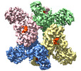 32382MC 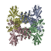 7wacC  7waeC 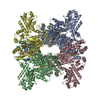 7wafC M: map data used to model this data C: citing same article ( |
|---|---|
| Similar structure data |
-Links
 PDBj
PDBj


- Assembly
| Deposited unit | 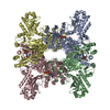
|
|---|---|
| 1 |
|
-Components
| #1: Protein | / Cyanophycin synthetase Mass: 99079.914 Da / Num. of mol.: 4 Source method: isolated from a genetically manipulated source Source: (gene. exp.) Trichodesmium erythraeum IMS101 (bacteria)Strain: IMS101 / Gene: Tery_1965 / Production host: Escherichia coli (E. coli)References: UniProt: Q113V7, cyanophycin synthase (L-aspartate-adding), cyanophycin synthase (L-arginine-adding)#2: Chemical | ChemComp-MG /   Mass: 24.305 Da / Num. of mol.: 4 / Source method: obtained synthetically / Formula: Mg / Feature type: SUBJECT OF INVESTIGATION Mass: 24.305 Da / Num. of mol.: 4 / Source method: obtained synthetically / Formula: Mg / Feature type: SUBJECT OF INVESTIGATION#3: Chemical | ChemComp-AGS /   Mass: 523.247 Da / Num. of mol.: 8 / Source method: obtained synthetically / Formula: C10H16N5O12P3S / Feature type: SUBJECT OF INVESTIGATION / Comment: ATP-gamma-S, energy-carrying molecule analogue*YM Mass: 523.247 Da / Num. of mol.: 8 / Source method: obtained synthetically / Formula: C10H16N5O12P3S / Feature type: SUBJECT OF INVESTIGATION / Comment: ATP-gamma-S, energy-carrying molecule analogue*YMHas ligand of interest | Y | |
|---|
-Experimental details
-Experiment
| Experiment | Method: ELECTRON MICROSCOPY |
|---|---|
| EM experiment | Aggregation state: PARTICLE / 3D reconstruction method: single particle reconstruction |
- Sample preparation
| Component | Name: TeCphA1 with ATPgammaS / Type: COMPLEX / Details: Homotetramer / Entity ID: #1 / Source: RECOMBINANT | ||||||||||||||||||||||||
|---|---|---|---|---|---|---|---|---|---|---|---|---|---|---|---|---|---|---|---|---|---|---|---|---|---|
| Molecular weight | Value: 0.40 MDa / Experimental value: NO | ||||||||||||||||||||||||
| Source (natural) | Organism: Trichodesmium erythraeum IMS101 (bacteria) | ||||||||||||||||||||||||
| Source (recombinant) | Organism: Escherichia coli (E. coli) / Cell: BL21(DE3) / Plasmid: pET-22b(+) | ||||||||||||||||||||||||
| Buffer solution | pH: 8.2 Details: The buffer contains 4 mM ATPgammaS, 5 mM aspartate, 0.5 mM arginine and 10 mM 4-mer cyanophycin peptide as ligands. | ||||||||||||||||||||||||
| Buffer component |
| ||||||||||||||||||||||||
| Specimen | Conc.: 0.2 mg/ml / Embedding applied: NO / Shadowing applied: NO / Staining applied: NO / Vitrification applied: YES / Details: This sample was mono-disperse. | ||||||||||||||||||||||||
| Specimen support | Details: The grid was washed by acetone prior to use. / Grid material: COPPER / Grid mesh size: 300 divisions/in. / Grid type: Quantifoil R1.2/1.3 | ||||||||||||||||||||||||
| Vitrification | Instrument: FEI VITROBOT MARK IV / Cryogen name: ETHANE / Humidity: 100 % / Chamber temperature: 291 K / Details: Blotting time was 15 seconds (blot force 10). |
- Electron microscopy imaging
| Experimental equipment |  Model: Talos Arctica / Image courtesy: FEI Company |
|---|---|
| Microscopy | Model: FEI TALOS ARCTICA |
| Electron gun | Electron source: FIELD EMISSION GUN / Accelerating voltage: 200 kV / Illumination mode: FLOOD BEAM |
| Electron lens | Mode: BRIGHT FIELDBright-field microscopy / Nominal magnification: 120000 X / Nominal defocus max: 2500 nm / Nominal defocus min: 1000 nm / Cs: 2.7 mm / C2 aperture diameter: 50 µm |
| Specimen holder | Cryogen: NITROGEN |
| Image recording | Average exposure time: 56.18 sec. / Electron dose: 50 e/Å2 / Detector mode: COUNTING / Film or detector model: FEI FALCON III (4k x 4k) / Num. of grids imaged: 1 / Num. of real images: 1730 |
- Processing
| Software | Name: PHENIX / Version: 1.19.2_4158: / Classification: refinement | |||||||||||||||||||||||||||||||||||||||||||||||||||||||||||||||||
|---|---|---|---|---|---|---|---|---|---|---|---|---|---|---|---|---|---|---|---|---|---|---|---|---|---|---|---|---|---|---|---|---|---|---|---|---|---|---|---|---|---|---|---|---|---|---|---|---|---|---|---|---|---|---|---|---|---|---|---|---|---|---|---|---|---|---|
| EM software |
| |||||||||||||||||||||||||||||||||||||||||||||||||||||||||||||||||
| CTF correction | Type: PHASE FLIPPING AND AMPLITUDE CORRECTION | |||||||||||||||||||||||||||||||||||||||||||||||||||||||||||||||||
| Particle selection | Num. of particles selected: 927603 | |||||||||||||||||||||||||||||||||||||||||||||||||||||||||||||||||
| Symmetry | Point symmetry: D2 (2x2 fold dihedral) | |||||||||||||||||||||||||||||||||||||||||||||||||||||||||||||||||
| 3D reconstruction | Resolution: 2.96 Å / Resolution method: FSC 0.143 CUT-OFF / Num. of particles: 49842 / Algorithm: FOURIER SPACE / Num. of class averages: 1 / Symmetry type: POINT | |||||||||||||||||||||||||||||||||||||||||||||||||||||||||||||||||
| Refine LS restraints |
|
