 Movie
Movie Controller


+ Open data

- Basic information
| Entry | Database: PDB / ID: 7pnf | ||||||
|---|---|---|---|---|---|---|---|
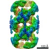
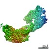
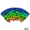
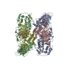
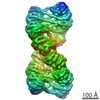
| Title | VWF Tubules of D1D2D'D3 domains | ||||||
 Components Components | von Willebrand factor | ||||||
 Keywords Keywords | BLOOD CLOTTING / VWF | ||||||
| Function / homology |  Function and homology informationWeibel-Palade body / Defective F8 binding to von Willebrand factor / hemostasis / Platelet Adhesion to exposed collagen / platelet alpha granule / positive regulation of intracellular signal transduction / GP1b-IX-V activation signalling / p130Cas linkage to MAPK signaling for integrins / cell-substrate adhesion / Defective F8 cleavage by thrombin ...Weibel-Palade body / Defective F8 binding to von Willebrand factor / hemostasis / Platelet Adhesion to exposed collagen / platelet alpha granule / positive regulation of intracellular signal transduction / GP1b-IX-V activation signalling / p130Cas linkage to MAPK signaling for integrins / cell-substrate adhesion / Defective F8 cleavage by thrombin / immunoglobulin binding / Platelet Aggregation (Plug Formation) / GRB2:SOS provides linkage to MAPK signaling for Integrins / Integrin cell surface interactions / collagen binding / Intrinsic Pathway of Fibrin Clot Formation / Integrin signaling / extracellular matrix / platelet alpha granule lumen / Signaling by high-kinase activity BRAF mutants / MAP2K and MAPK activation / platelet activation / response to wounding / Signaling by RAF1 mutants / Signaling by moderate kinase activity BRAF mutants / Paradoxical activation of RAF signaling by kinase inactive BRAF / Signaling downstream of RAS mutants / Signaling by BRAF and RAF1 fusions / blood coagulation / integrin binding / Platelet degranulation / protein-folding chaperone binding / collagen-containing extracellular matrix / protease binding / cell adhesion / endoplasmic reticulum / extracellular space / extracellular exosome / extracellular region / identical protein binding Function and homology informationWeibel-Palade body / Defective F8 binding to von Willebrand factor / hemostasis / Platelet Adhesion to exposed collagen / platelet alpha granule / positive regulation of intracellular signal transduction / GP1b-IX-V activation signalling / p130Cas linkage to MAPK signaling for integrins / cell-substrate adhesion / Defective F8 cleavage by thrombin ...Weibel-Palade body / Defective F8 binding to von Willebrand factor / hemostasis / Platelet Adhesion to exposed collagen / platelet alpha granule / positive regulation of intracellular signal transduction / GP1b-IX-V activation signalling / p130Cas linkage to MAPK signaling for integrins / cell-substrate adhesion / Defective F8 cleavage by thrombin / immunoglobulin binding / Platelet Aggregation (Plug Formation) / GRB2:SOS provides linkage to MAPK signaling for Integrins / Integrin cell surface interactions / collagen binding / Intrinsic Pathway of Fibrin Clot Formation / Integrin signaling / extracellular matrix / platelet alpha granule lumen / Signaling by high-kinase activity BRAF mutants / MAP2K and MAPK activation / platelet activation / response to wounding / Signaling by RAF1 mutants / Signaling by moderate kinase activity BRAF mutants / Paradoxical activation of RAF signaling by kinase inactive BRAF / Signaling downstream of RAS mutants / Signaling by BRAF and RAF1 fusions / blood coagulation / integrin binding / Platelet degranulation / protein-folding chaperone binding / collagen-containing extracellular matrix / protease binding / cell adhesion / endoplasmic reticulum / extracellular space / extracellular exosome / extracellular region / identical protein bindingSimilarity search - Function | ||||||
| Biological species |  Homo sapiens (human) Homo sapiens (human) | ||||||
| Method | ELECTRON MICROSCOPY / helical reconstruction / cryo EM / Resolution: 4.35 Å | ||||||
 Authors Authors | Javitt, G. / Fass, D. | ||||||
| Funding support | 1items
| ||||||
 Citation Citation | Journal: Proc Natl Acad Sci U S A / Year: 2022 Title: Helical self-assembly of a mucin segment suggests an evolutionary origin for von Willebrand factor tubules. Authors: Gabriel Javitt / Deborah Fass /  Abstract: The glycoprotein von Willebrand factor (VWF) contributes to hemostasis by stanching injuries in blood vessel walls. A distinctive feature of VWF is its assembly into long, helical tubules in ...The glycoprotein von Willebrand factor (VWF) contributes to hemostasis by stanching injuries in blood vessel walls. A distinctive feature of VWF is its assembly into long, helical tubules in endothelial cells prior to secretion. When VWF is released into the bloodstream, these tubules unfurl to release linear polymers that bind subendothelial collagen at wound sites, recruit platelets, and initiate the clotting cascade. VWF evolved from gel-forming mucins, the polymeric glycoproteins that coat and protect exposed epithelia. Despite the divergent function of VWF in blood vessel repair, sequence conservation and shared domain organization imply that VWF retained key aspects of the mucin bioassembly mechanism. Here, we show using cryo-electron microscopy that the ability to form tubules, a property hitherto thought to have arisen as a VWF adaptation to the vasculature, is a feature of the amino-terminal region of mucin. This segment of the human intestinal gel-forming mucin (MUC2) was found to self-assemble into tubules with a striking resemblance to those of VWF itself. To facilitate a comparison, we determined the residue-resolution structure of tubules formed by the homologous segment of VWF. The structures of the MUC2 and VWF tubules revealed the flexible joints and the intermolecular interactions required for tubule formation. Steric constraints in full-length MUC2 suggest that linear filaments, a previously observed supramolecular assembly form, are more likely than tubules to be the physiological mucin storage intermediate. Nevertheless, MUC2 tubules indicate a possible evolutionary origin for VWF tubules and elucidate design principles present in mucins and VWF. | ||||||
| History |
|
- Structure visualization
| Movie |
Movie viewer |
|---|---|
| Structure viewer | Molecule: MolmilJmol/JSmol |
- Downloads & links
-Download
| PDBx/mmCIF format | 7pnf.cif.gz | 712 KB | Display | PDBx/mmCIF format |
|---|---|---|---|---|
| PDB format | pdb7pnf.ent.gz | 610.3 KB | Display | PDB format |
| PDBx/mmJSON format | 7pnf.json.gz | Tree view | PDBx/mmJSON format | |
| Others |  Other downloads Other downloads |
-Validation report
| Arichive directory | https://data.pdbj.org/pub/pdb/validation_reports/pn/7pnfftp://data.pdbj.org/pub/pdb/validation_reports/pn/7pnf | HTTPS FTP |
|---|
-Related structure data
| Related structure data | 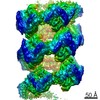 13547MC  7pmvC 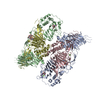 7povC 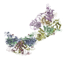 7pp6C C: citing same article ( M: map data used to model this data |
|---|---|
| Similar structure data |


-Links
 PDBj
PDBj


- Assembly
| Deposited unit | 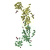
|
|---|---|
| 1 | x 18
|
-Components
| #1: Protein | / vWF Mass: 137345.641 Da / Num. of mol.: 2 Source method: isolated from a genetically manipulated source Source: (gene. exp.) Homo sapiens (human) / Gene: VWF, F8VWF / Production host: Homo sapiens (human) / References: UniProt: P04275#2: Polysaccharide | / Mass: 586.542 Da / Num. of mol.: 2Source method: isolated from a genetically manipulated source #3: Sugar | ChemComp-NAG / N-Acetylglucosamine  Type: D-saccharide, beta linking / Mass: 221.208 Da / Num. of mol.: 8 / Source method: obtained synthetically / Formula: C8H15NO6 Type: D-saccharide, beta linking / Mass: 221.208 Da / Num. of mol.: 8 / Source method: obtained synthetically / Formula: C8H15NO6#4: Chemical | ChemComp-CA /   Mass: 40.078 Da / Num. of mol.: 6 / Source method: obtained synthetically / Formula: Ca Mass: 40.078 Da / Num. of mol.: 6 / Source method: obtained synthetically / Formula: CaHas ligand of interest | N | |
|---|
-Experimental details
-Experiment
| Experiment | Method: ELECTRON MICROSCOPY |
|---|---|
| EM experiment | Aggregation state: HELICAL ARRAY / 3D reconstruction method: helical reconstruction |
- Sample preparation
| Component | Name: VWF tubule of domains D1D2D'D3 / Type: COMPLEX / Entity ID: #1 / Source: RECOMBINANT |
|---|---|
| Molecular weight | Experimental value: NO |
| Source (natural) | Organism: Homo sapiens (human) |
| Source (recombinant) | Organism: Homo sapiens (human) / Strain: HEK 293F / Plasmid: pCDNA3.1 |
| Buffer solution | pH: 6 |
| Specimen | Embedding applied: NO / Shadowing applied: NO / Staining applied: NO / Vitrification applied: YES |
| Vitrification | Cryogen name: ETHANE |
- Electron microscopy imaging
| Experimental equipment |  Model: Titan Krios / Image courtesy: FEI Company |
|---|---|
| Microscopy | Model: FEI TITAN KRIOS |
| Electron gun | Electron source: FIELD EMISSION GUN / Accelerating voltage: 300 kV / Illumination mode: FLOOD BEAM |
| Electron lens | Mode: BRIGHT FIELDBright-field microscopy |
| Image recording | Electron dose: 48 e/Å2 / Film or detector model: GATAN K3 BIOQUANTUM (6k x 4k) |
- Processing
| CTF correction | Type: PHASE FLIPPING AND AMPLITUDE CORRECTION |
|---|---|
| Helical symmerty | Angular rotation/subunit: 85.984 ° / Axial rise/subunit: 28.156 Å / Axial symmetry: D1 |
| 3D reconstruction | Resolution: 4.35 Å / Resolution method: FSC 0.143 CUT-OFF / Num. of particles: 88715 / Symmetry type: HELICAL |
