 Movie
Movie Controller


+ Open data

- Basic information
| Entry | Database: PDB / ID: 7mir | ||||||||||||
|---|---|---|---|---|---|---|---|---|---|---|---|---|---|
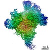
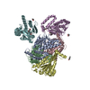
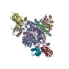
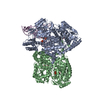
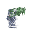
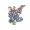
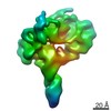
| Title | Cryo-EM structure of SidJ-SdeA-CaM reaction intermediate complex | ||||||||||||
 Components Components |
| ||||||||||||
 Keywords Keywords |  TRANSFERASE / HYDROLASE/LIGASE / SidJ / SdeA / CaM / complex / Intermediate / Acyl / Adenylate / Legionella / Ubiquitination / HYDROLASE-LIGASE complex TRANSFERASE / HYDROLASE/LIGASE / SidJ / SdeA / CaM / complex / Intermediate / Acyl / Adenylate / Legionella / Ubiquitination / HYDROLASE-LIGASE complex | ||||||||||||
| Function / homology |  Function and homology informationLigases / NAD+-protein-arginine ADP-ribosyltransferase / negative regulation of calcium ion transmembrane transporter activity / deNEDDylase activity / NAD+-protein-arginine ADP-ribosyltransferase activity / protein deneddylation / Transferases; Acyltransferases; Aminoacyltransferases / host cell / K63-linked deubiquitinase activity / negative regulation of calcium ion export across plasma membrane ...Ligases / NAD+-protein-arginine ADP-ribosyltransferase / negative regulation of calcium ion transmembrane transporter activity / deNEDDylase activity / NAD+-protein-arginine ADP-ribosyltransferase activity / protein deneddylation / Transferases; Acyltransferases; Aminoacyltransferases / host cell / K63-linked deubiquitinase activity / negative regulation of calcium ion export across plasma membrane / positive regulation of ryanodine-sensitive calcium-release channel activity / regulation of cell communication by electrical coupling involved in cardiac conduction / negative regulation of peptidyl-threonine phosphorylation / protein deubiquitination / protein phosphatase activator activity / positive regulation of cyclic-nucleotide phosphodiesterase activity / positive regulation of phosphoprotein phosphatase activity / ligase activity / adenylate cyclase binding / catalytic complex / detection of calcium ion / negative regulation of ryanodine-sensitive calcium-release channel activity / regulation of cardiac muscle contraction / calcium channel inhibitor activity / regulation of cardiac muscle contraction by regulation of the release of sequestered calcium ion / voltage-gated potassium channel complex / cysteine-type peptidase activity / regulation of release of sequestered calcium ion into cytosol by sarcoplasmic reticulum / regulation of calcium-mediated signaling / positive regulation of protein dephosphorylation / titin binding / positive regulation of protein autophosphorylation / sperm midpiece / calcium channel complex / substantia nigra development / adenylate cyclase activator activity / regulation of heart rate / nucleotidyltransferase activity / protein serine/threonine kinase activator activity / sarcomere / positive regulation of peptidyl-threonine phosphorylation / regulation of cytokinesis / spindle microtubule / positive regulation of protein serine/threonine kinase activity / spindle pole / response to calcium ion / calcium-dependent protein binding / G2/M transition of mitotic cell cycle / myelin sheath / transferase activity / vesicle / transmembrane transporter binding / Hydrolases; Acting on peptide bonds (peptidases); Cysteine endopeptidases / protein ubiquitination / G protein-coupled receptor signaling pathway / nucleotide binding / centrosome / calcium ion binding / protein kinase binding / protein-containing complex / proteolysis / extracellular region / membrane / metal ion binding / nucleus / plasma membrane / cytoplasm Function and homology informationLigases / NAD+-protein-arginine ADP-ribosyltransferase / negative regulation of calcium ion transmembrane transporter activity / deNEDDylase activity / NAD+-protein-arginine ADP-ribosyltransferase activity / protein deneddylation / Transferases; Acyltransferases; Aminoacyltransferases / host cell / K63-linked deubiquitinase activity / negative regulation of calcium ion export across plasma membrane ...Ligases / NAD+-protein-arginine ADP-ribosyltransferase / negative regulation of calcium ion transmembrane transporter activity / deNEDDylase activity / NAD+-protein-arginine ADP-ribosyltransferase activity / protein deneddylation / Transferases; Acyltransferases; Aminoacyltransferases / host cell / K63-linked deubiquitinase activity / negative regulation of calcium ion export across plasma membrane / positive regulation of ryanodine-sensitive calcium-release channel activity / regulation of cell communication by electrical coupling involved in cardiac conduction / negative regulation of peptidyl-threonine phosphorylation / protein deubiquitination / protein phosphatase activator activity / positive regulation of cyclic-nucleotide phosphodiesterase activity / positive regulation of phosphoprotein phosphatase activity / ligase activity / adenylate cyclase binding / catalytic complex / detection of calcium ion / negative regulation of ryanodine-sensitive calcium-release channel activity / regulation of cardiac muscle contraction / calcium channel inhibitor activity / regulation of cardiac muscle contraction by regulation of the release of sequestered calcium ion / voltage-gated potassium channel complex / cysteine-type peptidase activity / regulation of release of sequestered calcium ion into cytosol by sarcoplasmic reticulum / regulation of calcium-mediated signaling / positive regulation of protein dephosphorylation / titin binding / positive regulation of protein autophosphorylation / sperm midpiece / calcium channel complex / substantia nigra development / adenylate cyclase activator activity / regulation of heart rate / nucleotidyltransferase activity / protein serine/threonine kinase activator activity / sarcomere / positive regulation of peptidyl-threonine phosphorylation / regulation of cytokinesis / spindle microtubule / positive regulation of protein serine/threonine kinase activity / spindle pole / response to calcium ion / calcium-dependent protein binding / G2/M transition of mitotic cell cycle / myelin sheath / transferase activity / vesicle / transmembrane transporter binding / Hydrolases; Acting on peptide bonds (peptidases); Cysteine endopeptidases / protein ubiquitination / G protein-coupled receptor signaling pathway / nucleotide binding / centrosome / calcium ion binding / protein kinase binding / protein-containing complex / proteolysis / extracellular region / membrane / metal ion binding / nucleus / plasma membrane / cytoplasmSimilarity search - Function | ||||||||||||
| Biological species |   Legionella pneumophila (bacteria) Legionella pneumophila (bacteria) Homo sapiens (human) Homo sapiens (human) | ||||||||||||
| Method | ELECTRON MICROSCOPY / single particle reconstruction / cryo EM / Resolution: 2.5 Å | ||||||||||||
 Authors Authors | Osinski, A. / Black, M.H. / Pawlowski, K. / Chen, Z. / Li, Y. / Tagliabracci, V.S. | ||||||||||||
| Funding support |  United States, 3items United States, 3items
| ||||||||||||
 Citation Citation | Journal: Mol Cell / Year: 2021 Title: Structural and mechanistic basis for protein glutamylation by the kinase fold. Authors: Adam Osinski / Miles H Black / Krzysztof Pawłowski / Zhe Chen / Yang Li / Vincent S Tagliabracci /  Abstract: The kinase domain transfers phosphate from ATP to substrates. However, the Legionella effector SidJ adopts a kinase fold, yet catalyzes calmodulin (CaM)-dependent glutamylation to inactivate the SidE ...The kinase domain transfers phosphate from ATP to substrates. However, the Legionella effector SidJ adopts a kinase fold, yet catalyzes calmodulin (CaM)-dependent glutamylation to inactivate the SidE ubiquitin ligases. The structural and mechanistic basis in which the kinase domain catalyzes protein glutamylation is unknown. Here we present cryo-EM reconstructions of SidJ:CaM:SidE reaction intermediate complexes. We show that the kinase-like active site of SidJ adenylates an active-site Glu in SidE, resulting in the formation of a stable reaction intermediate complex. An insertion in the catalytic loop of the kinase domain positions the donor Glu near the acyl-adenylate for peptide bond formation. Our structural analysis led us to discover that the SidJ paralog SdjA is a glutamylase that differentially regulates the SidE ligases during Legionella infection. Our results uncover the structural and mechanistic basis in which the kinase fold catalyzes non-ribosomal amino acid ligations and reveal an unappreciated level of SidE-family regulation. | ||||||||||||
| History |
|
- Structure visualization
| Movie |
Movie viewer |
|---|---|
| Structure viewer | Molecule: MolmilJmol/JSmol |
- Downloads & links
-Download
| PDBx/mmCIF format | 7mir.cif.gz | 540.6 KB | Display | PDBx/mmCIF format |
|---|---|---|---|---|
| PDB format | pdb7mir.ent.gz | 453.4 KB | Display | PDB format |
| PDBx/mmJSON format | 7mir.json.gz | Tree view | PDBx/mmJSON format | |
| Others |  Other downloads Other downloads |
-Validation report
| Arichive directory | https://data.pdbj.org/pub/pdb/validation_reports/mi/7mirftp://data.pdbj.org/pub/pdb/validation_reports/mi/7mir | HTTPS FTP |
|---|
-Related structure data
| Related structure data | 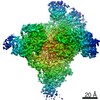 23862MC 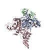 7misC M: map data used to model this data C: citing same article ( |
|---|---|
| Similar structure data |


-Links
 PDBj
PDBj


- Assembly
| Deposited unit | 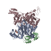
|
|---|---|
| 1 |
|
-Components
-Protein , 3 types, 3 molecules ABC
| #1: Protein | Mass: 87374.031 Da / Num. of mol.: 1 Source method: isolated from a genetically manipulated source Source: (gene. exp.) Legionella pneumophila (bacteria) / Gene: sidJ, lpg2155 / Production host: Escherichia coli (E. coli) / References: UniProt: Q5ZTK6, Ligases |
|---|---|
| #2: Protein | Mass: 16939.623 Da / Num. of mol.: 1 Source method: isolated from a genetically manipulated source Source: (gene. exp.) Homo sapiens (human) / Gene: CALM2, CAM2, CAMB / Production host: Escherichia coli (E. coli) / References: UniProt: P0DP24 |
| #3: Protein | Mass: 109120.219 Da / Num. of mol.: 1 Source method: isolated from a genetically manipulated source Source: (gene. exp.) Legionella pneumophila (bacteria) / Gene: sdeA, lpg2157 / Production host: Escherichia coli (E. coli)References: UniProt: Q5ZTK4, Hydrolases; Acting on peptide bonds (peptidases); Cysteine endopeptidases, Transferases; Acyltransferases; Aminoacyltransferases, NAD+-protein-arginine ADP-ribosyltransferase |
-Non-polymers , 4 types, 5 molecules 


| #4: Chemical | ChemComp-AMP / Adenosine monophosphate Mass: 347.221 Da / Num. of mol.: 1 / Source method: obtained synthetically / Formula: C10H14N5O7P / Feature type: SUBJECT OF INVESTIGATION / Comment: AMP*YM Mass: 347.221 Da / Num. of mol.: 1 / Source method: obtained synthetically / Formula: C10H14N5O7P / Feature type: SUBJECT OF INVESTIGATION / Comment: AMP*YM | ||||
|---|---|---|---|---|---|
| #5: Chemical |  Mass: 24.305 Da / Num. of mol.: 2 / Source method: obtained synthetically / Formula: Mg / Feature type: SUBJECT OF INVESTIGATION Mass: 24.305 Da / Num. of mol.: 2 / Source method: obtained synthetically / Formula: Mg / Feature type: SUBJECT OF INVESTIGATION#6: Chemical | ChemComp-ATP / | Adenosine triphosphate Mass: 507.181 Da / Num. of mol.: 1 / Source method: obtained synthetically / Formula: C10H16N5O13P3 / Feature type: SUBJECT OF INVESTIGATION / Comment: ATP, energy-carrying molecule*YM Mass: 507.181 Da / Num. of mol.: 1 / Source method: obtained synthetically / Formula: C10H16N5O13P3 / Feature type: SUBJECT OF INVESTIGATION / Comment: ATP, energy-carrying molecule*YM#7: Chemical | ChemComp-CA / |  Mass: 40.078 Da / Num. of mol.: 1 / Source method: obtained synthetically / Formula: Ca / Feature type: SUBJECT OF INVESTIGATION Mass: 40.078 Da / Num. of mol.: 1 / Source method: obtained synthetically / Formula: Ca / Feature type: SUBJECT OF INVESTIGATION |
-Details
| Has ligand of interest | Y |
|---|
-Experimental details
-Experiment
| Experiment | Method: ELECTRON MICROSCOPY |
|---|---|
| EM experiment | Aggregation state: PARTICLE / 3D reconstruction method: single particle reconstruction |
- Sample preparation
| Component |
| ||||||||||||||||||||||||
|---|---|---|---|---|---|---|---|---|---|---|---|---|---|---|---|---|---|---|---|---|---|---|---|---|---|
| Molecular weight | Experimental value: NO | ||||||||||||||||||||||||
| Source (natural) |
| ||||||||||||||||||||||||
| Source (recombinant) |
| ||||||||||||||||||||||||
| Buffer solution | pH: 6.5 Details: 25 mM Bis-tris pH 6.5, 100 mM NaCl, 1 mM TCEP, 2 mM MgCl2, 1 mM ATP | ||||||||||||||||||||||||
| Specimen | Conc.: 0.35 mg/ml / Embedding applied: NO / Shadowing applied: NO / Staining applied: NO / Vitrification applied: YES | ||||||||||||||||||||||||
| Vitrification | Instrument: FEI VITROBOT MARK IV / Cryogen name: ETHANE |
- Electron microscopy imaging
| Experimental equipment |  Model: Titan Krios / Image courtesy: FEI Company |
|---|---|
| Microscopy | Model: FEI TITAN KRIOS |
| Electron gun | Electron source: FIELD EMISSION GUN / Accelerating voltage: 300 kV / Illumination mode: FLOOD BEAM |
| Electron lens | Mode: BRIGHT FIELDBright-field microscopy |
| Image recording | Electron dose: 50 e/Å2 / Film or detector model: GATAN K3 BIOQUANTUM (6k x 4k) |
- Processing
| Software | Name: PHENIX / Version: 1.19.1_4122: / Classification: refinement | ||||||||||||||||||||||||
|---|---|---|---|---|---|---|---|---|---|---|---|---|---|---|---|---|---|---|---|---|---|---|---|---|---|
| CTF correction | Type: PHASE FLIPPING AND AMPLITUDE CORRECTION | ||||||||||||||||||||||||
| 3D reconstruction | Resolution: 2.5 Å / Resolution method: FSC 0.143 CUT-OFF / Num. of particles: 310154 / Symmetry type: POINT | ||||||||||||||||||||||||
| Refine LS restraints |
|
