 ムービー
ムービー コントローラー


+ データを開く

- 基本情報
基本情報
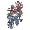
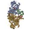
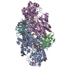
| 登録情報 | データベース: PDB / ID: 7kui | |||||||||
|---|---|---|---|---|---|---|---|---|---|---|
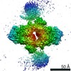
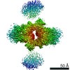
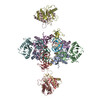
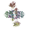
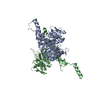
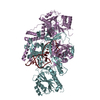
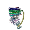
| タイトル | Cryo-EM structure of Rous sarcoma virus cleaved synaptic complex (CSC) with HIV-1 integrase strand transfer inhibitor MK-2048. CIC region of a cluster identified by 3-dimensional variability analysis in cryoSPARC. | |||||||||
 要素 要素 |
| |||||||||
 キーワード キーワード |  VIRAL PROTEIN (ウイルスタンパク質) / intasome / integrase-viral DNA complex / HYDROLASE-DNA-INHIBITOR complex VIRAL PROTEIN (ウイルスタンパク質) / intasome / integrase-viral DNA complex / HYDROLASE-DNA-INHIBITOR complex | |||||||||
| 機能・相同性 |  機能・相同性情報加水分解酵素; プロテアーゼ; ペプチド結合加水分解酵素; アスパラギン酸プロテアーゼ / リボヌクレアーゼH / DNA integration / 逆転写酵素 / viral genome integration into host DNA / establishment of integrated proviral latency / RNA-directed DNA polymerase activity / RNA-DNA hybrid ribonuclease activity / 転移酵素; リンを含む基を移すもの; 核酸を移すもの / viral nucleocapsid ...加水分解酵素; プロテアーゼ; ペプチド結合加水分解酵素; アスパラギン酸プロテアーゼ / リボヌクレアーゼH / DNA integration / 逆転写酵素 / viral genome integration into host DNA / establishment of integrated proviral latency / RNA-directed DNA polymerase activity / RNA-DNA hybrid ribonuclease activity / 転移酵素; リンを含む基を移すもの; 核酸を移すもの / viral nucleocapsid / DNA recombination / 加水分解酵素; エステル加水分解酵素 / DNAポリメラーゼ / aspartic-type endopeptidase activity / DNA-directed DNA polymerase activity / symbiont entry into host cell / タンパク質分解 / DNA binding / RNA binding / zinc ion binding 機能・相同性情報加水分解酵素; プロテアーゼ; ペプチド結合加水分解酵素; アスパラギン酸プロテアーゼ / リボヌクレアーゼH / DNA integration / 逆転写酵素 / viral genome integration into host DNA / establishment of integrated proviral latency / RNA-directed DNA polymerase activity / RNA-DNA hybrid ribonuclease activity / 転移酵素; リンを含む基を移すもの; 核酸を移すもの / viral nucleocapsid ...加水分解酵素; プロテアーゼ; ペプチド結合加水分解酵素; アスパラギン酸プロテアーゼ / リボヌクレアーゼH / DNA integration / 逆転写酵素 / viral genome integration into host DNA / establishment of integrated proviral latency / RNA-directed DNA polymerase activity / RNA-DNA hybrid ribonuclease activity / 転移酵素; リンを含む基を移すもの; 核酸を移すもの / viral nucleocapsid / DNA recombination / 加水分解酵素; エステル加水分解酵素 / DNAポリメラーゼ / aspartic-type endopeptidase activity / DNA-directed DNA polymerase activity / symbiont entry into host cell / タンパク質分解 / DNA binding / RNA binding / zinc ion binding類似検索 - 分子機能 | |||||||||
| 生物種 |  Rous sarcoma virus (ラウス肉腫ウイルス) Rous sarcoma virus (ラウス肉腫ウイルス) | |||||||||
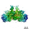
| 手法 | 電子顕微鏡法 / 単粒子再構成法 / クライオ電子顕微鏡法 / 解像度: 3.4 Å | |||||||||
 データ登録者 データ登録者 | Pandey, K.K. / Bera, S. / Shi, K. / Aihara, H. / Grandgenett, D.P. | |||||||||
| 資金援助 |  米国, 2件 米国, 2件
| |||||||||
 引用 引用 | ジャーナル: Commun Biol / 年: 2021 タイトル: Cryo-EM structure of the Rous sarcoma virus octameric cleaved synaptic complex intasome. 著者: Krishan K Pandey / Sibes Bera / Ke Shi / Michael J Rau / Amarachi V Oleru / James A J Fitzpatrick / Alan N Engelman / Hideki Aihara / Duane P Grandgenett / 要旨: Despite conserved catalytic integration mechanisms, retroviral intasomes composed of integrase (IN) and viral DNA possess diverse structures with variable numbers of IN subunits. To investigate ...Despite conserved catalytic integration mechanisms, retroviral intasomes composed of integrase (IN) and viral DNA possess diverse structures with variable numbers of IN subunits. To investigate intasome assembly mechanisms, we employed the Rous sarcoma virus (RSV) IN dimer that assembles a precursor tetrameric structure in transit to the mature octameric intasome. We determined the structure of RSV octameric intasome stabilized by a HIV-1 IN strand transfer inhibitor using single particle cryo-electron microscopy. The structure revealed significant flexibility of the two non-catalytic distal IN dimers along with previously unrecognized movement of the conserved intasome core, suggesting ordered conformational transitions between intermediates that may be important to capture the target DNA. Single amino acid substitutions within the IN C-terminal domain affected intasome assembly and function in vitro and infectivity of pseudotyped RSV virions. Unexpectedly, 17 C-terminal amino acids of IN were dispensable for virus infection despite regulating the transition of the tetrameric intasome to the octameric form in vitro. We speculate that this region may regulate the binding of highly flexible distal IN dimers to the intasome core to form the octameric complex. Our studies reveal key steps in the assembly of RSV intasomes. | |||||||||
| 履歴 |
|
- 構造の表示
構造の表示
| ムービー |
ムービービューア |
|---|---|
| 構造ビューア | 分子: MolmilJmol/JSmol |
- ダウンロードとリンク
-ダウンロード
| PDBx/mmCIF形式 | 7kui.cif.gz | 257.2 KB | 表示 | PDBx/mmCIF形式 |
|---|---|---|---|---|
| PDB形式 | pdb7kui.ent.gz | 198.1 KB | 表示 | PDB形式 |
| PDBx/mmJSON形式 | 7kui.json.gz | ツリー表示 | PDBx/mmJSON形式 | |
| その他 |  その他のダウンロード その他のダウンロード |
-検証レポート
| アーカイブディレクトリ | https://data.pdbj.org/pub/pdb/validation_reports/ku/7kuiftp://data.pdbj.org/pub/pdb/validation_reports/ku/7kui | HTTPS FTP |
|---|
-関連構造データ

-リンク
 PDBj
PDBj


- 集合体
集合体
| 登録構造単位 | 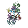
|
|---|---|
| 1 |
|
-要素
| #1: タンパク質 | インテグラーゼ 分子量: 30926.582 Da / 分子数: 8 / 由来タイプ: 組換発現 由来: (組換発現) Rous sarcoma virus (strain Schmidt-Ruppin A) (ラウス肉腫ウイルス)株: Schmidt-Ruppin A / 遺伝子: gag-pol / 発現宿主:  Escherichia coli BL21(DE3) (大腸菌) / 株 (発現宿主): pLysS Escherichia coli BL21(DE3) (大腸菌) / 株 (発現宿主): pLysS参照: UniProt: P03354, 転移酵素; リンを含む基を移すもの; 核酸を移すもの, 加水分解酵素; エステル加水分解酵素#2: DNA鎖 | 分子量: 5520.600 Da / 分子数: 2 / 由来タイプ: 合成 由来: (合成) Rous sarcoma virus (strain Schmidt-Ruppin A) (ラウス肉腫ウイルス)#3: DNA鎖 | 分子量: 4899.232 Da / 分子数: 2 / 由来タイプ: 合成 由来: (合成) Rous sarcoma virus (strain Schmidt-Ruppin A) (ラウス肉腫ウイルス)#4: 化合物 |   分子量: 65.409 Da / 分子数: 2 / 由来タイプ: 合成 / 式: Zn 分子量: 65.409 Da / 分子数: 2 / 由来タイプ: 合成 / 式: Zn#5: 化合物 | MK-2048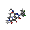  分子量: 461.874 Da / 分子数: 2 / 由来タイプ: 合成 / 式: C21H21ClFN5O4 / タイプ: SUBJECT OF INVESTIGATION / コメント: 阻害剤*YM 分子量: 461.874 Da / 分子数: 2 / 由来タイプ: 合成 / 式: C21H21ClFN5O4 / タイプ: SUBJECT OF INVESTIGATION / コメント: 阻害剤*YM研究の焦点であるリガンドがあるか | Y | |
|---|
-実験情報
-実験
| 実験 | 手法: 電子顕微鏡法 |
|---|---|
| EM実験 | 試料の集合状態: PARTICLE / 3次元再構成法: 単粒子再構成法 |
- 試料調製
試料調製
| 構成要素 | 名称: CIC region of a cluster identified by 3-dimensional variability analysis in cryoSPARC of a cleaved synaptic complex (CSC) formed with Rous sarcoma virus integrase and viral DNA in presence of ...名称: CIC region of a cluster identified by 3-dimensional variability analysis in cryoSPARC of a cleaved synaptic complex (CSC) formed with Rous sarcoma virus integrase and viral DNA in presence of HIV-1 integrase strand inhibitor MK-2048 タイプ: COMPLEX / Entity ID: #1-#3 / 由来: RECOMBINANT |
|---|---|
| 分子量 | 実験値: NO |
| 由来(天然) | 生物種: Rous sarcoma virus (strain Schmidt-Ruppin A) (ラウス肉腫ウイルス) |
| 由来(組換発現) | 生物種: Escherichia coli BL21(DE3) (大腸菌) / 株: pLysS |
| 緩衝液 | pH: 7.5 |
| 試料 | 濃度: 0.5 mg/ml / 包埋: NO / シャドウイング: NO / 染色: NO / 凍結: YES |
| 試料支持 | グリッドの材料: COPPER / グリッドのサイズ: 300 divisions/in. / グリッドのタイプ: Quantifoil R2/2 |
| 急速凍結 | 装置: FEI VITROBOT MARK IV / 凍結剤: ETHANE / 湿度: 100 % / 凍結前の試料温度: 277 K |
- 電子顕微鏡撮影
| 実験機器 |  モデル: Titan Krios / 画像提供: FEI Company |
|---|---|
| 顕微鏡 | モデル: TFS KRIOS |
| 電子銃 | 電子線源: FIELD EMISSION GUN / 加速電圧: 300 kV / 照射モード: FLOOD BEAM |
| 電子レンズ | モード: BRIGHT FIELDBright-field microscopy / 倍率(公称値): 105000 X / Cs: 0.01 mm / アライメント法: BASIC |
| 試料ホルダ | 凍結剤: NITROGEN 試料ホルダーモデル: FEI TITAN KRIOS AUTOGRID HOLDER |
| 撮影 | 電子線照射量: 66 e/Å2 / 検出モード: SUPER-RESOLUTION フィルム・検出器のモデル: GATAN K2 SUMMIT (4k x 4k) 実像数: 5187 |
| 電子光学装置 | エネルギーフィルター名称: GIF Bioquantum / エネルギーフィルタースリット幅: 20 eV |
| 画像スキャン | 動画フレーム数/画像: 40 |
- 解析
解析
| ソフトウェア | 名称: PHENIX / バージョン: 1.19rc5_4047: / 分類: 精密化 | ||||||||||||||||||||||||
|---|---|---|---|---|---|---|---|---|---|---|---|---|---|---|---|---|---|---|---|---|---|---|---|---|---|
| EMソフトウェア |
| ||||||||||||||||||||||||
| CTF補正 | タイプ: NONE | ||||||||||||||||||||||||
| 粒子像の選択 | 選択した粒子像数: 1811357 | ||||||||||||||||||||||||
| 3次元再構成 | 解像度: 3.4 Å / 解像度の算出法: FSC 0.143 CUT-OFF / 粒子像の数: 64449 詳細: Refined map is for one cluster identified by 3-dimensional variability analysis done in cryoSPARC. The model represents the CIC region. 対称性のタイプ: POINT | ||||||||||||||||||||||||
| 原子モデル構築 | B value: 30 / プロトコル: RIGID BODY FIT / 空間: REAL / Target criteria: correlation coefficient | ||||||||||||||||||||||||
| 原子モデル構築 | PDB-ID: 7KU7 Accession code: 7KU7 / Source name: PDB / タイプ: experimental model | ||||||||||||||||||||||||
| 拘束条件 |
|
