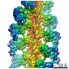
 Movie
Movie Controller

[English] 日本語
 Yorodumi
- PDB-7bkc: Formate dehydrogenase - heterodisulfide reductase - formylmethano... -
+ Open data

- Basic information
| Entry | Database: PDB / ID: 7bkc | |||||||||||||||
|---|---|---|---|---|---|---|---|---|---|---|---|---|---|---|---|---|
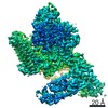
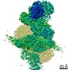
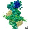
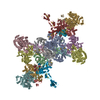
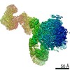
| Title | Formate dehydrogenase - heterodisulfide reductase - formylmethanofuran dehydrogenase complex from Methanospirillum hungatei (dimeric, composite structure) | |||||||||||||||
 Components Components |
| |||||||||||||||
 Keywords Keywords |  OXIDOREDUCTASE / methanogenesis / flavin-based electron bifurcation / CO2-fixation / formate dehydrogenase OXIDOREDUCTASE / methanogenesis / flavin-based electron bifurcation / CO2-fixation / formate dehydrogenase | |||||||||||||||
| Function / homology |  Function and homology informationformylmethanofuran dehydrogenase / formylmethanofuran dehydrogenase / formylmethanofuran dehydrogenase activity / Oxidoreductases; Acting on the aldehyde or oxo group of donors; With unknown physiological acceptors / CoB--CoM heterodisulfide reductase activity / methanogenesis, from carbon dioxide / formate metabolic process / Oxidoreductases; Acting on a sulfur group of donors / methanogenesis / formate dehydrogenase (NAD+) activity ...formylmethanofuran dehydrogenase / formylmethanofuran dehydrogenase / formylmethanofuran dehydrogenase activity / Oxidoreductases; Acting on the aldehyde or oxo group of donors; With unknown physiological acceptors / CoB--CoM heterodisulfide reductase activity / methanogenesis, from carbon dioxide / formate metabolic process / Oxidoreductases; Acting on a sulfur group of donors / methanogenesis / formate dehydrogenase (NAD+) activity / hydrolase activity, acting on carbon-nitrogen (but not peptide) bonds / molybdopterin cofactor binding / iron-sulfur cluster binding / transition metal ion binding / 4 iron, 4 sulfur cluster binding / oxidoreductase activity / metal ion binding Function and homology informationformylmethanofuran dehydrogenase / formylmethanofuran dehydrogenase / formylmethanofuran dehydrogenase activity / Oxidoreductases; Acting on the aldehyde or oxo group of donors; With unknown physiological acceptors / CoB--CoM heterodisulfide reductase activity / methanogenesis, from carbon dioxide / formate metabolic process / Oxidoreductases; Acting on a sulfur group of donors / methanogenesis / formate dehydrogenase (NAD+) activity ...formylmethanofuran dehydrogenase / formylmethanofuran dehydrogenase / formylmethanofuran dehydrogenase activity / Oxidoreductases; Acting on the aldehyde or oxo group of donors; With unknown physiological acceptors / CoB--CoM heterodisulfide reductase activity / methanogenesis, from carbon dioxide / formate metabolic process / Oxidoreductases; Acting on a sulfur group of donors / methanogenesis / formate dehydrogenase (NAD+) activity / hydrolase activity, acting on carbon-nitrogen (but not peptide) bonds / molybdopterin cofactor binding / iron-sulfur cluster binding / transition metal ion binding / 4 iron, 4 sulfur cluster binding / oxidoreductase activity / metal ion bindingSimilarity search - Function | |||||||||||||||
| Biological species |  Methanospirillum hungatei JF-1 (archaea) Methanospirillum hungatei JF-1 (archaea) | |||||||||||||||
| Method | ELECTRON MICROSCOPY / single particle reconstruction / cryo EM / Resolution: 3 Å | |||||||||||||||
 Authors Authors | Pfeil-Gardiner, O. / Watanabe, T. / Shima, S. / Murphy, B.J. | |||||||||||||||
| Funding support |  Germany, Germany,  Japan, 4items Japan, 4items
| |||||||||||||||
 Citation Citation | Journal: Science / Year: 2021 Title: Three-megadalton complex of methanogenic electron-bifurcating and CO-fixing enzymes. Authors: Tomohiro Watanabe / Olivia Pfeil-Gardiner / Jörg Kahnt / Jürgen Koch / Seigo Shima / Bonnie J Murphy / Abstract: The first reaction of the methanogenic pathway from carbon dioxide (CO) is the reduction and condensation of CO to formyl-methanofuran, catalyzed by formyl-methanofuran dehydrogenase (Fmd). Strongly ...The first reaction of the methanogenic pathway from carbon dioxide (CO) is the reduction and condensation of CO to formyl-methanofuran, catalyzed by formyl-methanofuran dehydrogenase (Fmd). Strongly reducing electrons for this reaction are generated by heterodisulfide reductase (Hdr) in complex with hydrogenase or formate dehydrogenase (Fdh) using a flavin-based electron-bifurcation mechanism. Here, we report enzymological and structural characterizations of Fdh-Hdr-Fmd complexes from . The complexes catalyze this reaction using electrons from formate and the reduced form of the electron carrier F. Conformational changes in HdrA mediate electron bifurcation, and polyferredoxin FmdF directly transfers electrons to the CO reduction site, as evidenced by methanofuran-dependent flavin-based electron bifurcation even without free ferredoxin, a diffusible electron carrier between Hdr and Fmd. Conservation of Hdr and Fmd structures suggests that this complex is common among hydrogenotrophic methanogens. #1: Journal: Comput. Cryst. Newsl. / Year: 2013Title: New tool: phenix.real_space_refine Authors: Afonine, P.V. / Headd, J.J. / Terwilliger, T.C. / Adams, P.D. | |||||||||||||||
| History |
|
- Structure visualization
| Movie |
Movie viewer |
|---|---|
| Structure viewer | Molecule: MolmilJmol/JSmol |
- Downloads & links
-Download
| PDBx/mmCIF format | 7bkc.cif.gz | 1.4 MB | Display | PDBx/mmCIF format |
|---|---|---|---|---|
| PDB format | pdb7bkc.ent.gz | 1.2 MB | Display | PDB format |
| PDBx/mmJSON format | 7bkc.json.gz | Tree view | PDBx/mmJSON format | |
| Others |  Other downloads Other downloads |
-Validation report
| Arichive directory | https://data.pdbj.org/pub/pdb/validation_reports/bk/7bkcftp://data.pdbj.org/pub/pdb/validation_reports/bk/7bkc | HTTPS FTP |
|---|
-Related structure data
| Related structure data |  12209MC  7bkbC  7bkdC  7bkeC M: map data used to model this data C: citing same article ( |
|---|---|
| Similar structure data |


-Links
 PDBj
PDBj


- Assembly
| Deposited unit | 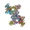
|
|---|---|
| 1 |
|
-Components
-Protein , 5 types, 10 molecules AaFfEeDdIi
| #1: Protein | Mass: 72885.062 Da / Num. of mol.: 2 / Source method: isolated from a natural source / Source: (natural) Methanospirillum hungatei JF-1 (archaea)References: UniProt: Q2FKZ1, Oxidoreductases; Acting on a sulfur group of donors#2: Protein | Mass: 15692.425 Da / Num. of mol.: 2 / Source method: isolated from a natural source / Source: (natural) Methanospirillum hungatei JF-1 (archaea) / References: UniProt: Q2FKZ0#3: Protein | Mass: 45639.895 Da / Num. of mol.: 2 / Source method: isolated from a natural source / Source: (natural) Methanospirillum hungatei JF-1 (archaea)References: UniProt: Q2FME3, Oxidoreductases; Acting on the aldehyde or oxo group of donors; With unknown physiological acceptors#6: Protein | Mass: 75911.445 Da / Num. of mol.: 2 / Source method: isolated from a natural source / Source: (natural) Methanospirillum hungatei JF-1 (archaea) / References: UniProt: Q2FRK1, formate dehydrogenase#7: Protein | Mass: 28696.902 Da / Num. of mol.: 2 / Source method: isolated from a natural source / Source: (natural) Methanospirillum hungatei JF-1 (archaea)References: UniProt: Q2FRL8, formylmethanofuran dehydrogenase |
|---|
-CoB--CoM heterodisulfide reductase subunit ... , 2 types, 4 molecules CcBb
| #4: Protein | CoB—CoM heterodisulfide reductase Mass: 21706.963 Da / Num. of mol.: 2 / Source method: isolated from a natural source / Source: (natural) Methanospirillum hungatei JF-1 (archaea) / References: UniProt: Q2FKZ3#5: Protein | CoB—CoM heterodisulfide reductaseMass: 32930.938 Da / Num. of mol.: 2 / Source method: isolated from a natural source / Source: (natural) Methanospirillum hungatei JF-1 (archaea) / References: UniProt: Q2FKZ2 |
|---|
-Formylmethanofuran dehydrogenase, subunit ... , 5 types, 12 molecules LlGgJjMKmkHh
| #8: Protein | Mass: 16388.020 Da / Num. of mol.: 2 / Source method: isolated from a natural source / Source: (natural) Methanospirillum hungatei JF-1 (archaea)References: UniProt: Q2FKZ5, formylmethanofuran dehydrogenase#9: Protein | Mass: 63636.211 Da / Num. of mol.: 2 / Source method: isolated from a natural source / Source: (natural) Methanospirillum hungatei JF-1 (archaea)References: UniProt: Q2FRL9, formylmethanofuran dehydrogenase#10: Protein | Mass: 15157.499 Da / Num. of mol.: 2 / Source method: isolated from a natural source / Source: (natural) Methanospirillum hungatei JF-1 (archaea)References: UniProt: Q2FRF6, formylmethanofuran dehydrogenase#11: Protein | Mass: 42216.734 Da / Num. of mol.: 4 / Source method: isolated from a natural source / Source: (natural) Methanospirillum hungatei JF-1 (archaea)References: UniProt: Q2FKZ4, formylmethanofuran dehydrogenase#12: Protein | Mass: 49371.586 Da / Num. of mol.: 2 / Source method: isolated from a natural source / Source: (natural) Methanospirillum hungatei JF-1 (archaea)References: UniProt: Q2FRM0, formylmethanofuran dehydrogenase |
|---|
-Non-polymers , 7 types, 68 molecules 


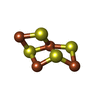


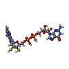
| #13: Chemical | ChemComp-SF4 / Iron–sulfur cluster Mass: 351.640 Da / Num. of mol.: 48 / Source method: obtained synthetically / Formula: Fe4S4 Mass: 351.640 Da / Num. of mol.: 48 / Source method: obtained synthetically / Formula: Fe4S4#14: Chemical | ChemComp-FAD / Flavin adenine dinucleotide Mass: 785.550 Da / Num. of mol.: 4 / Source method: obtained synthetically / Formula: C27H33N9O15P2 / Feature type: SUBJECT OF INVESTIGATION / Comment: FAD*YM Mass: 785.550 Da / Num. of mol.: 4 / Source method: obtained synthetically / Formula: C27H33N9O15P2 / Feature type: SUBJECT OF INVESTIGATION / Comment: FAD*YM#15: Chemical | Iron–sulfur cluster Mass: 175.820 Da / Num. of mol.: 2 / Source method: obtained synthetically / Formula: Fe2S2 Mass: 175.820 Da / Num. of mol.: 2 / Source method: obtained synthetically / Formula: Fe2S2#16: Chemical | ChemComp-9S8 /  Mass: 351.640 Da / Num. of mol.: 4 / Source method: obtained synthetically / Formula: Fe4S4 / Feature type: SUBJECT OF INVESTIGATION Mass: 351.640 Da / Num. of mol.: 4 / Source method: obtained synthetically / Formula: Fe4S4 / Feature type: SUBJECT OF INVESTIGATION#17: Chemical | ChemComp-ZN /  Mass: 65.409 Da / Num. of mol.: 4 / Source method: obtained synthetically / Formula: Zn Mass: 65.409 Da / Num. of mol.: 4 / Source method: obtained synthetically / Formula: Zn#18: Chemical |  Mass: 95.940 Da / Num. of mol.: 2 / Source method: obtained synthetically / Formula: Mo Mass: 95.940 Da / Num. of mol.: 2 / Source method: obtained synthetically / Formula: Mo#19: Chemical | ChemComp-MGD /  Mass: 740.557 Da / Num. of mol.: 4 / Source method: obtained synthetically / Formula: C20H26N10O13P2S2 / Feature type: SUBJECT OF INVESTIGATION Mass: 740.557 Da / Num. of mol.: 4 / Source method: obtained synthetically / Formula: C20H26N10O13P2S2 / Feature type: SUBJECT OF INVESTIGATION |
|---|
-Details
| Has ligand of interest | Y |
|---|
-Experimental details
-Experiment
| Experiment | Method: ELECTRON MICROSCOPY |
|---|---|
| EM experiment | Aggregation state: PARTICLE / 3D reconstruction method: single particle reconstruction |
- Sample preparation
| Component | Name: Dimeric formate dehydrogenase - heterodisulfide reductase - formylmethanofuran dehydrogenase complex Type: COMPLEX / Entity ID: #1-#12 / Source: NATURAL |
|---|---|
| Molecular weight | Value: 0.948 MDa / Experimental value: NO |
| Source (natural) | Organism: Methanospirillum hungatei JF-1 (archaea) / Cellular location: cytoplasm |
| Buffer solution | pH: 7.6 |
| Buffer component | Conc.: 25 mM / Name: Tris-HClTris / Formula: Tris-HClTris |
| Specimen | Conc.: 0.5 mg/ml / Embedding applied: NO / Shadowing applied: NO / Staining applied: NO / Vitrification applied: YES Details: Preparation in an anaerobic tent (O2 < 20 ppm at all times, nearly always < 2ppm) |
| Specimen support | Details: 15mA / Grid material: COPPER / Grid mesh size: 300 divisions/in. / Grid type: C-flat-2/1 |
| Vitrification | Instrument: HOMEMADE PLUNGER / Cryogen name: ETHANE / Humidity: 70 % / Chamber temperature: 293 K |
- Electron microscopy imaging
| Experimental equipment |  Model: Titan Krios / Image courtesy: FEI Company |
|---|---|
| Microscopy | Model: FEI TITAN KRIOS |
| Electron gun | Electron source: FIELD EMISSION GUN / Accelerating voltage: 300 kV / Illumination mode: FLOOD BEAM |
| Electron lens | Mode: BRIGHT FIELDBright-field microscopy / Cs: 2.7 mm / C2 aperture diameter: 70 µm / Alignment procedure: COMA FREE |
| Specimen holder | Cryogen: NITROGEN / Specimen holder model: FEI TITAN KRIOS AUTOGRID HOLDER |
| Image recording | Electron dose: 50 e/Å2 / Film or detector model: GATAN K3 BIOQUANTUM (6k x 4k) / Num. of real images: 8745 |
| EM imaging optics | Energyfilter name: GIF Bioquantum / Energyfilter slit width: 20 eV |
- Processing
| Software | Name: PHENIX / Classification: refinement | ||||||||||||||||||||||||||||||||
|---|---|---|---|---|---|---|---|---|---|---|---|---|---|---|---|---|---|---|---|---|---|---|---|---|---|---|---|---|---|---|---|---|---|
| EM software |
| ||||||||||||||||||||||||||||||||
| Image processing | Details: Recorded in counted mode | ||||||||||||||||||||||||||||||||
| CTF correction | Type: PHASE FLIPPING AND AMPLITUDE CORRECTION | ||||||||||||||||||||||||||||||||
| Symmetry | Point symmetry: C2 (2 fold cyclic) | ||||||||||||||||||||||||||||||||
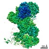
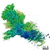
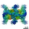
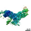
| 3D reconstruction | Resolution: 3 Å / Resolution method: FSC 0.143 CUT-OFF / Num. of particles: 1239454 Details: As this is a composite map, the resolution is estimated from the resolutions for masked refinements of several regions, which range from 2.6 to 3.7 A. Symmetry type: POINT | ||||||||||||||||||||||||||||||||
| Atomic model building | Protocol: OTHER | ||||||||||||||||||||||||||||||||
| Refinement | Cross valid method: THROUGHOUT | ||||||||||||||||||||||||||||||||
| Displacement parameters | Biso max: 96.67 Å2 / Biso mean: 46.8325 Å2 / Biso min: 6.51 Å2 | ||||||||||||||||||||||||||||||||
| Refine LS restraints |
|
