 Movie
Movie Controller

[English] 日本語
 Yorodumi
- PDB-7bgd: Staphylococcus aureus 30S ribosomal subunit in presence of spermi... -
+ Open data

- Basic information
| Entry | Database: PDB / ID: 7bgd | |||||||||
|---|---|---|---|---|---|---|---|---|---|---|
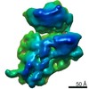
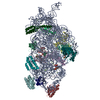
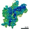
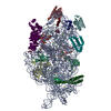
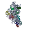
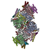
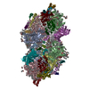
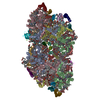
| Title | Staphylococcus aureus 30S ribosomal subunit in presence of spermidine (body only) | |||||||||
 Components Components |
| |||||||||
 Keywords Keywords |  RIBOSOME / Pathogen / small ribosomal subunit / spermidine RIBOSOME / Pathogen / small ribosomal subunit / spermidine | |||||||||
| Function / homology |  Function and homology informationribosomal small subunit biogenesis / small ribosomal subunit rRNA binding / cytosolic small ribosomal subunit / small ribosomal subunit / tRNA binding / rRNA binding / ribosome / structural constituent of ribosome / ribonucleoprotein complex / translation ...ribosomal small subunit biogenesis / small ribosomal subunit rRNA binding / cytosolic small ribosomal subunit / small ribosomal subunit / tRNA binding / rRNA binding / ribosome / structural constituent of ribosome / ribonucleoprotein complex / translation / cytosol / cytoplasm Function and homology informationribosomal small subunit biogenesis / small ribosomal subunit rRNA binding / cytosolic small ribosomal subunit / small ribosomal subunit / tRNA binding / rRNA binding / ribosome / structural constituent of ribosome / ribonucleoprotein complex / translation ...ribosomal small subunit biogenesis / small ribosomal subunit rRNA binding / cytosolic small ribosomal subunit / small ribosomal subunit / tRNA binding / rRNA binding / ribosome / structural constituent of ribosome / ribonucleoprotein complex / translation / cytosol / cytoplasmSimilarity search - Function | |||||||||
| Biological species |   Staphylococcus aureus (bacteria) Staphylococcus aureus (bacteria) | |||||||||
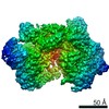
| Method | ELECTRON MICROSCOPY / single particle reconstruction / cryo EM / Resolution: 3.2 Å | |||||||||
 Authors Authors | Belinite, M. / Khusainov, I. / Marzi, S. / Romby, P. / Yusupov, M. / Hashem, Y. | |||||||||
| Funding support |  France, 2items France, 2items
| |||||||||
 Citation Citation | Journal: Front Mol Biosci / Year: 2021 Title: Stabilization of Ribosomal RNA of the Small Subunit by Spermidine in . Authors: Margarita Belinite / Iskander Khusainov / Heddy Soufari / Stefano Marzi / Pascale Romby / Marat Yusupov / Yaser Hashem /  Abstract: Cryo-electron microscopy is now used as a method of choice in structural biology for studying protein synthesis, a process mediated by the ribosome machinery. In order to achieve high-resolution ...Cryo-electron microscopy is now used as a method of choice in structural biology for studying protein synthesis, a process mediated by the ribosome machinery. In order to achieve high-resolution structures using this approach, one needs to obtain homogeneous and stable samples, which requires optimization of ribosome purification in a species-dependent manner. This is especially critical for the bacterial small ribosomal subunit that tends to be unstable in the absence of ligands. Here, we report a protocol for purification of stable 30 S from the Gram-positive bacterium and its cryo-EM structures: in presence of spermidine at a resolution ranging between 3.4 and 3.6 Å and in its absence at 5.3 Å. Using biochemical characterization and cryo-EM, we demonstrate the importance of spermidine for stabilization of the 30 S preserving favorable conformation of the helix 44. | |||||||||
| History |
|
- Structure visualization
| Movie |
Movie viewer |
|---|---|
| Structure viewer | Molecule: MolmilJmol/JSmol |
- Downloads & links
-Download
| PDBx/mmCIF format | 7bgd.cif.gz | 809.7 KB | Display | PDBx/mmCIF format |
|---|---|---|---|---|
| PDB format | pdb7bgd.ent.gz | 625.5 KB | Display | PDB format |
| PDBx/mmJSON format | 7bgd.json.gz | Tree view | PDBx/mmJSON format | |
| Others |  Other downloads Other downloads |
-Validation report
| Arichive directory | https://data.pdbj.org/pub/pdb/validation_reports/bg/7bgdftp://data.pdbj.org/pub/pdb/validation_reports/bg/7bgd | HTTPS FTP |
|---|
-Related structure data
| Related structure data |  12178MC 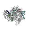 7bgeC  7kwgC M: map data used to model this data C: citing same article ( |
|---|---|
| Similar structure data |


-Links
 PDBj
PDBj


- Assembly
| Deposited unit | 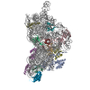
|
|---|---|
| 1 |
|
-Components
-RNA chain , 2 types, 2 molecules Aa
| #1: RNA chain | Messenger RNA Mass: 10392.300 Da / Num. of mol.: 1 Source method: isolated from a genetically manipulated source Source: (gene. exp.) Staphylococcus aureus (bacteria) / Strain: NCTC 8325 / Production host: Escherichia coli (E. coli) |
|---|---|
| #2: RNA chain | Mass: 503218.031 Da / Num. of mol.: 1 / Source method: isolated from a natural source / Source: (natural) Staphylococcus aureus (bacteria) / Strain: NCTC 8325 |
-30S ribosomal protein ... , 12 types, 12 molecules bdefhklopqrt
| #3: Protein | Mass: 29136.369 Da / Num. of mol.: 1 / Source method: isolated from a natural source Source: (natural) Staphylococcus aureus (strain NCTC 8325) (bacteria)Strain: NCTC 8325 / References: UniProt: Q2FZ25 |
|---|---|
| #4: Protein | Mass: 23051.416 Da / Num. of mol.: 1 / Source method: isolated from a natural source Source: (natural) Staphylococcus aureus (strain NCTC 8325) (bacteria)Strain: NCTC 8325 / References: UniProt: Q2FXK6 |
| #5: Protein | Mass: 17770.512 Da / Num. of mol.: 1 / Source method: isolated from a natural source Source: (natural) Staphylococcus aureus (strain NCTC 8325) (bacteria)Strain: NCTC 8325 / References: UniProt: Q2FW23 |
| #6: Protein | Mass: 11613.146 Da / Num. of mol.: 1 / Source method: isolated from a natural source Source: (natural) Staphylococcus aureus (strain NCTC 8325) (bacteria)Strain: NCTC 8325 / References: UniProt: Q2G113 |
| #7: Protein | Mass: 14854.315 Da / Num. of mol.: 1 / Source method: isolated from a natural source Source: (natural) Staphylococcus aureus (strain NCTC 8325) (bacteria)Strain: NCTC 8325 / References: UniProt: Q2FW20 |
| #8: Protein | Mass: 13907.978 Da / Num. of mol.: 1 / Source method: isolated from a natural source Source: (natural) Staphylococcus aureus (strain NCTC 8325) (bacteria)Strain: NCTC 8325 / References: UniProt: Q2FW31 |
| #9: Protein | Mass: 15320.870 Da / Num. of mol.: 1 / Source method: isolated from a natural source Source: (natural) Staphylococcus aureus (strain NCTC 8325) (bacteria)Strain: NCTC 8325 / References: UniProt: P0A0H0 |
| #10: Protein | Mass: 10634.330 Da / Num. of mol.: 1 / Source method: isolated from a natural source Source: (natural) Staphylococcus aureus (strain NCTC 8325) (bacteria)Strain: NCTC 8325 / References: UniProt: Q2G2Q1 |
| #11: Protein | Mass: 10253.886 Da / Num. of mol.: 1 / Source method: isolated from a natural source Source: (natural) Staphylococcus aureus (strain NCTC 8325) (bacteria)Strain: NCTC 8325 / References: UniProt: Q2FZ45 |
| #12: Protein | Mass: 10196.888 Da / Num. of mol.: 1 / Source method: isolated from a natural source Source: (natural) Staphylococcus aureus (strain NCTC 8325) (bacteria)Strain: NCTC 8325 / References: UniProt: Q2FW15 |
| #13: Protein | Mass: 9332.018 Da / Num. of mol.: 1 / Source method: isolated from a natural source Source: (natural) Staphylococcus aureus (strain NCTC 8325) (bacteria)Strain: NCTC 8325 / References: UniProt: Q2G111 |
| #14: Protein | Mass: 9039.472 Da / Num. of mol.: 1 / Source method: isolated from a natural source Source: (natural) Staphylococcus aureus (strain NCTC 8325) (bacteria)Strain: NCTC 8325 / References: UniProt: Q2FXY6 |
-Experimental details
-Experiment
| Experiment | Method: ELECTRON MICROSCOPY |
|---|---|
| EM experiment | Aggregation state: PARTICLE / 3D reconstruction method: single particle reconstruction |
- Sample preparation
| Component |
| ||||||||||||||||||||||||
|---|---|---|---|---|---|---|---|---|---|---|---|---|---|---|---|---|---|---|---|---|---|---|---|---|---|
| Source (natural) |
| ||||||||||||||||||||||||
| Source (recombinant) | Organism: Escherichia coli (E. coli) | ||||||||||||||||||||||||
| Buffer solution | pH: 7.5 | ||||||||||||||||||||||||
| Specimen | Embedding applied: NO / Shadowing applied: NO / Staining applied: NO / Vitrification applied: YES | ||||||||||||||||||||||||
| Vitrification | Instrument: FEI VITROBOT MARK IV / Cryogen name: ETHANE |
- Electron microscopy imaging
| Experimental equipment |  Model: Talos Arctica / Image courtesy: FEI Company |
|---|---|
| Microscopy | Model: FEI TALOS ARCTICA |
| Electron gun | Electron source: FIELD EMISSION GUN / Accelerating voltage: 200 kV / Illumination mode: OTHER |
| Electron lens | Mode: BRIGHT FIELDBright-field microscopy |
| Image recording | Electron dose: 3 e/Å2 / Film or detector model: FEI FALCON III (4k x 4k) |
- Processing
| EM software | Name: Gctf / Version: 1.06 / Category: CTF correction |
|---|---|
| CTF correction | Type: PHASE FLIPPING AND AMPLITUDE CORRECTION |
| 3D reconstruction | Resolution: 3.2 Å / Resolution method: FSC 0.143 CUT-OFF / Num. of particles: 529602 / Symmetry type: POINT |
