 Movie
Movie Controller


+ Open data

- Basic information
| Entry | Database: PDB / ID: 6wwt | |||||||||||||||||||||||||||||||||
|---|---|---|---|---|---|---|---|---|---|---|---|---|---|---|---|---|---|---|---|---|---|---|---|---|---|---|---|---|---|---|---|---|---|---|
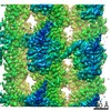
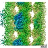
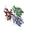
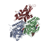
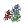
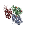
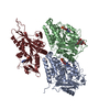
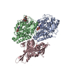
| Title | Apo KIF14[391-735] in complex with a microtubule | |||||||||||||||||||||||||||||||||
 Components Components |
| |||||||||||||||||||||||||||||||||
 Keywords Keywords |  MOTOR PROTEIN / KIF14 / kinesin / motility / microtubule / tubulin MOTOR PROTEIN / KIF14 / kinesin / motility / microtubule / tubulin | |||||||||||||||||||||||||||||||||
| Function / homology |  Function and homology information Function and homology informationcerebellar granular layer structural organization / regulation of cell maturation / cerebellar Purkinje cell layer structural organization / RHO GTPases activate CIT / RND2 GTPase cycle / negative regulation of integrin activation / RND1 GTPase cycle / cell proliferation in forebrain / regulation of Rap protein signal transduction / cerebellar cortex development ...cerebellar granular layer structural organization / regulation of cell maturation / cerebellar Purkinje cell layer structural organization / RHO GTPases activate CIT / RND2 GTPase cycle / negative regulation of integrin activation / RND1 GTPase cycle / cell proliferation in forebrain / regulation of Rap protein signal transduction / cerebellar cortex development / olfactory bulb development / Microtubule-dependent trafficking of connexons from Golgi to the plasma membrane / Hedgehog 'off' state / Cilium Assembly / Intraflagellar transport / COPI-dependent Golgi-to-ER retrograde traffic / Carboxyterminal post-translational modifications of tubulin / RHOH GTPase cycle / Sealing of the nuclear envelope (NE) by ESCRT-III / Kinesins / PKR-mediated signaling / Resolution of Sister Chromatid Cohesion / Mitotic Prometaphase / EML4 and NUDC in mitotic spindle formation / Separation of Sister Chromatids / The role of GTSE1 in G2/M progression after G2 checkpoint / Aggrephagy / plus-end-directed microtubule motor activity / Flemming body / RHO GTPases activate IQGAPs / RHO GTPases Activate Formins / Recruitment of NuMA to mitotic centrosomes / HSP90 chaperone cycle for steroid hormone receptors (SHR) in the presence of ligand / MHC class II antigen presentation / COPI-mediated anterograde transport / regulation of myelination / microtubule motor activity / kinesin complex / mitotic metaphase chromosome alignment / SCF-dependent proteasomal ubiquitin-dependent protein catabolic process / activation of protein kinase activity / microtubule-based movement / positive regulation of cytokinesis / regulation of G1/S transition of mitotic cell cycle / spindle midzone / microtubule-based process / regulation of cell adhesion / regulation of neuron apoptotic process / regulation of cell migration / regulation of G2/M transition of mitotic cell cycle / tubulin binding / substrate adhesion-dependent cell spreading / PDZ domain binding / regulation of cell growth / hippocampus development / Hydrolases; Acting on acid anhydrides; Acting on GTP to facilitate cellular and subcellular movement / establishment of protein localization / structural constituent of cytoskeleton / cerebral cortex development / microtubule cytoskeleton organization / microtubule cytoskeleton / mitotic cell cycle / midbody / microtubule binding / proteasome-mediated ubiquitin-dependent protein catabolic process / microtubule / negative regulation of neuron apoptotic process / cell division / GTPase activity / positive regulation of cell population proliferation / GTP binding / negative regulation of apoptotic process / protein kinase binding / ATP hydrolysis activity / ATP binding / membrane / metal ion binding / nucleus / plasma membrane / cytosol / cytoplasmSimilarity search - Function | |||||||||||||||||||||||||||||||||
| Biological species |  Mus musculus (house mouse)Sus scrofa (pig) Mus musculus (house mouse)Sus scrofa (pig) | |||||||||||||||||||||||||||||||||
| Method | ELECTRON MICROSCOPY / helical reconstruction / cryo EM / Resolution: 3.2 Å | |||||||||||||||||||||||||||||||||
 Authors Authors | Benoit, M.P.M.H. / Asenjo, A.B. / Paydar, M. / Dhakal, S. / Kwok, B. / Sosa, H. | |||||||||||||||||||||||||||||||||
| Funding support |  United States, United States,  Canada, 10items Canada, 10items
| |||||||||||||||||||||||||||||||||
 Citation Citation | Journal: Nat Commun / Year: 2021 Title: Structural basis of mechano-chemical coupling by the mitotic kinesin KIF14. Authors: Matthieu P M H Benoit / Ana B Asenjo / Mohammadjavad Paydar / Sabin Dhakal / Benjamin H Kwok / Hernando Sosa / Abstract: KIF14 is a mitotic kinesin whose malfunction is associated with cerebral and renal developmental defects and several cancers. Like other kinesins, KIF14 couples ATP hydrolysis and microtubule binding ...KIF14 is a mitotic kinesin whose malfunction is associated with cerebral and renal developmental defects and several cancers. Like other kinesins, KIF14 couples ATP hydrolysis and microtubule binding to the generation of mechanical work, but the coupling mechanism between these processes is still not fully clear. Here we report 20 high-resolution (2.7-3.9 Å) cryo-electron microscopy KIF14-microtubule structures with complementary functional assays. Analysis procedures were implemented to separate coexisting conformations of microtubule-bound monomeric and dimeric KIF14 constructs. The data provide a comprehensive view of the microtubule and nucleotide induced KIF14 conformational changes. It shows that: 1) microtubule binding, the nucleotide species, and the neck-linker domain govern the transition between three major conformations of the motor domain; 2) an undocked neck-linker prevents the nucleotide-binding pocket to fully close and dampens ATP hydrolysis; 3) 13 neck-linker residues are required to assume a stable docked conformation; 4) the neck-linker position controls the hydrolysis rather than the nucleotide binding step; 5) the two motor domains of KIF14 dimers adopt distinct conformations when bound to the microtubule; and 6) the formation of the two-heads-bound-state introduces structural changes in both motor domains of KIF14 dimers. These observations provide the structural basis for a coordinated chemo-mechanical kinesin translocation model. | |||||||||||||||||||||||||||||||||
| History |
|
- Structure visualization
| Movie |
Movie viewer |
|---|---|
| Structure viewer | Molecule: MolmilJmol/JSmol |
- Downloads & links
-Download
| PDBx/mmCIF format | 6wwt.cif.gz | 225.8 KB | Display | PDBx/mmCIF format |
|---|---|---|---|---|
| PDB format | pdb6wwt.ent.gz | 176.1 KB | Display | PDB format |
| PDBx/mmJSON format | 6wwt.json.gz | Tree view | PDBx/mmJSON format | |
| Others |  Other downloads Other downloads |
-Validation report
| Arichive directory | https://data.pdbj.org/pub/pdb/validation_reports/ww/6wwtftp://data.pdbj.org/pub/pdb/validation_reports/ww/6wwt | HTTPS FTP |
|---|
-Related structure data
| Related structure data |  21947MC  6wweC  6wwfC  6wwgC  6wwhC  6wwiC  6wwjC  6wwkC  6wwlC  6wwmC  6wwnC  6wwoC  6wwpC  6wwqC  6wwrC  6wwsC  6wwuC  6wwvC  7lvqC  7lvrC M: map data used to model this data C: citing same article ( |
|---|---|
| Similar structure data |


-Links
 PDBj
PDBj


- Assembly
| Deposited unit | 
|
|---|---|
| 1 |
|
-Components
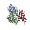
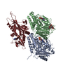
-Protein , 3 types, 3 molecules ABK
| #1: Protein | Mass: 50204.445 Da / Num. of mol.: 1 / Source method: isolated from a natural source / Source: (natural) Sus scrofa (pig) / Organ: Brain / References: UniProt: Q2XVP4 |
|---|---|
| #2: Protein | Mass: 49999.887 Da / Num. of mol.: 1 / Source method: isolated from a natural source / Source: (natural) Sus scrofa (pig) / Organ: Brain / References: UniProt: A0A287AGU7 |
| #3: Protein | Mass: 38898.695 Da / Num. of mol.: 1 / Fragment: UNP residues 391-735 Source method: isolated from a genetically manipulated source Source: (gene. exp.) Mus musculus (house mouse) / Gene: Kif14 / Production host:  Escherichia coli (E. coli) / References: UniProt: L0N7N1 Escherichia coli (E. coli) / References: UniProt: L0N7N1 |
-Non-polymers , 4 types, 4 molecules 


| #4: Chemical | ChemComp-GTP / Guanosine triphosphate Mass: 523.180 Da / Num. of mol.: 1 / Source method: obtained synthetically / Formula: C10H16N5O14P3 / Comment: GTP, energy-carrying molecule*YM Mass: 523.180 Da / Num. of mol.: 1 / Source method: obtained synthetically / Formula: C10H16N5O14P3 / Comment: GTP, energy-carrying molecule*YM |
|---|---|
| #5: Chemical | ChemComp-MG /  Mass: 24.305 Da / Num. of mol.: 1 / Source method: obtained synthetically / Formula: Mg Mass: 24.305 Da / Num. of mol.: 1 / Source method: obtained synthetically / Formula: Mg |
| #6: Chemical | ChemComp-GDP / Guanosine diphosphate Type: RNA linking / Mass: 443.201 Da / Num. of mol.: 1 / Source method: obtained synthetically / Formula: C10H15N5O11P2 / Comment: GDP, energy-carrying molecule*YM Type: RNA linking / Mass: 443.201 Da / Num. of mol.: 1 / Source method: obtained synthetically / Formula: C10H15N5O11P2 / Comment: GDP, energy-carrying molecule*YM |
| #7: Chemical | ChemComp-TA1 / Paclitaxel Mass: 853.906 Da / Num. of mol.: 1 / Source method: obtained synthetically / Formula: C47H51NO14 / Comment: medication, chemotherapy*YM Mass: 853.906 Da / Num. of mol.: 1 / Source method: obtained synthetically / Formula: C47H51NO14 / Comment: medication, chemotherapy*YM |
-Details
| Has ligand of interest | N |
|---|
-Experimental details
-Experiment
| Experiment | Method: ELECTRON MICROSCOPY |
|---|---|
| EM experiment | Aggregation state: FILAMENT / 3D reconstruction method: helical reconstruction |
- Sample preparation
| Component |
| ||||||||||||||||||||||||
|---|---|---|---|---|---|---|---|---|---|---|---|---|---|---|---|---|---|---|---|---|---|---|---|---|---|
| Molecular weight | Experimental value: NO | ||||||||||||||||||||||||
| Source (natural) |
| ||||||||||||||||||||||||
| Source (recombinant) | Organism: Escherichia coli (E. coli) | ||||||||||||||||||||||||
| Buffer solution | pH: 6.8 | ||||||||||||||||||||||||
| Buffer component |
| ||||||||||||||||||||||||
| Specimen | Embedding applied: YES / Shadowing applied: NO / Staining applied: NO / Vitrification applied: YES | ||||||||||||||||||||||||
| Specimen support | Grid type: Quantifoil R2/2 | ||||||||||||||||||||||||
| EM embedding | Material: i | ||||||||||||||||||||||||
| Vitrification | Instrument: FEI VITROBOT MARK IV / Cryogen name: ETHANE |
- Electron microscopy imaging
| Experimental equipment |  Model: Titan Krios / Image courtesy: FEI Company |
|---|---|
| Microscopy | Model: FEI TITAN KRIOS |
| Electron gun | Electron source: FIELD EMISSION GUN / Accelerating voltage: 300 kV / Illumination mode: FLOOD BEAM |
| Electron lens | Mode: BRIGHT FIELDBright-field microscopy / Nominal magnification: 22500 X / Calibrated magnification: 46598 X / Alignment procedure: COMA FREE |
| Specimen holder | Cryogen: NITROGEN / Specimen holder model: FEI TITAN KRIOS AUTOGRID HOLDER |
| Image recording | Electron dose: 69.9 e/Å2 / Detector mode: COUNTING / Film or detector model: GATAN K2 SUMMIT (4k x 4k) |
| Image scans | Movie frames/image: 50 / Used frames/image: 1-50 |
- Processing
| EM software |
| |||||||||||||||||||||||||||||||||||||||||||||||||||||||
|---|---|---|---|---|---|---|---|---|---|---|---|---|---|---|---|---|---|---|---|---|---|---|---|---|---|---|---|---|---|---|---|---|---|---|---|---|---|---|---|---|---|---|---|---|---|---|---|---|---|---|---|---|---|---|---|---|
| CTF correction | Type: PHASE FLIPPING AND AMPLITUDE CORRECTION | |||||||||||||||||||||||||||||||||||||||||||||||||||||||
| Helical symmerty | Angular rotation/subunit: 168.09 ° / Axial rise/subunit: 5.46 Å / Axial symmetry: C1 | |||||||||||||||||||||||||||||||||||||||||||||||||||||||
| Particle selection | Details: manual picking of filaments | |||||||||||||||||||||||||||||||||||||||||||||||||||||||
| 3D reconstruction | Resolution: 3.2 Å / Resolution method: FSC 0.143 CUT-OFF / Num. of particles: 101569 Details: 2 half datasets containing one distinct half of each filament were refined independently. Symmetry type: HELICAL | |||||||||||||||||||||||||||||||||||||||||||||||||||||||
| Atomic model building | Protocol: FLEXIBLE FIT / Space: REAL |
