 Movie
Movie Controller


+ Open data

- Basic information
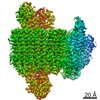
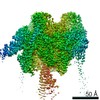
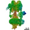
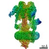
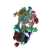
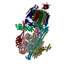
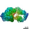
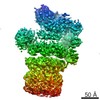
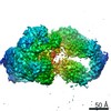
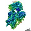
| Entry | Database: PDB / ID: 6wm2 | ||||||
|---|---|---|---|---|---|---|---|
| Title | Human V-ATPase in state 1 with SidK and ADP | ||||||
 Components Components |
| ||||||
 Keywords Keywords |  MEMBRANE PROTEIN / V-ATPase / proton pump MEMBRANE PROTEIN / V-ATPase / proton pump | ||||||
| Function / homology |  Function and homology information Function and homology informationproton-transporting two-sector ATPase complex / Blockage of phagosome acidification / Ion channel transport / eye pigmentation / central nervous system maturation / intracellular pH reduction / transporter activator activity / rostrocaudal neural tube patterning / cellular response to increased oxygen levels / Nef Mediated CD8 Down-regulation ...proton-transporting two-sector ATPase complex / Blockage of phagosome acidification / Ion channel transport / eye pigmentation / central nervous system maturation / intracellular pH reduction / transporter activator activity / rostrocaudal neural tube patterning / cellular response to increased oxygen levels / Nef Mediated CD8 Down-regulation / positive regulation of transforming growth factor beta1 production / ATPase-coupled ion transmembrane transporter activity / synaptic vesicle lumen acidification / endosome to plasma membrane protein transport / proton-transporting V-type ATPase, V0 domain / extrinsic component of synaptic vesicle membrane / Golgi lumen acidification / plasma membrane proton-transporting V-type ATPase complex / Transferrin endocytosis and recycling / lysosomal lumen acidification / clathrin-coated vesicle membrane / vacuolar proton-transporting V-type ATPase, V0 domain / endosomal lumen acidification / vacuolar proton-transporting V-type ATPase, V1 domain / vacuolar transport / proton-transporting V-type ATPase complex / XBP1(S) activates chaperone genes / Amino acids regulate mTORC1 / vacuolar proton-transporting V-type ATPase complex / head morphogenesis / protein localization to cilium / vacuolar acidification / ROS and RNS production in phagocytes / Nef Mediated CD4 Down-regulation / dendritic spine membrane / regulation of cellular pH / osteoclast development / azurophil granule membrane / transmembrane transporter complex / ATPase activator activity / autophagosome membrane / microvillus / regulation of MAPK cascade / tertiary granule membrane / ficolin-1-rich granule membrane / proton transmembrane transporter activity / cilium assembly / positive regulation of Wnt signaling pathway / RHOA GTPase cycle / angiotensin maturation / regulation of macroautophagy / Metabolism of Angiotensinogen to Angiotensins / specific granule membrane / enzyme regulator activity / axon terminus / ATP metabolic process / H+-transporting two-sector ATPase / RNA endonuclease activity / ruffle / Insulin receptor recycling / proton transmembrane transport / proton-transporting ATPase activity, rotational mechanism / endoplasmic reticulum-Golgi intermediate compartment membrane / proton-transporting ATP synthase activity, rotational mechanism / receptor-mediated endocytosis / secretory granule membrane / secretory granule / transmembrane transport / cilium / synaptic vesicle membrane / small GTPase binding / endocytosis / phagocytic vesicle membrane / melanosome / positive regulation of canonical Wnt signaling pathway / presynapse / apical part of cell / signaling receptor activity / ATPase binding / postsynaptic membrane / intracellular iron ion homeostasis / receptor-mediated endocytosis of virus by host cell / Hydrolases; Acting on ester bonds / lysosome / early endosome / endosome membrane / endosome / nuclear speck / apical plasma membrane / lysosomal membrane / external side of plasma membrane / axon / Golgi membrane / intracellular membrane-bounded organelle / focal adhesion / centrosome / ubiquitin protein ligase binding / Neutrophil degranulation / protein-containing complex binding / endoplasmic reticulum membraneSimilarity search - Function | ||||||
| Biological species |  Homo sapiens (human) Homo sapiens (human)  Legionella pneumophila (bacteria) Legionella pneumophila (bacteria) | ||||||
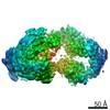
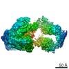
| Method | ELECTRON MICROSCOPY / single particle reconstruction / cryo EM / Resolution: 3.1 Å | ||||||
 Authors Authors | Wang, L. / Wu, H. / Fu, T.M. | ||||||
 Citation Citation | Journal: Mol Cell / Year: 2020 Title: Structures of a Complete Human V-ATPase Reveal Mechanisms of Its Assembly. Authors: Longfei Wang / Di Wu / Carol V Robinson / Hao Wu / Tian-Min Fu /   Abstract: Vesicular- or vacuolar-type adenosine triphosphatases (V-ATPases) are ATP-driven proton pumps comprised of a cytoplasmic V complex for ATP hydrolysis and a membrane-embedded V complex for proton ...Vesicular- or vacuolar-type adenosine triphosphatases (V-ATPases) are ATP-driven proton pumps comprised of a cytoplasmic V complex for ATP hydrolysis and a membrane-embedded V complex for proton transfer. They play important roles in acidification of intracellular vesicles, organelles, and the extracellular milieu in eukaryotes. Here, we report cryoelectron microscopy structures of human V-ATPase in three rotational states at up to 2.9-Å resolution. Aided by mass spectrometry, we build all known protein subunits with associated N-linked glycans and identify glycolipids and phospholipids in the V complex. We define ATP6AP1 as a structural hub for V complex assembly because it connects to multiple V subunits and phospholipids in the c-ring. The glycolipids and the glycosylated V subunits form a luminal glycan coat critical for V-ATPase folding, localization, and stability. This study identifies mechanisms of V-ATPase assembly and biogenesis that rely on the integrated roles of ATP6AP1, glycans, and lipids. | ||||||
| History |
|
- Structure visualization
| Movie |
Movie viewer |
|---|---|
| Structure viewer | Molecule: MolmilJmol/JSmol |
- Downloads & links
-Download
| PDBx/mmCIF format | 6wm2.cif.gz | 1.6 MB | Display | PDBx/mmCIF format |
|---|---|---|---|---|
| PDB format | pdb6wm2.ent.gz | 1.3 MB | Display | PDB format |
| PDBx/mmJSON format | 6wm2.json.gz | Tree view | PDBx/mmJSON format | |
| Others |  Other downloads Other downloads |
-Validation report
| Arichive directory | https://data.pdbj.org/pub/pdb/validation_reports/wm/6wm2ftp://data.pdbj.org/pub/pdb/validation_reports/wm/6wm2 | HTTPS FTP |
|---|
-Related structure data
| Related structure data | 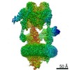 21847MC 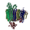 6wlwC 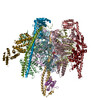 6wlzC 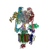 6wm3C  6wm4C M: map data used to model this data C: citing same article ( |
|---|---|
| Similar structure data | |
| EM raw data | EMPIAR-11132 (Title: Cryo-EM structures of human V-ATPase / Data size: 8.4 TB Data #1: Unaligned multi frame micrographs of human V-ATPase in complex with SidK [micrographs - multiframe]) |
-Links
 PDBj
PDBj


- Assembly
| Deposited unit | 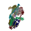
|
|---|---|
| 1 |
|
-Components
-V-type proton ATPase ... , 14 types, 30 molecules HIJKLMOPRABCDEFGN0123456789QSU
| #1: Protein | Mass: 26183.346 Da / Num. of mol.: 3 / Source method: isolated from a natural source / Source: (natural) Homo sapiens (human) / References: UniProt: P36543#2: Protein | Mass: 13781.547 Da / Num. of mol.: 3 / Source method: isolated from a natural source / Source: (natural) Homo sapiens (human) / References: UniProt: O75348#3: Protein | | Mass: 43999.500 Da / Num. of mol.: 1 / Source method: isolated from a natural source / Source: (natural) Homo sapiens (human) / References: UniProt: P21283#4: Protein | | Mass: 55949.949 Da / Num. of mol.: 1 / Source method: isolated from a natural source / Source: (natural) Homo sapiens (human) / References: UniProt: Q9UI12#5: Protein | | Mass: 96512.414 Da / Num. of mol.: 1 / Source method: isolated from a natural source / Source: (natural) Homo sapiens (human) / References: UniProt: Q93050#6: Protein | Mass: 68379.875 Da / Num. of mol.: 3 / Source method: isolated from a natural source / Source: (natural) Homo sapiens (human)References: UniProt: P38606, H+-transporting two-sector ATPase#7: Protein | Mass: 56561.500 Da / Num. of mol.: 3 / Source method: isolated from a natural source / Source: (natural) Homo sapiens (human) / References: UniProt: P21281#9: Protein | | Mass: 28311.918 Da / Num. of mol.: 1 / Source method: isolated from a natural source / Source: (natural) Homo sapiens (human) / References: UniProt: Q9Y5K8#10: Protein | | Mass: 13388.210 Da / Num. of mol.: 1 / Source method: isolated from a natural source / Source: (natural) Homo sapiens (human) / References: UniProt: Q16864#11: Protein | | Mass: 21418.213 Da / Num. of mol.: 1 / Source method: isolated from a natural source / Source: (natural) Homo sapiens (human) / References: UniProt: Q99437#12: Protein | Mass: 15743.655 Da / Num. of mol.: 9 / Source method: isolated from a natural source / Source: (natural) Homo sapiens (human) / References: UniProt: P27449#13: Protein | | Mass: 40369.949 Da / Num. of mol.: 1 / Source method: isolated from a natural source / Source: (natural) Homo sapiens (human) / References: UniProt: P61421#14: Protein | | Mass: 9380.329 Da / Num. of mol.: 1 / Source method: isolated from a natural source / Source: (natural) Homo sapiens (human) / References: UniProt: O15342#16: Protein | | Mass: 52067.480 Da / Num. of mol.: 1 / Source method: isolated from a natural source / Source: (natural) Homo sapiens (human) / References: UniProt: Q15904 |
|---|
-Protein , 3 types, 5 molecules XYZTV
| #8: Protein | Mass: 65505.297 Da / Num. of mol.: 3 / Source method: isolated from a natural source / Source: (natural) Legionella pneumophila (bacteria) / References: UniProt: A0A4T1L9X6, UniProt: Q5ZWW6*PLUS#15: Protein | | Mass: 15435.220 Da / Num. of mol.: 1 / Source method: isolated from a natural source / Source: (natural) Homo sapiens (human)References: UniProt: Q6P5S7, Hydrolases; Acting on ester bonds#17: Protein | | / ATPase H(+)-transporting lysosomal accessory protein 2 / ATPase H(+)-transporting lysosomal- ...ATPase H(+)-transporting lysosomal accessory protein 2 / ATPase H(+)-transporting lysosomal-interacting protein 2 / ER-localized type I transmembrane adapter / Embryonic liver differentiation factor 10 / N14F / Renin/prorenin receptor / Vacuolar ATP synthase membrane sector-associated protein M8-9 / V-ATPase M8.9 subunitMass: 39045.855 Da / Num. of mol.: 1 / Source method: isolated from a natural source / Source: (natural) Homo sapiens (human) / References: UniProt: O75787 |
|---|
-Sugars , 5 types, 10 molecules 
| #18: Polysaccharide | beta-D-galactopyranose-(1-3)-2-acetamido-2-deoxy-beta-D-galactopyranose-(1-4)-[N-acetyl-alpha- ...beta-D-galactopyranose-(1-3)-2-acetamido-2-deoxy-beta-D-galactopyranose-(1-4)-[N-acetyl-alpha-neuraminic acid-(2-3)]beta-D-galactopyranose-(1-4)-beta-D-glucopyranose / Mass: 998.885 Da / Num. of mol.: 1 Source method: isolated from a genetically manipulated source | ||||
|---|---|---|---|---|---|
| #19: Polysaccharide | beta-D-galactopyranose-(1-3)-2-acetamido-2-deoxy-beta-D-galactopyranose-(1-4)-[N-acetyl-alpha- ...beta-D-galactopyranose-(1-3)-2-acetamido-2-deoxy-beta-D-galactopyranose-(1-4)-[N-acetyl-alpha-neuraminic acid-(2-3)]beta-D-galactopyranose / Mass: 836.744 Da / Num. of mol.: 1 Source method: isolated from a genetically manipulated source | ||||
| #20: Polysaccharide | 2-acetamido-2-deoxy-beta-D-glucopyranose-(1-4)-2-acetamido-2-deoxy-beta-D-glucopyranose / Mass: 424.401 Da / Num. of mol.: 5Source method: isolated from a genetically manipulated source #21: Polysaccharide | alpha-D-glucopyranose-(1-2)-alpha-D-glucopyranose-(1-3)-alpha-D-glucopyranose-(1-3)-alpha-D- ...alpha-D-glucopyranose-(1-2)-alpha-D-glucopyranose-(1-3)-alpha-D-glucopyranose-(1-3)-alpha-D-mannopyranose-(1-2)-alpha-D-mannopyranose-(1-2)-alpha-D-mannopyranose-(1-3)-[alpha-D-mannopyranose-(1-6)-alpha-D-mannopyranose-(1-6)]beta-D-mannopyranose-(1-4)-2-acetamido-2-deoxy-beta-D-glucopyranose-(1-4)-2-acetamido-2-deoxy-beta-D-glucopyranose | / Mass: 1883.668 Da / Num. of mol.: 1 / Source method: obtained synthetically#26: Sugar | N-Acetylglucosamine Type: D-saccharide, beta linking / Mass: 221.208 Da / Num. of mol.: 2 / Source method: obtained synthetically / Formula: C8H15NO6 Type: D-saccharide, beta linking / Mass: 221.208 Da / Num. of mol.: 2 / Source method: obtained synthetically / Formula: C8H15NO6 |
-Non-polymers , 7 types, 31 molecules 
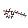

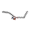
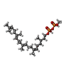

| #22: Chemical | ChemComp-CLR / Cholesterol Mass: 386.654 Da / Num. of mol.: 4 / Source method: obtained synthetically / Formula: C27H46O Mass: 386.654 Da / Num. of mol.: 4 / Source method: obtained synthetically / Formula: C27H46O#23: Chemical | ChemComp-PSF / | Phosphatidylserine Mass: 455.437 Da / Num. of mol.: 1 / Source method: obtained synthetically / Formula: C18H34NO10P Mass: 455.437 Da / Num. of mol.: 1 / Source method: obtained synthetically / Formula: C18H34NO10P#24: Chemical | ChemComp-PTY / Phosphatidylethanolamine Mass: 734.039 Da / Num. of mol.: 13 / Source method: obtained synthetically / Formula: C40H80NO8P / Comment: phospholipid*YM Mass: 734.039 Da / Num. of mol.: 13 / Source method: obtained synthetically / Formula: C40H80NO8P / Comment: phospholipid*YM#25: Chemical | ChemComp-WSS /  Mass: 787.121 Da / Num. of mol.: 10 Mass: 787.121 Da / Num. of mol.: 10Source method: isolated from a genetically manipulated source Formula: C44H85NO8P #27: Chemical | ChemComp-WJP / |  Mass: 534.603 Da / Num. of mol.: 1 / Source method: obtained synthetically / Formula: C26H48O7P2 / Feature type: SUBJECT OF INVESTIGATION Mass: 534.603 Da / Num. of mol.: 1 / Source method: obtained synthetically / Formula: C26H48O7P2 / Feature type: SUBJECT OF INVESTIGATION#28: Chemical | ChemComp-ADP / | Adenosine diphosphate Mass: 427.201 Da / Num. of mol.: 1 / Source method: obtained synthetically / Formula: C10H15N5O10P2 / Comment: ADP, energy-carrying molecule*YM Mass: 427.201 Da / Num. of mol.: 1 / Source method: obtained synthetically / Formula: C10H15N5O10P2 / Comment: ADP, energy-carrying molecule*YM#29: Chemical | ChemComp-WJS / ( |  Mass: 497.391 Da / Num. of mol.: 1 / Source method: obtained synthetically / Formula: C24H20NO9P Mass: 497.391 Da / Num. of mol.: 1 / Source method: obtained synthetically / Formula: C24H20NO9P |
|---|
-Details
| Has ligand of interest | Y |
|---|
-Experimental details
-Experiment
| Experiment | Method: ELECTRON MICROSCOPY |
|---|---|
| EM experiment | Aggregation state: PARTICLE / 3D reconstruction method: single particle reconstruction |
- Sample preparation
| Component | Name: Human V-ATPase in state 1 with SidK and ADP / Type: COMPLEX / Entity ID: #1-#17 / Source: RECOMBINANT |
|---|---|
| Source (natural) | Organism: Homo sapiens (human) |
| Source (recombinant) | Organism: Homo sapiens (human) |
| Buffer solution | pH: 7.4 |
| Specimen | Embedding applied: NO / Shadowing applied: NO / Staining applied: NO / Vitrification applied: YES |
| Vitrification | Cryogen name: ETHANE |
- Electron microscopy imaging
| Experimental equipment |  Model: Titan Krios / Image courtesy: FEI Company |
|---|---|
| Microscopy | Model: FEI TITAN KRIOS |
| Electron gun | Electron source: FIELD EMISSION GUN / Accelerating voltage: 300 kV / Illumination mode: SPOT SCAN |
| Electron lens | Mode: BRIGHT FIELDBright-field microscopy |
| Image recording | Electron dose: 50.1 e/Å2 / Film or detector model: GATAN K3 (6k x 4k) |
- Processing
| Software | Name: PHENIX / Version: 1.17.1_3660: / Classification: refinement | ||||||||||||||||||||||||
|---|---|---|---|---|---|---|---|---|---|---|---|---|---|---|---|---|---|---|---|---|---|---|---|---|---|
| CTF correction | Type: NONE | ||||||||||||||||||||||||
| 3D reconstruction | Resolution: 3.1 Å / Resolution method: FSC 0.143 CUT-OFF / Num. of particles: 1000000 / Symmetry type: POINT | ||||||||||||||||||||||||
| Refine LS restraints |
|
