 Movie
Movie Controller

[English] 日本語
 Yorodumi
- PDB-6vm0: Full length Glycine receptor reconstituted in lipid nanodisc in G... -
+ Open data

- Basic information
| Entry | Database: PDB / ID: 6vm0 | |||||||||
|---|---|---|---|---|---|---|---|---|---|---|
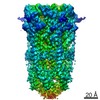
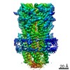
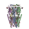
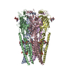
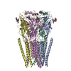
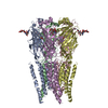
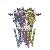
| Title | Full length Glycine receptor reconstituted in lipid nanodisc in Gly/IVM-conformation (State-1) | |||||||||
 Components Components | Glycine receptor subunit alphaZ1 | |||||||||
 Keywords Keywords | MEMBRANE PROTEIN / Ions Ligands Receptors / Glycine receptor Recombinant Proteins Glycine | |||||||||
| Function / homology |  Function and homology information Function and homology informationNeurotransmitter receptors and postsynaptic signal transmission / extracellularly glycine-gated ion channel activity / extracellularly glycine-gated chloride channel activity / synaptic transmission, glycinergic / cellular response to ethanol / cellular response to zinc ion / regulation of neuron differentiation / glycine binding / chloride channel complex / ligand-gated monoatomic ion channel activity ...Neurotransmitter receptors and postsynaptic signal transmission / extracellularly glycine-gated ion channel activity / extracellularly glycine-gated chloride channel activity / synaptic transmission, glycinergic / cellular response to ethanol / cellular response to zinc ion / regulation of neuron differentiation / glycine binding / chloride channel complex / ligand-gated monoatomic ion channel activity / neuropeptide signaling pathway / response to amino acid / monoatomic ion transport / chloride transmembrane transport / central nervous system development / transmitter-gated monoatomic ion channel activity involved in regulation of postsynaptic membrane potential / cellular response to amino acid stimulus / transmembrane signaling receptor activity / perikaryon / postsynaptic membrane / neuron projection / synapse / dendrite / zinc ion binding / membrane / plasma membraneSimilarity search - Function | |||||||||
| Biological species |  Danio rerio (zebrafish) Danio rerio (zebrafish) | |||||||||
| Method | ELECTRON MICROSCOPY / single particle reconstruction / cryo EM / Resolution: 3.14 Å | |||||||||
 Authors Authors | Kumar, A. / Basak, S. / Chakrapani, S. | |||||||||
| Funding support |  United States, 2items United States, 2items
| |||||||||
 Citation Citation | Journal: Nat Commun / Year: 2020 Title: Mechanisms of activation and desensitization of full-length glycine receptor in lipid nanodiscs. Authors: Arvind Kumar / Sandip Basak / Shanlin Rao / Yvonne Gicheru / Megan L Mayer / Mark S P Sansom / Sudha Chakrapani /  Abstract: Glycinergic synapses play a central role in motor control and pain processing in the central nervous system. Glycine receptors (GlyRs) are key players in mediating fast inhibitory neurotransmission ...Glycinergic synapses play a central role in motor control and pain processing in the central nervous system. Glycine receptors (GlyRs) are key players in mediating fast inhibitory neurotransmission at these synapses. While previous high-resolution structures have provided insights into the molecular architecture of GlyR, several mechanistic questions pertaining to channel function are still unanswered. Here, we present Cryo-EM structures of the full-length GlyR protein complex reconstituted into lipid nanodiscs that are captured in the unliganded (closed), glycine-bound (open and desensitized), and allosteric modulator-bound conformations. A comparison of these states reveals global conformational changes underlying GlyR channel gating and modulation. The functional state assignments were validated by molecular dynamics simulations, and the observed permeation events are in agreement with the anion selectivity and conductance of GlyR. These studies provide the structural basis for gating, ion selectivity, and single-channel conductance properties of GlyR in a lipid environment. #1: Journal: Nature / Year: 2015Title: Glycine receptor mechanism elucidated by electron cryo-microscopy. Authors: Juan Du / Wei Lü / Shenping Wu / Yifan Cheng / Eric Gouaux / Abstract: The strychnine-sensitive glycine receptor (GlyR) mediates inhibitory synaptic transmission in the spinal cord and brainstem and is linked to neurological disorders, including autism and hyperekplexia. ...The strychnine-sensitive glycine receptor (GlyR) mediates inhibitory synaptic transmission in the spinal cord and brainstem and is linked to neurological disorders, including autism and hyperekplexia. Understanding of molecular mechanisms and pharmacology of glycine receptors has been hindered by a lack of high-resolution structures. Here we report electron cryo-microscopy structures of the zebrafish α1 GlyR with strychnine, glycine, or glycine and ivermectin (glycine/ivermectin). Strychnine arrests the receptor in an antagonist-bound closed ion channel state, glycine stabilizes the receptor in an agonist-bound open channel state, and the glycine/ivermectin complex adopts a potentially desensitized or partially open state. Relative to the glycine-bound state, strychnine expands the agonist-binding pocket via outward movement of the C loop, promotes rearrangement of the extracellular and transmembrane domain 'wrist' interface, and leads to rotation of the transmembrane domain towards the pore axis, occluding the ion conduction pathway. These structures illuminate the GlyR mechanism and define a rubric to interpret structures of Cys-loop receptors. | |||||||||
| History |
|
- Structure visualization
| Movie |
Movie viewer |
|---|---|
| Structure viewer | Molecule: MolmilJmol/JSmol |
- Downloads & links
-Download
| PDBx/mmCIF format | 6vm0.cif.gz | 342.4 KB | Display | PDBx/mmCIF format |
|---|---|---|---|---|
| PDB format | pdb6vm0.ent.gz | 281.7 KB | Display | PDB format |
| PDBx/mmJSON format | 6vm0.json.gz | Tree view | PDBx/mmJSON format | |
| Others |  Other downloads Other downloads |
-Validation report
| Arichive directory | https://data.pdbj.org/pub/pdb/validation_reports/vm/6vm0ftp://data.pdbj.org/pub/pdb/validation_reports/vm/6vm0 | HTTPS FTP |
|---|
-Related structure data
| Related structure data | 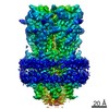 21234MC 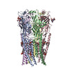 6ubsC  6ubtC 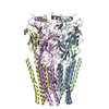 6ud3C 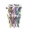 6vm2C  6vm3C M: map data used to model this data C: citing same article ( |
|---|---|
| Similar structure data |


-Links
 PDBj
PDBj


- Assembly
| Deposited unit | 
|
|---|---|
| 1 |
|
-Components
| #1: Protein | Mass: 50821.711 Da / Num. of mol.: 5 Source method: isolated from a genetically manipulated source Source: (gene. exp.) Danio rerio (zebrafish) / Gene: glra1 / Production host:   Spodoptera frugiperda (fall armyworm) / References: UniProt: O93430 Spodoptera frugiperda (fall armyworm) / References: UniProt: O93430#2: Polysaccharide | 2-acetamido-2-deoxy-beta-D-glucopyranose-(1-4)-2-acetamido-2-deoxy-beta-D-glucopyranose / Mass: 424.401 Da / Num. of mol.: 5Source method: isolated from a genetically manipulated source #3: Chemical | ChemComp-PIO / [(   Mass: 746.566 Da / Num. of mol.: 5 Mass: 746.566 Da / Num. of mol.: 5Source method: isolated from a genetically manipulated source Formula: C25H49O19P3 #4: Chemical | ChemComp-GLY / Glycine  Type: peptide linking / Mass: 75.067 Da / Num. of mol.: 5 / Source method: obtained synthetically / Formula: C2H5NO2 / Feature type: SUBJECT OF INVESTIGATION Type: peptide linking / Mass: 75.067 Da / Num. of mol.: 5 / Source method: obtained synthetically / Formula: C2H5NO2 / Feature type: SUBJECT OF INVESTIGATION#5: Chemical | ChemComp-IVM / ( Ivermectin  Mass: 875.093 Da / Num. of mol.: 5 Mass: 875.093 Da / Num. of mol.: 5Source method: isolated from a genetically manipulated source Formula: C48H74O14 / Feature type: SUBJECT OF INVESTIGATION / Comment: antiparasitic*YM Has ligand of interest | Y | |
|---|
-Experimental details
-Experiment
| Experiment | Method: ELECTRON MICROSCOPY |
|---|---|
| EM experiment | Aggregation state: PARTICLE / 3D reconstruction method: single particle reconstruction |
- Sample preparation
| Component | Name: Glycine receptor subunit alpha Z1 / Type: COMPLEX / Entity ID: #1 / Source: RECOMBINANT | |||||||||||||||
|---|---|---|---|---|---|---|---|---|---|---|---|---|---|---|---|---|
| Molecular weight | Value: 250 kDa/nm / Experimental value: NO | |||||||||||||||
| Source (natural) | Organism: Danio rerio (zebrafish) | |||||||||||||||
| Source (recombinant) | Organism: Spodoptera frugiperda (fall armyworm) | |||||||||||||||
| Buffer solution | pH: 8 | |||||||||||||||
| Buffer component |
| |||||||||||||||
| Specimen | Conc.: 0.1 mg/ml / Embedding applied: NO / Shadowing applied: NO / Staining applied: NO / Vitrification applied: YES Details: Full length Zebrafish GlyR alpha1 homopentamer reconsituted in Nanodisc | |||||||||||||||
| Vitrification | Instrument: FEI VITROBOT MARK IV / Cryogen name: ETHANE / Humidity: 100 % / Chamber temperature: 4 K Details: 3.5 ul of 0.1 mg/ml protein solution was applied on a grid in the Vitrobot MkIV chamber set to 100% RH at 4 degC for 30s and then blotted for 2 s and plunged |
- Electron microscopy imaging
| Experimental equipment |  Model: Titan Krios / Image courtesy: FEI Company |
|---|---|
| Microscopy | Model: FEI TITAN KRIOS |
| Electron gun | Electron source: FIELD EMISSION GUN / Accelerating voltage: 300 kV / Illumination mode: FLOOD BEAM |
| Electron lens | Mode: BRIGHT FIELDBright-field microscopy / Nominal magnification: 130000 X / Nominal defocus max: 2500 nm / Nominal defocus min: 1000 nm / Cs: 2.7 mm / C2 aperture diameter: 100 µm / Alignment procedure: COMA FREE |
| Specimen holder | Cryogen: NITROGEN / Specimen holder model: FEI TITAN KRIOS AUTOGRID HOLDER |
| Image recording | Average exposure time: 9 sec. / Electron dose: 40 e/Å2 / Detector mode: SUPER-RESOLUTION / Film or detector model: GATAN K3 BIOQUANTUM (6k x 4k) / Num. of grids imaged: 8 / Num. of real images: 9700 |
| EM imaging optics | Energyfilter name: GIF Bioquantum / Energyfilter slit width: 20 eV |
| Image scans | Movie frames/image: 40 |
- Processing
| Software | Name: PHENIX / Version: 1.17.1_3660: / Classification: refinement | ||||||||||||||||||||||||
|---|---|---|---|---|---|---|---|---|---|---|---|---|---|---|---|---|---|---|---|---|---|---|---|---|---|
| EM software |
| ||||||||||||||||||||||||
| CTF correction | Type: PHASE FLIPPING AND AMPLITUDE CORRECTION | ||||||||||||||||||||||||
| Symmetry | Point symmetry: C5 (5 fold cyclic) | ||||||||||||||||||||||||
| 3D reconstruction | Resolution: 3.14 Å / Resolution method: FSC 0.143 CUT-OFF / Num. of particles: 19600 / Num. of class averages: 1 / Symmetry type: POINT | ||||||||||||||||||||||||
| Refine LS restraints |
|
