 Movie
Movie Controller

[English] 日本語
 Yorodumi
- PDB-6s7o: Cryo-EM structure of human oligosaccharyltransferase complex OST-A -
+ Open data

- Basic information
| Entry | Database: PDB / ID: 6s7o | |||||||||
|---|---|---|---|---|---|---|---|---|---|---|
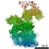
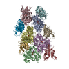
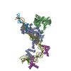
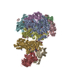
| Title | Cryo-EM structure of human oligosaccharyltransferase complex OST-A | |||||||||
 Components Components |
| |||||||||
 Keywords Keywords |  TRANSFERASE / N-glycosylation / Oligosaccharyltransferase / OSTA TRANSFERASE / N-glycosylation / Oligosaccharyltransferase / OSTA | |||||||||
| Function / homology |  Function and homology informationoligosaccharyltransferase complex binding / oligosaccharyltransferase I complex / oligosaccharyltransferase III complex / Asparagine N-linked glycosylation / membrane-bounded organelle / co-translational protein modification / oligosaccharyltransferase complex / dolichyl-diphosphooligosaccharide-protein glycotransferase / dolichyl-diphosphooligosaccharide-protein glycotransferase activity / protein N-linked glycosylation via asparagine ...oligosaccharyltransferase complex binding / oligosaccharyltransferase I complex / oligosaccharyltransferase III complex / Asparagine N-linked glycosylation / membrane-bounded organelle / co-translational protein modification / oligosaccharyltransferase complex / dolichyl-diphosphooligosaccharide-protein glycotransferase / dolichyl-diphosphooligosaccharide-protein glycotransferase activity / protein N-linked glycosylation via asparagine / protein N-linked glycosylation / epithelial cell apoptotic process / azurophil granule membrane / protein glycosylation / blastocyst development / Advanced glycosylation endproduct receptor signaling / SRP-dependent cotranslational protein targeting to membrane / enzyme activator activity / rough endoplasmic reticulum / T cell activation / response to endoplasmic reticulum stress / post-translational protein modification / response to cytokine / protein modification process / regulation of protein stability / melanosome / transferase activity / Maturation of spike protein / nuclear body / inflammatory response / intracellular membrane-bounded organelle / apoptotic process / Neutrophil degranulation / endoplasmic reticulum membrane / negative regulation of apoptotic process / endoplasmic reticulum / protein-containing complex / RNA binding / membrane / metal ion binding / plasma membrane / cytosol / cytoplasm Function and homology informationoligosaccharyltransferase complex binding / oligosaccharyltransferase I complex / oligosaccharyltransferase III complex / Asparagine N-linked glycosylation / membrane-bounded organelle / co-translational protein modification / oligosaccharyltransferase complex / dolichyl-diphosphooligosaccharide-protein glycotransferase / dolichyl-diphosphooligosaccharide-protein glycotransferase activity / protein N-linked glycosylation via asparagine ...oligosaccharyltransferase complex binding / oligosaccharyltransferase I complex / oligosaccharyltransferase III complex / Asparagine N-linked glycosylation / membrane-bounded organelle / co-translational protein modification / oligosaccharyltransferase complex / dolichyl-diphosphooligosaccharide-protein glycotransferase / dolichyl-diphosphooligosaccharide-protein glycotransferase activity / protein N-linked glycosylation via asparagine / protein N-linked glycosylation / epithelial cell apoptotic process / azurophil granule membrane / protein glycosylation / blastocyst development / Advanced glycosylation endproduct receptor signaling / SRP-dependent cotranslational protein targeting to membrane / enzyme activator activity / rough endoplasmic reticulum / T cell activation / response to endoplasmic reticulum stress / post-translational protein modification / response to cytokine / protein modification process / regulation of protein stability / melanosome / transferase activity / Maturation of spike protein / nuclear body / inflammatory response / intracellular membrane-bounded organelle / apoptotic process / Neutrophil degranulation / endoplasmic reticulum membrane / negative regulation of apoptotic process / endoplasmic reticulum / protein-containing complex / RNA binding / membrane / metal ion binding / plasma membrane / cytosol / cytoplasmSimilarity search - Function | |||||||||
| Biological species |  Homo sapiens (human) Homo sapiens (human) | |||||||||
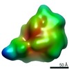
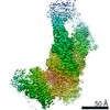
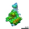
| Method | ELECTRON MICROSCOPY / single particle reconstruction / cryo EM / Resolution: 3.5 Å | |||||||||
 Authors Authors | Ramirez, A.S. / Kowal, J. / Locher, K.P. | |||||||||
| Funding support |  Switzerland, 1items Switzerland, 1items
| |||||||||
 Citation Citation | Journal: Science / Year: 2019 Title: Cryo-electron microscopy structures of human oligosaccharyltransferase complexes OST-A and OST-B. Authors: Ana S Ramírez / Julia Kowal / Kaspar P Locher / Abstract: Oligosaccharyltransferase (OST) catalyzes the transfer of a high-mannose glycan onto secretory proteins in the endoplasmic reticulum. Mammals express two distinct OST complexes that act in a ...Oligosaccharyltransferase (OST) catalyzes the transfer of a high-mannose glycan onto secretory proteins in the endoplasmic reticulum. Mammals express two distinct OST complexes that act in a cotranslational (OST-A) or posttranslocational (OST-B) manner. Here, we present high-resolution cryo-electron microscopy structures of human OST-A and OST-B. Although they have similar overall architectures, structural differences in the catalytic subunits STT3A and STT3B facilitate contacts to distinct OST subunits, DC2 in OST-A and MAGT1 in OST-B. In OST-A, interactions with TMEM258 and STT3A allow ribophorin-I to form a four-helix bundle that can bind to a translating ribosome, whereas the equivalent region is disordered in OST-B. We observed an acceptor peptide and dolichylphosphate bound to STT3B, but only dolichylphosphate in STT3A, suggesting distinct affinities of the two OST complexes for protein substrates. | |||||||||
| History |
|
- Structure visualization
| Movie |
Movie viewer |
|---|---|
| Structure viewer | Molecule: MolmilJmol/JSmol |
- Downloads & links
-Download
| PDBx/mmCIF format | 6s7o.cif.gz | 418.9 KB | Display | PDBx/mmCIF format |
|---|---|---|---|---|
| PDB format | pdb6s7o.ent.gz | 337.2 KB | Display | PDB format |
| PDBx/mmJSON format | 6s7o.json.gz | Tree view | PDBx/mmJSON format | |
| Others |  Other downloads Other downloads |
-Validation report
| Arichive directory | https://data.pdbj.org/pub/pdb/validation_reports/s7/6s7oftp://data.pdbj.org/pub/pdb/validation_reports/s7/6s7o | HTTPS FTP |
|---|
-Related structure data
| Related structure data | 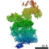 10110MC 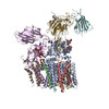 6s7tC M: map data used to model this data C: citing same article ( |
|---|---|
| Similar structure data |
-Links
 PDBj
PDBj


- Assembly
| Deposited unit | 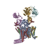
|
|---|---|
| 1 |
|
-Components
-Dolichyl-diphosphooligosaccharide--protein glycosyltransferase subunit ... , 5 types, 5 molecules ABDEF
| #1: Protein | Mass: 80607.320 Da / Num. of mol.: 1 / Source method: isolated from a natural source / Source: (natural) Homo sapiens (human)References: UniProt: P46977, dolichyl-diphosphooligosaccharide-protein glycotransferase |
|---|---|
| #2: Protein/peptide | Mass: 4196.004 Da / Num. of mol.: 1 / Source method: isolated from a natural source / Source: (natural) Homo sapiens (human) / References: UniProt: P0C6T2 |
| #4: Protein | Mass: 12503.631 Da / Num. of mol.: 1 / Source method: isolated from a natural source / Source: (natural) Homo sapiens (human) / References: UniProt: P61803 |
| #5: Protein | Mass: 68656.156 Da / Num. of mol.: 1 / Source method: isolated from a natural source / Source: (natural) Homo sapiens (human) / References: UniProt: P04843 |
| #6: Protein | Mass: 69347.508 Da / Num. of mol.: 1 / Source method: isolated from a natural source / Source: (natural) Homo sapiens (human) / References: UniProt: P04844 |
-Protein , 3 types, 3 molecules CGH
| #3: Protein | Transmembrane protein / Dolichyl-diphosphooligosaccharide--protein glycosyltransferase subunit TMEM258 / Oligosaccharyl ...Dolichyl-diphosphooligosaccharide--protein glycosyltransferase subunit TMEM258 / Oligosaccharyl transferase subunit TMEM258 Mass: 9083.804 Da / Num. of mol.: 1 / Source method: isolated from a natural source / Source: (natural) Homo sapiens (human) / References: UniProt: P61165 |
|---|---|
| #7: Protein | Mass: 50754.438 Da / Num. of mol.: 1 / Source method: isolated from a natural source / Source: (natural) Homo sapiens (human) / References: UniProt: A0A024RAD5, UniProt: P39656*PLUS |
| #8: Protein | Oligosaccharyltransferase / Hydrophobic protein HSF-28 Mass: 16844.215 Da / Num. of mol.: 1 Source method: isolated from a genetically manipulated source Source: (gene. exp.) Homo sapiens (human) / Gene: OSTC, DC2, HDCMD45P, HSPC307 / Production host: Homo sapiens (human) / References: UniProt: Q9NRP0 |
-Sugars , 3 types, 3 molecules
| #9: Polysaccharide | beta-D-mannopyranose-(1-4)-2-acetamido-2-deoxy-beta-D-glucopyranose-(1-4)-2-acetamido-2-deoxy-beta- ...beta-D-mannopyranose-(1-4)-2-acetamido-2-deoxy-beta-D-glucopyranose-(1-4)-2-acetamido-2-deoxy-beta-D-glucopyranose / Mass: 586.542 Da / Num. of mol.: 1 Source method: isolated from a genetically manipulated source |
|---|---|
| #10: Polysaccharide | alpha-D-mannopyranose-(1-2)-alpha-D-mannopyranose-(1-2)-alpha-D-mannopyranose-(1-3)-[alpha-D- ...alpha-D-mannopyranose-(1-2)-alpha-D-mannopyranose-(1-2)-alpha-D-mannopyranose-(1-3)-[alpha-D-mannopyranose-(1-6)-alpha-D-mannopyranose-(1-6)]beta-D-mannopyranose-(1-4)-2-acetamido-2-deoxy-beta-D-glucopyranose-(1-4)-2-acetamido-2-deoxy-beta-D-glucopyranose / Mass: 1397.245 Da / Num. of mol.: 1 Source method: isolated from a genetically manipulated source |
| #11: Polysaccharide | alpha-D-mannopyranose-(1-2)-alpha-D-mannopyranose-(1-3)-[alpha-D-mannopyranose-(1-3)-[alpha-D- ...alpha-D-mannopyranose-(1-2)-alpha-D-mannopyranose-(1-3)-[alpha-D-mannopyranose-(1-3)-[alpha-D-mannopyranose-(1-6)]alpha-D-mannopyranose-(1-6)]beta-D-mannopyranose-(1-4)-2-acetamido-2-deoxy-beta-D-glucopyranose-(1-4)-2-acetamido-2-deoxy-beta-D-glucopyranose / Mass: 1397.245 Da / Num. of mol.: 1 Source method: isolated from a genetically manipulated source |
-Non-polymers , 4 types, 19 molecules 

| #12: Chemical | ChemComp-KZB / (  Mass: 610.776 Da / Num. of mol.: 9 Mass: 610.776 Da / Num. of mol.: 9Source method: isolated from a genetically manipulated source Formula: C33H54O10 #13: Chemical | ChemComp-EGY / (  Mass: 636.861 Da / Num. of mol.: 7 Mass: 636.861 Da / Num. of mol.: 7Source method: isolated from a genetically manipulated source Formula: C33H67NO8P / Comment: phospholipid*YM #14: Chemical |  Mass: 24.305 Da / Num. of mol.: 2 Mass: 24.305 Da / Num. of mol.: 2Source method: isolated from a genetically manipulated source Formula: Mg #15: Chemical | ChemComp-KZE / [( |  Mass: 508.713 Da / Num. of mol.: 1 Mass: 508.713 Da / Num. of mol.: 1Source method: isolated from a genetically manipulated source Formula: C30H53O4P |
|---|
-Details
| Has ligand of interest | N |
|---|
-Experimental details
-Experiment
| Experiment | Method: ELECTRON MICROSCOPY |
|---|---|
| EM experiment | Aggregation state: PARTICLE / 3D reconstruction method: single particle reconstruction |
- Sample preparation
| Component |
| ||||||||||||||||||||||||
|---|---|---|---|---|---|---|---|---|---|---|---|---|---|---|---|---|---|---|---|---|---|---|---|---|---|
| Molecular weight | Experimental value: NO | ||||||||||||||||||||||||
| Source (natural) |
| ||||||||||||||||||||||||
| Source (recombinant) | Organism: Homo sapiens (human) | ||||||||||||||||||||||||
| Buffer solution | pH: 7.5 | ||||||||||||||||||||||||
| Specimen | Embedding applied: NO / Shadowing applied: NO / Staining applied: NO / Vitrification applied: YES | ||||||||||||||||||||||||
| Vitrification | Cryogen name: ETHANE-PROPANE |
- Electron microscopy imaging
| Experimental equipment |  Model: Titan Krios / Image courtesy: FEI Company |
|---|---|
| Microscopy | Model: FEI TITAN KRIOS |
| Electron gun | Electron source: FIELD EMISSION GUN / Accelerating voltage: 300 kV / Illumination mode: FLOOD BEAM |
| Electron lens | Mode: BRIGHT FIELDBright-field microscopy / Cs: 2.7 mm / Alignment procedure: COMA FREE |
| Specimen holder | Cryogen: NITROGEN / Specimen holder model: FEI TITAN KRIOS AUTOGRID HOLDER |
| Image recording | Average exposure time: 8 sec. / Electron dose: 68 e/Å2 / Detector mode: COUNTING / Film or detector model: GATAN K2 SUMMIT (4k x 4k) / Num. of grids imaged: 3 / Num. of real images: 6035 |
| EM imaging optics | Energyfilter name: GIF Quantum LS / Energyfilter slit width: 20 eV |
| Image scans | Movie frames/image: 40 |
- Processing
| EM software |
| ||||||||||||||||||||||||||||
|---|---|---|---|---|---|---|---|---|---|---|---|---|---|---|---|---|---|---|---|---|---|---|---|---|---|---|---|---|---|
| CTF correction | Type: PHASE FLIPPING AND AMPLITUDE CORRECTION | ||||||||||||||||||||||||||||
| Particle selection | Num. of particles selected: 424817 | ||||||||||||||||||||||||||||
| Symmetry | Point symmetry: C1 (asymmetric) | ||||||||||||||||||||||||||||
| 3D reconstruction | Resolution: 3.5 Å / Resolution method: FSC 0.143 CUT-OFF / Num. of particles: 156950 / Symmetry type: POINT |
