 Movie
Movie Controller


+ Open data

- Basic information
| Entry | Database: PDB / ID: 6pr5 | ||||||
|---|---|---|---|---|---|---|---|
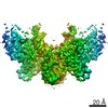
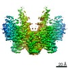
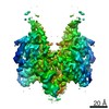
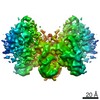
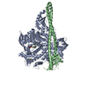
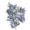
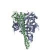
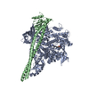
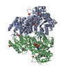
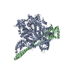
| Title | Cryo-EM structure of HzTransib strand transfer complex (STC) | ||||||
 Components Components |
| ||||||
 Keywords Keywords | RECOMBINATION/DNA / RAG-like transposase / DDE family enzyme /  Transib / Strand transfer. / RECOMBINATION / RECOMBINATION-DNA complex Transib / Strand transfer. / RECOMBINATION / RECOMBINATION-DNA complex | ||||||
| Function / homology | DNA / DNA (> 10) / Putative DNA-mediated transposase Function and homology information Function and homology information | ||||||
| Biological species |  Helicoverpa zea (corn earworm) Helicoverpa zea (corn earworm) | ||||||
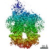
| Method | ELECTRON MICROSCOPY / single particle reconstruction / cryo EM / Resolution: 3.3 Å | ||||||
 Authors Authors | Liu, C. / Yang, Y. / Schatz, D.G. | ||||||
| Funding support |  United States, 1items United States, 1items
| ||||||
 Citation Citation | Journal: Nature / Year: 2019 Title: Structures of a RAG-like transposase during cut-and-paste transposition. Authors: Chang Liu / Yang Yang / David G Schatz / Abstract: Transposons have had a pivotal role in genome evolution and are believed to be the evolutionary progenitors of the RAG1-RAG2 recombinase, an essential component of the adaptive immune system in jawed ...Transposons have had a pivotal role in genome evolution and are believed to be the evolutionary progenitors of the RAG1-RAG2 recombinase, an essential component of the adaptive immune system in jawed vertebrates. Here we report one crystal structure and five cryo-electron microscopy structures of Transib, a RAG1-like transposase from Helicoverpa zea, that capture the entire transposition process from the apo enzyme to the terminal strand transfer complex with transposon ends covalently joined to target DNA, at resolutions of 3.0-4.6 Å. These structures reveal a butterfly-shaped complex that undergoes two cycles of marked conformational changes in which the 'wings' of the transposase unfurl to bind substrate DNA, close to execute cleavage, open to release the flanking DNA and close again to capture and attack target DNA. Transib possesses unique structural elements that compensate for the absence of a RAG2 partner, including a loop that interacts with the transposition target site and an accordion-like C-terminal tail that elongates and contracts to help to control the opening and closing of the enzyme and assembly of the active site. Our findings reveal the detailed reaction pathway of a eukaryotic cut-and-paste transposase and illuminate some of the earliest steps in the evolution of the RAG recombinase. | ||||||
| History |
|
- Structure visualization
| Movie |
Movie viewer |
|---|---|
| Structure viewer | Molecule: MolmilJmol/JSmol |
- Downloads & links
-Download
| PDBx/mmCIF format | 6pr5.cif.gz | 240.8 KB | Display | PDBx/mmCIF format |
|---|---|---|---|---|
| PDB format | pdb6pr5.ent.gz | 183.6 KB | Display | PDB format |
| PDBx/mmJSON format | 6pr5.json.gz | Tree view | PDBx/mmJSON format | |
| Others |  Other downloads Other downloads |
-Validation report
| Arichive directory | https://data.pdbj.org/pub/pdb/validation_reports/pr/6pr5ftp://data.pdbj.org/pub/pdb/validation_reports/pr/6pr5 | HTTPS FTP |
|---|
-Related structure data
| Related structure data | 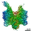 20457MC 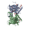 6pqnC 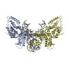 6pqrC 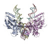 6pquC 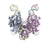 6pqxC 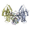 6pqyC C: citing same article ( M: map data used to model this data |
|---|---|
| Similar structure data |

-Links
 PDBj
PDBj


- Assembly
| Deposited unit | 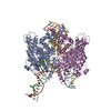
|
|---|---|
| 1 |
|
-Components
-Protein , 1 types, 2 molecules AE
| #1: Protein | Mass: 56582.734 Da / Num. of mol.: 2 Source method: isolated from a genetically manipulated source Source: (gene. exp.) Helicoverpa zea (corn earworm) / Production host:  Spodoptera frugiperda (fall armyworm) / References: UniProt: B0F0C5 Spodoptera frugiperda (fall armyworm) / References: UniProt: B0F0C5 |
|---|
-DNA chain , 5 types, 6 molecules BCGDFH
| #2: DNA chain | Mass: 5491.566 Da / Num. of mol.: 1 / Source method: obtained synthetically / Details: Target DNA 5' flank / Source: (synth.) Helicoverpa zea (corn earworm) | ||||||
|---|---|---|---|---|---|---|---|
| #3: DNA chain | Mass: 4956.244 Da / Num. of mol.: 2 / Source method: obtained synthetically / Details: Non-transferred strand of transposon end DNA / Source: (synth.) Helicoverpa zea (corn earworm)#4: DNA chain | | Mass: 9238.951 Da / Num. of mol.: 1 / Source method: obtained synthetically / Details: Strand transfer product forward strand / Source: (synth.) Helicoverpa zea (corn earworm)#5: DNA chain | | Mass: 2696.783 Da / Num. of mol.: 1 / Source method: obtained synthetically / Details: Target DNA 3' flank / Source: (synth.) Helicoverpa zea (corn earworm)#6: DNA chain | | Mass: 11935.659 Da / Num. of mol.: 1 / Source method: obtained synthetically / Details: Strand transfer product reverse strand / Source: (synth.) Helicoverpa zea (corn earworm) |
-Non-polymers , 2 types, 6 molecules 

| #7: Chemical | ChemComp-MG /  Mass: 24.305 Da / Num. of mol.: 4 / Source method: obtained synthetically / Formula: Mg Mass: 24.305 Da / Num. of mol.: 4 / Source method: obtained synthetically / Formula: Mg#8: Chemical |  Mass: 65.409 Da / Num. of mol.: 2 / Source method: obtained synthetically / Formula: Zn Mass: 65.409 Da / Num. of mol.: 2 / Source method: obtained synthetically / Formula: Zn |
|---|
-Details
| Has ligand of interest | N |
|---|
-Experimental details
-Experiment
| Experiment | Method: ELECTRON MICROSCOPY |
|---|---|
| EM experiment | Aggregation state: PARTICLE / 3D reconstruction method: single particle reconstruction |
- Sample preparation
| Component | Name: Strand transfer complex of HzTransib with transposon ends covalently linked to target DNA. Type: COMPLEX / Entity ID: #1-#6 / Source: RECOMBINANT | |||||||||||||||||||||||||
|---|---|---|---|---|---|---|---|---|---|---|---|---|---|---|---|---|---|---|---|---|---|---|---|---|---|---|
| Molecular weight | Experimental value: NO | |||||||||||||||||||||||||
| Source (natural) | Organism: Helicoverpa zea (corn earworm) | |||||||||||||||||||||||||
| Source (recombinant) | Organism: Spodoptera frugiperda (fall armyworm) / Cell: Sf9 | |||||||||||||||||||||||||
| Buffer solution | pH: 7.5 Details: Solutions were made fresh from concentrated and filtered to avoid microbial contamination. | |||||||||||||||||||||||||
| Buffer component |
| |||||||||||||||||||||||||
| Specimen | Conc.: 0.3 mg/ml / Embedding applied: NO / Shadowing applied: NO / Staining applied: NO / Vitrification applied: YES Details: Recombinantly expressed HzTransib transposase was mixed with chemically synthesized TIR substrate DNA. The complex was further purified on size-exclusion chromatography column. The final ...Details: Recombinantly expressed HzTransib transposase was mixed with chemically synthesized TIR substrate DNA. The complex was further purified on size-exclusion chromatography column. The final complex was monodisperse. | |||||||||||||||||||||||||
| Vitrification | Instrument: FEI VITROBOT MARK IV / Cryogen name: ETHANE / Humidity: 100 % / Chamber temperature: 296 K / Details: Blot for 3 seconds before plunging |
- Electron microscopy imaging
| Experimental equipment |  Model: Titan Krios / Image courtesy: FEI Company |
|---|---|
| Microscopy | Model: FEI TITAN KRIOS / Details: Preliminary grid screening was performed manually. |
| Electron gun | Electron source: FIELD EMISSION GUN / Accelerating voltage: 300 kV / Illumination mode: FLOOD BEAM |
| Electron lens | Mode: BRIGHT FIELDBright-field microscopy / Nominal magnification: 130000 X / Nominal defocus max: 2400 nm / Nominal defocus min: 1400 nm / Cs: 2.7 mm / Alignment procedure: COMA FREE |
| Specimen holder | Cryogen: NITROGEN / Specimen holder model: FEI TITAN KRIOS AUTOGRID HOLDER |
| Image recording | Average exposure time: 8 sec. / Electron dose: 54.4 e/Å2 / Detector mode: SUPER-RESOLUTION / Film or detector model: GATAN K2 SUMMIT (4k x 4k) Details: Images were collected in movie-mode at 5 frames per second. |
| EM imaging optics | Energyfilter name: GIF Quantum LS / Energyfilter slit width: 20 eV |
| Image scans | Movie frames/image: 40 / Used frames/image: 1-40 |
- Processing
| Software | Name: PHENIX / Version: 1.15.2_3472: / Classification: refinement | ||||||||||||||||||||||||||||||||||||
|---|---|---|---|---|---|---|---|---|---|---|---|---|---|---|---|---|---|---|---|---|---|---|---|---|---|---|---|---|---|---|---|---|---|---|---|---|---|
| EM software |
| ||||||||||||||||||||||||||||||||||||
| CTF correction | Type: PHASE FLIPPING AND AMPLITUDE CORRECTION | ||||||||||||||||||||||||||||||||||||
| Symmetry | Point symmetry: C1 (asymmetric) | ||||||||||||||||||||||||||||||||||||
| 3D reconstruction | Resolution: 3.3 Å / Resolution method: FSC 0.143 CUT-OFF / Num. of particles: 43661 / Num. of class averages: 1 / Symmetry type: POINT | ||||||||||||||||||||||||||||||||||||
| Atomic model building | Protocol: RIGID BODY FIT / Space: REAL / Target criteria: Correlation coefficient Details: Initial local fitting was done using UCSF Chimera, then manually adjusted and rebuilt in Coot. Final model was refined using Phenix real-space refinement. | ||||||||||||||||||||||||||||||||||||
| Atomic model building | PDB-ID: 6PQN Pdb chain-ID: A / Accession code: 6PQN / Pdb chain residue range: 21-501 / Source name: PDB / Type: experimental model | ||||||||||||||||||||||||||||||||||||
| Refine LS restraints |
|
