National Institutes of Health/National Institute Of Allergy and Infectious Diseases (NIH/NIAID)
AI039657
United States
National Institutes of Health/National Cancer Institute (NIH/NCI)
CA116087
United States
National Institutes of Health/National Institute of General Medical Sciences (NIH/NIGMS)
T32GM08230
United States
National Institutes of Health/National Institute of General Medical Sciences (NIH/NIGMS)
R35GM118089
United States
National Institutes of Health/National Cancer Institute (NIH/NCI)
T32CA119925
United States
Other government
5I01BX000627
United States
National Institutes of Health/National Institute Of Allergy and Infectious Diseases (NIH/NIAID)
F31AI112324
United States
Citation
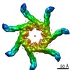
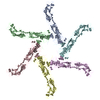
Journal: J Mol Biol / Year: 2019 Title: Cryo-EM Analysis Reveals Structural Basis of Helicobacter pylori VacA Toxin Oligomerization. Authors: Min Su / Amanda L Erwin / Anne M Campbell / Tasia M Pyburn / Lauren E Salay / Jessica L Hanks / D Borden Lacy / David L Akey / Timothy L Cover / Melanie D Ohi / Abstract: Helicobacter pylori colonizes the human stomach and contributes to the development of gastric cancer and peptic ulcer disease. H. pylori secretes a pore-forming toxin called vacuolating cytotoxin A ...Helicobacter pylori colonizes the human stomach and contributes to the development of gastric cancer and peptic ulcer disease. H. pylori secretes a pore-forming toxin called vacuolating cytotoxin A (VacA), which contains two domains (p33 and p55) and assembles into oligomeric structures. Using single-particle cryo-electron microscopy, we have determined low-resolution structures of a VacA dodecamer and heptamer, as well as a 3.8-Å structure of the VacA hexamer. These analyses show that VacA p88 consists predominantly of a right-handed beta-helix that extends from the p55 domain into the p33 domain. We map the regions of p33 and p55 involved in hexamer assembly, model how interactions between protomers support heptamer formation, and identify surfaces of VacA that likely contact membrane. This work provides structural insights into the process of VacA oligomerization and identifies regions of VacA protomers that are predicted to contact the host cell surface during channel formation.
In the structure databanks used in Yorodumi, some data are registered as the other names, "COVID-19 virus" and "2019-nCoV". Here are the details of the virus and the list of structure data.
Jan 31, 2019. EMDB accession codes are about to change! (news from PDBe EMDB page)
EMDB accession codes are about to change! (news from PDBe EMDB page)
The allocation of 4 digits for EMDB accession codes will soon come to an end. Whilst these codes will remain in use, new EMDB accession codes will include an additional digit and will expand incrementally as the available range of codes is exhausted. The current 4-digit format prefixed with “EMD-” (i.e. EMD-XXXX) will advance to a 5-digit format (i.e. EMD-XXXXX), and so on. It is currently estimated that the 4-digit codes will be depleted around Spring 2019, at which point the 5-digit format will come into force.
The EM Navigator/Yorodumi systems omit the EMD- prefix.
Related info.:Q: What is EMD? / ID/Accession-code notation in Yorodumi/EM Navigator
Yorodumi is a browser for structure data from EMDB, PDB, SASBDB, etc.
This page is also the successor to EM Navigator detail page, and also detail information page/front-end page for Omokage search.
The word "yorodu" (or yorozu) is an old Japanese word meaning "ten thousand". "mi" (miru) is to see.
Related info.:EMDB / PDB / SASBDB / Comparison of 3 databanks / Yorodumi Search / Aug 31, 2016. New EM Navigator & Yorodumi / Yorodumi Papers / Jmol/JSmol / Function and homology information / Changes in new EM Navigator and Yorodumi
 Movie
Movie Controller


 Open data
 Basic information
 Components
 Keywords
 TOXIN / VacA
 Function and homology information

 Authors
 United States, 7items
  Citation
 Structure visualization
 Downloads & links
 Other downloads
 PDBj
PDBj Assembly
 Sample preparation
 Electron microscopy imaging
 Processing