 Movie
Movie Controller

[English] 日本語
 Yorodumi
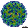
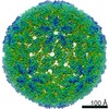
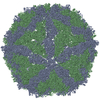
- PDB-6nj8: Encapsulin iron storage compartment from Quasibacillus thermotolerans -
+ Open data

- Basic information
| Entry | Database: PDB / ID: 6nj8 | ||||||
|---|---|---|---|---|---|---|---|
| Title | Encapsulin iron storage compartment from Quasibacillus thermotolerans | ||||||
 Components Components |
| ||||||
 Keywords Keywords | METAL TRANSPORT /  encapsulin / iron storage / IMEF / icosahedral encapsulin / iron storage / IMEF / icosahedral | ||||||
| Function / homology | Type 1 encapsulin shell protein / Encapsulating protein for peroxidase / encapsulin nanocompartment / iron ion transport / intracellular iron ion homeostasis / Type 1 encapsulin shell protein Function and homology information Function and homology information | ||||||
| Biological species |  | ||||||
| Method | ELECTRON MICROSCOPY / single particle reconstruction / cryo EM / Resolution: 3.85 Å | ||||||
 Authors Authors | Orlando, B.J. / Giessen, T.W. / Chambers, M.G. / Liao, M. / Silver, P.A. | ||||||
| Funding support |  Germany, 1items Germany, 1items
| ||||||
 Citation Citation | Journal: Elife / Year: 2019 Title: Large protein organelles form a new iron sequestration system with high storage capacity. Authors: Tobias W Giessen / Benjamin J Orlando / Andrew A Verdegaal / Melissa G Chambers / Jules Gardener / David C Bell / Gabriel Birrane / Maofu Liao / Pamela A Silver /  Abstract: Iron storage proteins are essential for cellular iron homeostasis and redox balance. Ferritin proteins are the major storage units for bioavailable forms of iron. Some organisms lack ferritins, and ...Iron storage proteins are essential for cellular iron homeostasis and redox balance. Ferritin proteins are the major storage units for bioavailable forms of iron. Some organisms lack ferritins, and it is not known how they store iron. Encapsulins, a class of protein-based organelles, have recently been implicated in microbial iron and redox metabolism. Here, we report the structural and mechanistic characterization of a 42 nm two-component encapsulin-based iron storage compartment from . Using cryo-electron microscopy and x-ray crystallography, we reveal the assembly principles of a thermostable T = 4 shell topology and its catalytic ferroxidase cargo and show interactions underlying cargo-shell co-assembly. This compartment has an exceptionally large iron storage capacity storing over 23,000 iron atoms. Our results reveal a new approach for survival in diverse habitats with limited or fluctuating iron availability via an iron storage system able to store 10 to 20 times more iron than ferritin. | ||||||
| History |
|
- Structure visualization
| Movie |
Movie viewer |
|---|---|
| Structure viewer | Molecule: MolmilJmol/JSmol |
- Downloads & links
-Download
| PDBx/mmCIF format | 6nj8.cif.gz | 204.2 KB | Display | PDBx/mmCIF format |
|---|---|---|---|---|
| PDB format | pdb6nj8.ent.gz | 166.9 KB | Display | PDB format |
| PDBx/mmJSON format | 6nj8.json.gz | Tree view | PDBx/mmJSON format | |
| Others |  Other downloads Other downloads |
-Validation report
| Arichive directory | https://data.pdbj.org/pub/pdb/validation_reports/nj/6nj8ftp://data.pdbj.org/pub/pdb/validation_reports/nj/6nj8 | HTTPS FTP |
|---|
-Related structure data
| Related structure data |  9383MC 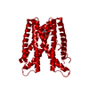 6n63C C: citing same article ( M: map data used to model this data |
|---|---|
| Similar structure data |

-Links
 PDBj
PDBj
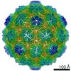
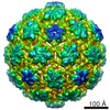
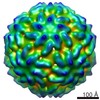
- Assembly
| Deposited unit | 
|
|---|---|
| 1 | x 60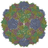
|
| 2 |
|
| 3 | x 5
|
| 4 | x 6
|
| 5 | 
|
| Symmetry | Point symmetry: (Schoenflies symbol: I (icosahedral)) |
-Components
| #1: Protein | Mass: 32239.459 Da / Num. of mol.: 4 Source method: isolated from a genetically manipulated source Source: (gene. exp.) Gene: QY95_01592 / Production host: Escherichia coli (E. coli) / References: UniProt: A0A0F5HPP7#2: Protein/peptide | Mass: 716.824 Da / Num. of mol.: 3 Source method: isolated from a genetically manipulated source Source: (gene. exp.) Escherichia coli (E. coli) |
|---|
-Experimental details
-Experiment
| Experiment | Method: ELECTRON MICROSCOPY |
|---|---|
| EM experiment | Aggregation state: PARTICLE / 3D reconstruction method: single particle reconstruction |
- Sample preparation
| Component | Name: Encapsulin iron storage compartment from Quasibacillus thermotolerans Type: COMPLEX / Entity ID: all / Source: RECOMBINANT |
|---|---|
| Molecular weight | Value: 9.6 MDa / Experimental value: NO |
| Source (natural) | Organism: |
| Source (recombinant) | Organism: Escherichia coli (E. coli) |
| Buffer solution | pH: 8 |
| Specimen | Conc.: 1.5 mg/ml / Embedding applied: NO / Shadowing applied: NO / Staining applied: NO / Vitrification applied: YES |
| Specimen support | Grid type: Quantifoil R1.2/1.3 |
| Vitrification | Instrument: GATAN CRYOPLUNGE 3 / Cryogen name: ETHANE / Humidity: 90 % |
- Electron microscopy imaging
| Experimental equipment |  Model: Tecnai F20 / Image courtesy: FEI Company |
|---|---|
| Microscopy | Model: FEI TECNAI F20 |
| Electron gun | Electron source: FIELD EMISSION GUN / Accelerating voltage: 200 kV / Illumination mode: FLOOD BEAM |
| Electron lens | Mode: BRIGHT FIELDBright-field microscopy |
| Image recording | Average exposure time: 7.2 sec. / Electron dose: 44 e/Å2 / Detector mode: SUPER-RESOLUTION / Film or detector model: GATAN K2 SUMMIT (4k x 4k) / Num. of real images: 601 |
- Processing
| EM software |
| ||||||||||||||||
|---|---|---|---|---|---|---|---|---|---|---|---|---|---|---|---|---|---|
| CTF correction | Type: PHASE FLIPPING AND AMPLITUDE CORRECTION | ||||||||||||||||
| Symmetry | Point symmetry: I (icosahedral) | ||||||||||||||||
| 3D reconstruction | Resolution: 3.85 Å / Resolution method: FSC 0.143 CUT-OFF / Num. of particles: 18995 / Symmetry type: POINT |
