 Movie
Movie Controller


+ Open data

- Basic information
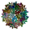
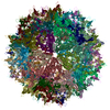
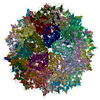
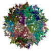
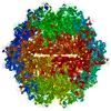
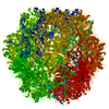
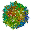
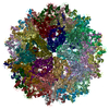
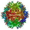
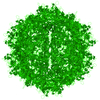
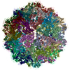
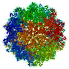
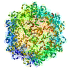
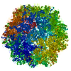
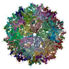
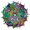
| Entry | Database: PDB / ID: 6ih9 | ||||||
|---|---|---|---|---|---|---|---|
| Title | Adeno-Associated Virus 2 at 2.8 ang | ||||||
 Components Components | Capsid protein VP1 | ||||||
 Keywords Keywords | VIRUS / AAV2 | ||||||
| Function / homology |  Function and homology information Function and homology informationpermeabilization of host organelle membrane involved in viral entry into host cell / symbiont entry into host cell via permeabilization of inner membrane / host cell nucleolus / T=1 icosahedral viral capsid / clathrin-dependent endocytosis of virus by host cell / virion attachment to host cell / structural molecule activity Similarity search - Function | ||||||
| Biological species |  Adeno-associated virus 2 Adeno-associated virus 2 | ||||||
| Method | ELECTRON MICROSCOPY / single particle reconstruction / cryo EM / Resolution: 2.8 Å | ||||||
 Authors Authors | Lou, Z.Y. / Zhang, R. | ||||||
| Funding support |  China, 1items China, 1items
| ||||||
 Citation Citation | Journal: Nat Microbiol / Year: 2019 Title: Adeno-associated virus 2 bound to its cellular receptor AAVR. Authors: Ran Zhang / Lin Cao / Mengtian Cui / Zixian Sun / Mingxu Hu / Rouxuan Zhang / William Stuart / Xiaochu Zhao / Zirui Yang / Xueming Li / Yuna Sun / Shentao Li / Wei Ding / Zhiyong Lou / Zihe Rao /  Abstract: Adeno-associated virus (AAV) is a leading vector for virus-based gene therapy. The receptor for AAV (AAVR; also named KIAA0319L) was recently identified, and the precise characterization of AAV-AAVR ...Adeno-associated virus (AAV) is a leading vector for virus-based gene therapy. The receptor for AAV (AAVR; also named KIAA0319L) was recently identified, and the precise characterization of AAV-AAVR recognition is in immediate demand. Taking advantage of a particle-filtering algorithm, we report here the cryo-electron microscopy structure of the AAV2-AAVR complex at 2.8 Å resolution. This structure reveals that of the five Ig-like polycystic kidney disease (PKD) domains in AAVR, PKD2 binds directly to the spike region of the AAV2 capsid adjacent to the icosahedral three-fold axis. Residues in strands B and E, and the BC loop of AAVR PKD2 interact directly with the AAV2 capsid. The interacting residues in the AAV2 capsid are mainly in AAV-featured variable regions. Mutagenesis of the amino acids at the AAV2-AAVR interface reduces binding activity and viral infectivity. Our findings provide insights into the biology of AAV entry with high-resolution details, providing opportunities for the development of new AAV vectors for gene therapy. | ||||||
| History |
|
- Structure visualization
| Movie |
Movie viewer |
|---|---|
| Structure viewer | Molecule: MolmilJmol/JSmol |
- Downloads & links
-Download
| PDBx/mmCIF format | 6ih9.cif.gz | 105.1 KB | Display | PDBx/mmCIF format |
|---|---|---|---|---|
| PDB format | pdb6ih9.ent.gz | 78.6 KB | Display | PDB format |
| PDBx/mmJSON format | 6ih9.json.gz | Tree view | PDBx/mmJSON format | |
| Others |  Other downloads Other downloads |
-Validation report
| Arichive directory | https://data.pdbj.org/pub/pdb/validation_reports/ih/6ih9ftp://data.pdbj.org/pub/pdb/validation_reports/ih/6ih9 | HTTPS FTP |
|---|
-Related structure data
| Related structure data | 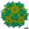 9671MC 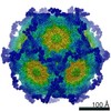 9672C  6ihbC M: map data used to model this data C: citing same article ( |
|---|---|
| Similar structure data |


-Links
 PDBj
PDBj


- Assembly
| Deposited unit | 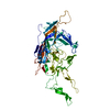
|
|---|---|
| 1 | x 60
|
| 2 |
|
| 3 | x 5
|
| 4 | x 6
|
| 5 | 
|
| Symmetry | Point symmetry: (Schoenflies symbol: I (icosahedral)) |
-Components
| #1: Protein | Mass: 58644.492 Da / Num. of mol.: 1 / Source method: isolated from a natural source Source: (natural) Adeno-associated virus 2 (isolate Srivastava/1982)Strain: isolate Srivastava/1982 / References: UniProt: P03135 |
|---|
-Experimental details
-Experiment
| Experiment | Method: ELECTRON MICROSCOPY |
|---|---|
| EM experiment | Aggregation state: PARTICLE / 3D reconstruction method: single particle reconstruction |
- Sample preparation
| Component | Name: Ao-associated virus 2 (isolate Srivastava/1982) / Type: VIRUS / Entity ID: all / Source: NATURAL |
|---|---|
| Molecular weight | Experimental value: NO |
| Source (natural) | Organism: Ao-associated virus 2 (isolate Srivastava/1982) |
| Details of virus | Empty: YES / Enveloped: NO / Isolate: STRAIN / Type: VIRION |
| Virus shell | Diameter: 280 nm |
| Buffer solution | pH: 7.4 |
| Specimen | Conc.: 1 mg/ml / Embedding applied: NO / Shadowing applied: NO / Staining applied: NO / Vitrification applied: YES Details: The AAV2 particles are purified fron 293T cell, with the yeild of approximately 10^12vg per 10L. |
| Vitrification | Instrument: FEI VITROBOT MARK I / Cryogen name: ETHANE / Humidity: 100 % |
- Electron microscopy imaging
| Experimental equipment |  Model: Talos Arctica / Image courtesy: FEI Company |
|---|---|
| Microscopy | Model: FEI TECNAI ARCTICA |
| Electron gun | Electron source: FIELD EMISSION GUN / Accelerating voltage: 200 kV / Illumination mode: FLOOD BEAM |
| Electron lens | Mode: BRIGHT FIELDBright-field microscopy / Cs: 2.7 mm / C2 aperture diameter: 70 µm |
| Image recording | Average exposure time: 1.2 sec. / Electron dose: 1.53 e/Å2 / Film or detector model: FEI FALCON II (4k x 4k) / Num. of grids imaged: 1 |
| Image scans | Movie frames/image: 19 / Used frames/image: 1-19 |
- Processing
| Software | Name: PHENIX / Version: 1.11.1_2575: / Classification: refinement | ||||||||||||||||||||||||
|---|---|---|---|---|---|---|---|---|---|---|---|---|---|---|---|---|---|---|---|---|---|---|---|---|---|
| EM software |
| ||||||||||||||||||||||||
| CTF correction | Type: PHASE FLIPPING AND AMPLITUDE CORRECTION | ||||||||||||||||||||||||
| 3D reconstruction | Resolution: 2.8 Å / Resolution method: FSC 0.143 CUT-OFF / Num. of particles: 14434 / Symmetry type: POINT | ||||||||||||||||||||||||
| Refine LS restraints |
|
