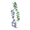
 Movie
Movie Controller


+ Open data

- Basic information
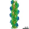
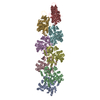
| Entry | Database: PDB / ID: 6bqw | ||||||||||||
|---|---|---|---|---|---|---|---|---|---|---|---|---|---|
| Title | AlfA Filament bound to AMPPNP | ||||||||||||
 Components Components | Bacterial actin AlfA | ||||||||||||
 Keywords Keywords |  CYTOSOLIC PROTEIN / actin / plasmid segregation / filament CYTOSOLIC PROTEIN / actin / plasmid segregation / filament | ||||||||||||
| Function / homology | Actin-like protein, N-terminal / Actin like proteins N terminal domain / ATPase, nucleotide binding domain / PHOSPHOAMINOPHOSPHONIC ACID-ADENYLATE ESTER / Actin-like protein N-terminal domain-containing protein Function and homology information Function and homology information | ||||||||||||
| Biological species |  Bacillus subtilis (bacteria) Bacillus subtilis (bacteria) | ||||||||||||
| Method | ELECTRON MICROSCOPY / helical reconstruction / cryo EM / Resolution: 4.2 Å | ||||||||||||
 Authors Authors | Usluer, G.D. / Kollman, J.M. / DiMaio, F. | ||||||||||||
| Funding support |  United States, United States,  Canada, Canada,  Turkey, 3items Turkey, 3items
| ||||||||||||
 Citation Citation | Journal: Proc Natl Acad Sci U S A / Year: 2018 Title: Cryo-EM structure of the bacterial actin AlfA reveals unique assembly and ATP-binding interactions and the absence of a conserved subdomain. Authors: Gülsima D Usluer / Frank DiMaio / Shun Kai Yang / Jesse M Hansen / Jessica K Polka / R Dyche Mullins / Justin M Kollman / Abstract: Bacterial actins are an evolutionarily diverse family of ATP-dependent filaments built from protomers with a conserved structural fold. Actin-based segregation systems are encoded on many bacterial ...Bacterial actins are an evolutionarily diverse family of ATP-dependent filaments built from protomers with a conserved structural fold. Actin-based segregation systems are encoded on many bacterial plasmids and function to partition plasmids into daughter cells. The bacterial actin AlfA segregates plasmids by a mechanism distinct from other partition systems, dependent on its unique dynamic properties. Here, we report the near-atomic resolution electron cryo-microscopy structure of the AlfA filament, which reveals a strikingly divergent filament architecture resulting from the loss of a subdomain conserved in all other actins and a mode of ATP binding. Its unusual assembly interfaces and nucleotide interactions provide insight into AlfA dynamics, and expand the range of evolutionary variation accessible to actin quaternary structure. | ||||||||||||
| History |
|
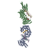
- Structure visualization
| Movie |
Movie viewer |
|---|---|
| Structure viewer | Molecule: MolmilJmol/JSmol |
- Downloads & links
-Download
| PDBx/mmCIF format | 6bqw.cif.gz | 421.4 KB | Display | PDBx/mmCIF format |
|---|---|---|---|---|
| PDB format | pdb6bqw.ent.gz | 340 KB | Display | PDB format |
| PDBx/mmJSON format | 6bqw.json.gz | Tree view | PDBx/mmJSON format | |
| Others |  Other downloads Other downloads |
-Validation report
| Arichive directory | https://data.pdbj.org/pub/pdb/validation_reports/bq/6bqwftp://data.pdbj.org/pub/pdb/validation_reports/bq/6bqw | HTTPS FTP |
|---|
-Related structure data
| Related structure data |  7134MC M: map data used to model this data C: citing same article ( |
|---|---|
| Similar structure data |


-Links
 PDBj
PDBj


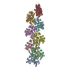
- Assembly
| Deposited unit | 
|
|---|---|
| 1 |
|
-Components
| #1: Protein | Mass: 31150.414 Da / Num. of mol.: 9 Source method: isolated from a genetically manipulated source Source: (gene. exp.) Bacillus subtilis (bacteria) / Production host: Escherichia coli (E. coli) / References: UniProt: O52947#2: Chemical | ChemComp-ANP /   Mass: 506.196 Da / Num. of mol.: 9 / Source method: obtained synthetically / Formula: C10H17N6O12P3 / Comment: AMP-PNP, energy-carrying molecule analogue*YM Mass: 506.196 Da / Num. of mol.: 9 / Source method: obtained synthetically / Formula: C10H17N6O12P3 / Comment: AMP-PNP, energy-carrying molecule analogue*YM |
|---|
-Experimental details
-Experiment
| Experiment | Method: ELECTRON MICROSCOPY |
|---|---|
| EM experiment | Aggregation state: FILAMENT / 3D reconstruction method: helical reconstruction |
- Sample preparation
| Component | Name: AlfA filament / Type: COMPLEX Details: Each protomer is bound to the non-hydrolyzable nucleotide analog AMPPNP Entity ID: #1 / Source: RECOMBINANT |
|---|---|
| Molecular weight | Value: 12750 kDa/nm / Experimental value: NO |
| Source (natural) | Organism: Bacillus subtilis (bacteria) |
| Source (recombinant) | Organism: Escherichia coli (E. coli) |
| Buffer solution | pH: 7.5 |
| Specimen | Conc.: 0.16 mg/ml / Embedding applied: NO / Shadowing applied: NO / Staining applied: NO / Vitrification applied: YES Details: Filaments were assembled in 5 mM AMPPNP for 15 minutes at room temperature |
| Vitrification | Cryogen name: ETHANE |
- Electron microscopy imaging
| Experimental equipment |  Model: Titan Krios / Image courtesy: FEI Company |
|---|---|
| Microscopy | Model: FEI TITAN KRIOS |
| Electron gun | Electron source: FIELD EMISSION GUN / Accelerating voltage: 300 kV / Illumination mode: FLOOD BEAM |
| Electron lens | Mode: BRIGHT FIELDBright-field microscopy |
| Specimen holder | Cryogen: NITROGEN / Specimen holder model: FEI TITAN KRIOS AUTOGRID HOLDER |
| Image recording | Electron dose: 72 e/Å2 / Detector mode: SUPER-RESOLUTION / Film or detector model: GATAN K2 SUMMIT (4k x 4k) |
- Processing
| EM software |
| ||||||||||||||||
|---|---|---|---|---|---|---|---|---|---|---|---|---|---|---|---|---|---|
| CTF correction | Type: PHASE FLIPPING AND AMPLITUDE CORRECTION | ||||||||||||||||
| Helical symmerty | Angular rotation/subunit: 157.7 ° / Axial rise/subunit: 24.4 Å / Axial symmetry: C1 | ||||||||||||||||
| 3D reconstruction | Resolution: 4.2 Å / Resolution method: FSC 0.143 CUT-OFF / Num. of particles: 113222 / Symmetry type: HELICAL | ||||||||||||||||
| Atomic model building | Protocol: FLEXIBLE FIT / Space: REAL |
