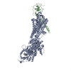
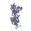
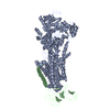
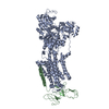
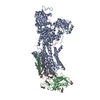
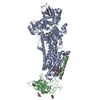
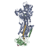
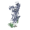
[日本語] English
- PDB-4ux1: Cryo-EM structure of antagonist-bound E2P gastric H,K-ATPase (SCH... -

+
データを開く


IDまたはキーワード:

読み込み中...

-
基本情報

登録情報
データベース: PDB / ID: 4ux1
タイトルCryo-EM structure of antagonist-bound E2P gastric H,K-ATPase (SCH.E2. AlF)
要素
  • POTASSIUM-TRANSPORTING ATPASE ALPHA CHAIN 1
  • POTASSIUM-TRANSPORTING ATPASE SUBUNIT BETA
キーワードTRANSPORT PROTEIN (運搬体タンパク質) / POTASSIUM-TRANSPORTING ATPASE
機能・相同性
機能・相同性情報


H+/K+-exchanging ATPase / potassium:proton exchanging ATPase complex / P-type potassium:proton transporter activity / Ion transport by P-type ATPases / P-type sodium:potassium-exchanging transporter activity / sodium:potassium-exchanging ATPase complex / sodium ion export across plasma membrane / intracellular potassium ion homeostasis / intracellular sodium ion homeostasis / potassium ion import across plasma membrane ...H+/K+-exchanging ATPase / potassium:proton exchanging ATPase complex / P-type potassium:proton transporter activity / Ion transport by P-type ATPases / P-type sodium:potassium-exchanging transporter activity / sodium:potassium-exchanging ATPase complex / sodium ion export across plasma membrane / intracellular potassium ion homeostasis / intracellular sodium ion homeostasis / potassium ion import across plasma membrane / ATPase activator activity / potassium ion binding / potassium ion transmembrane transport / proton transmembrane transport / 細胞接着 / apical plasma membrane / magnesium ion binding / ATP hydrolysis activity / ATP binding / 細胞膜
類似検索 - 分子機能
Gastric H+/K+-transporter P-type ATPase, N-terminal / Gastric H+/K+-ATPase, N terminal domain / Sodium/potassium-transporting ATPase subunit beta / Sodium/potassium-transporting ATPase subunit beta superfamily / Sodium / potassium ATPase beta chain / Sodium and potassium ATPases beta subunits signature 1. / Sodium and potassium ATPases beta subunits signature 2. / P-type ATPase subfamily IIC, subunit alpha / haloacid dehalogenase-like hydrolase / Cation-transporting P-type ATPase, C-terminal ...Gastric H+/K+-transporter P-type ATPase, N-terminal / Gastric H+/K+-ATPase, N terminal domain / Sodium/potassium-transporting ATPase subunit beta / Sodium/potassium-transporting ATPase subunit beta superfamily / Sodium / potassium ATPase beta chain / Sodium and potassium ATPases beta subunits signature 1. / Sodium and potassium ATPases beta subunits signature 2. / P-type ATPase subfamily IIC, subunit alpha / haloacid dehalogenase-like hydrolase / Cation-transporting P-type ATPase, C-terminal / Cation transporting ATPase, C-terminus / Cation transporter/ATPase, N-terminus / Cation-transporting P-type ATPase, N-terminal / Cation transporter/ATPase, N-terminus / Cation transport ATPase (P-type) / E1-E2 ATPase / P-type ATPase, haloacid dehalogenase domain / P-type ATPase, phosphorylation site / P-type ATPase, cytoplasmic domain N / E1-E2 ATPases phosphorylation site. / P-type ATPase, A domain superfamily / P-type ATPase / P-type ATPase, transmembrane domain superfamily / haloacid dehalogenase-like hydrolase / HAD superfamily / HAD-like superfamily
類似検索 - ドメイン・相同性
Potassium-transporting ATPase subunit beta / Potassium-transporting ATPase alpha chain 1
類似検索 - 構成要素
生物種SUS SCROFA (ブタ)
手法電子線結晶学 / クライオ電子顕微鏡法 / 解像度: 8 Å
データ登録者Abe, K. / Tani, K. / Fujiyoshi, Y.
引用ジャーナル: J Biol Chem / : 2014
タイトル: Systematic comparison of molecular conformations of H+,K+-ATPase reveals an important contribution of the A-M2 linker for the luminal gating.
著者: Kazuhiro Abe / Kazutoshi Tani / Yoshinori Fujiyoshi /
要旨: Gastric H(+),K(+)-ATPase, an ATP-driven proton pump responsible for gastric acidification, is a molecular target for anti-ulcer drugs. Here we show its cryo-electron microscopy (EM) structure in an ...Gastric H(+),K(+)-ATPase, an ATP-driven proton pump responsible for gastric acidification, is a molecular target for anti-ulcer drugs. Here we show its cryo-electron microscopy (EM) structure in an E2P analog state, bound to magnesium fluoride (MgF), and its K(+)-competitive antagonist SCH28080, determined at 7 Å resolution by electron crystallography of two-dimensional crystals. Systematic comparison with other E2P-related cryo-EM structures revealed that the molecular conformation in the (SCH)E2·MgF state is remarkably distinguishable. Although the azimuthal position of the A domain of the (SCH)E2·MgF state is similar to that in the E2·AlF (aluminum fluoride) state, in which the transmembrane luminal gate is closed, the arrangement of transmembrane helices in the (SCH)E2·MgF state shows a luminal-open conformation imposed on by bound SCH28080 at its luminal cavity, based on observations of the structure in the SCH28080-bound E2·BeF (beryllium fluoride) state. The molecular conformation of the (SCH)E2·MgF state thus represents a mixed overall structure in which its cytoplasmic and luminal half appear to be independently modulated by a phosphate analog and an antagonist bound to the respective parts of the enzyme. Comparison of the molecular conformations revealed that the linker region connecting the A domain and the transmembrane helix 2 (A-M2 linker) mediates the regulation of luminal gating. The mechanistic rationale underlying luminal gating observed in H(+),K(+)-ATPase is consistent with that observed in sarcoplasmic reticulum Ca(2+)-ATPase and other P-type ATPases and is most likely conserved for the P-type ATPase family in general.
履歴
登録2014年8月18日登録サイト: PDBE / 処理サイト: PDBE
改定 1.02014年9月17日Provider: repository / タイプ: Initial release
改定 1.12014年9月24日Group: Database references
改定 1.22014年10月1日Group: Database references
改定 1.32014年11月12日Group: Database references

-
構造の表示

ムービー
  • 登録構造単位
  • Jmolによる作画
  • ダウンロード
  • 単純化した表面モデル + あてはめた原子モデル
  • マップデータ: EMDB-2759
  • Jmolによる作画
  • ダウンロード
  • EMマップとの重ね合わせ
  • マップデータ: EMDB-2759
  • UCSF Chimeraによる作画
  • ダウンロード
  • EMマップとの重ね合わせ
  • マップデータ: EMDB-2759
  • UCSF Chimeraによる作画
  • ダウンロード
ムービービューア
構造ビューア分子:
MolmilJmol/JSmol

ダウンロードとリンク

-
集合体

登録構造単位
A: POTASSIUM-TRANSPORTING ATPASE ALPHA CHAIN 1
B: POTASSIUM-TRANSPORTING ATPASE SUBUNIT BETA


分子量 (理論値)分子数
合計 (水以外)147,5142
ポリマ-147,5142
非ポリマー00
0
1


  • 登録構造と同一
  • ソフトウェアが定義した集合体
タイプ名称対称操作
identity operation1_555x,y,z1
手法PISA
単位格子
Length a, b, c (Å)140.900, 111.300, 320.000
Angle α, β, γ (deg.)90.00, 90.00, 90.00
Int Tables number19
Space group name H-MP212121

-
要素

#1: タンパク質 POTASSIUM-TRANSPORTING ATPASE ALPHA CHAIN 1 / GASTRIC H(+)/K(+) ATPASE SUBUNIT ALPHA / PROTON PUMP / H / K-ATPASE


分子量: 114399.688 Da / 分子数: 1 / 由来タイプ: 天然 / 由来: (天然) SUS SCROFA (ブタ) / 参照: UniProt: P19156, H+/K+-exchanging ATPase
#2: タンパク質 POTASSIUM-TRANSPORTING ATPASE SUBUNIT BETA / GASTRIC H(+)/K(+) ATPASE SUBUNIT BETA / PROTON PUMP BETA CHA IN / GP60-90 / H / K-ATPASE


分子量: 33113.844 Da / 分子数: 1 / 由来タイプ: 天然 / 由来: (天然) SUS SCROFA (ブタ) / 参照: UniProt: P18434

-
実験情報

-
実験

実験手法: 電子線結晶学 / 使用した結晶の数: 267
EM実験試料の集合状態: 2D ARRAY / 3次元再構成法: 電子線結晶学

-
試料調製

構成要素名称: antagonist-bound E2P gastric H,K-ATPase / タイプ: COMPLEX
緩衝液pH: 4.8
試料包埋: NO / シャドウイング: NO / 染色: NO / 凍結: YES
急速凍結装置: LEICA KF80 / 凍結剤: NITROGEN
結晶化pH: 4.8 / 詳細: pH 4.8

-
データ収集

顕微鏡モデル: JEOL KYOTO-3000SFF / 日付: 2013年10月29日
電子銃電子線源: FIELD EMISSION GUN / 加速電圧: 300 kV / 照射モード: FLOOD BEAM
電子レンズモード: BRIGHT FIELDBright-field microscopy / 倍率(公称値): 40000 X / 最大 デフォーカス(公称値): 2997 nm / 最小 デフォーカス(公称値): 808 nm
撮影電子線照射量: 20 e/Å2 / フィルム・検出器のモデル: KODAK SO-163 FILM
回折平均測定温度: 4 K
検出器日付: 2013年10月29日
放射波長相対比: 1
反射解像度: 8→129 Å / Num. obs: 35798 / % possible obs: 52.6 % / Biso Wilson estimate: 32.4 Å2

-
解析

ソフトウェア
名称バージョン分類
MRCモデル構築
CNS1.3精密化
MRCSUITEデータスケーリング
MRC位相決定
3次元再構成解像度: 8 Å / 解像度の算出法: DIFFRACTION PATTERN/LAYERLINES / 対称性のタイプ: 2D CRYSTAL
精密化解像度: 8→128.95 Å / Rfactor Rfree error: 0.043 / Data cutoff high absF: 876135.35 / Data cutoff low absF: 0 / Isotropic thermal model: RESTRAINED / 交差検証法: THROUGHOUT / σ(F): 0
詳細: BULK SOLVENT MODEL USED SUBMISSION BASED ON EXPERIMENTAL DATA FROM EMDB EMD-2759. (DEPOSITION ID: 12786).
Rfactor反射数%反射Selection details
Rfree0.516 146 4.9 %RANDOM
Rwork0.507 ---
obs0.507 2994 52.6 %-
溶媒の処理溶媒モデル: FLAT MODEL / Bsol: 4098.69 Å2 / ksol: 0.1 e/Å3
原子変位パラメータBiso mean: 0 Å2
Baniso -1Baniso -2Baniso -3
1-25.76 Å20 Å20 Å2
2--37.87 Å20 Å2
3----63.64 Å2
Refine analyze
FreeObs
Luzzati coordinate error1.4 Å2.23 Å
Luzzati d res low-10 Å
Luzzati sigma a0.2 Å-2.74 Å
精密化ステップサイクル: LAST / 解像度: 8→128.95 Å
タンパク質核酸リガンド溶媒全体
原子数9161 0 0 0 9161
拘束条件
Refine-IDタイプDev idealDev ideal target
ELECTRON CRYSTALLOGRAPHYc_bond_d0.024
ELECTRON CRYSTALLOGRAPHYc_bond_d_na
ELECTRON CRYSTALLOGRAPHYc_bond_d_prot
ELECTRON CRYSTALLOGRAPHYc_angle_d
ELECTRON CRYSTALLOGRAPHYc_angle_d_na
ELECTRON CRYSTALLOGRAPHYc_angle_d_prot
ELECTRON CRYSTALLOGRAPHYc_angle_deg2.8
ELECTRON CRYSTALLOGRAPHYc_angle_deg_na
ELECTRON CRYSTALLOGRAPHYc_angle_deg_prot
ELECTRON CRYSTALLOGRAPHYc_dihedral_angle_d25.1
ELECTRON CRYSTALLOGRAPHYc_dihedral_angle_d_na
ELECTRON CRYSTALLOGRAPHYc_dihedral_angle_d_prot
ELECTRON CRYSTALLOGRAPHYc_improper_angle_d5.51
ELECTRON CRYSTALLOGRAPHYc_improper_angle_d_na
ELECTRON CRYSTALLOGRAPHYc_improper_angle_d_prot
ELECTRON CRYSTALLOGRAPHYc_mcbond_it01.5
ELECTRON CRYSTALLOGRAPHYc_mcangle_it02
ELECTRON CRYSTALLOGRAPHYc_scbond_it02
ELECTRON CRYSTALLOGRAPHYc_scangle_it02.5
Refine LS restraints NCSNCS model details: NONE
LS精密化 シェル解像度: 8→8.5 Å / Rfactor Rfree error: 0.224 / Total num. of bins used: 6
Rfactor反射数%反射
Rfree0.633 8 3 %
Rwork0.51 262 -
obs--29.4 %
Xplor fileSerial no: 1 / Param file: PROTEIN_REP.PARAM / Topol file: PROTEIN.TOP

+
万見について

-
お知らせ

-
2022年2月9日: EMDBエントリの付随情報ファイルのフォーマットが新しくなりました

EMDBエントリの付随情報ファイルのフォーマットが新しくなりました

  • EMDBのヘッダファイルのバージョン3が、公式のフォーマットとなりました。
  • これまでは公式だったバージョン1.9は、アーカイブから削除されます。

関連情報:EMDBヘッダ

外部リンク:wwPDBはEMDBデータモデルのバージョン3へ移行します

-
2020年8月12日: 新型コロナ情報

新型コロナ情報

URL: https://pdbj.org/emnavi/covid19.php

新ページ: EM Navigatorに新型コロナウイルスの特設ページを開設しました。

関連情報:Covid-19情報 / 2020年3月5日: 新型コロナウイルスの構造データ

+
2020年3月5日: 新型コロナウイルスの構造データ

新型コロナウイルスの構造データ

関連情報:万見生物種 / 2020年8月12日: 新型コロナ情報

外部リンク:COVID-19特集ページ - PDBj / 今月の分子2020年2月:コロナウイルスプロテーアーゼ

+
2019年1月31日: EMDBのIDの桁数の変更

EMDBのIDの桁数の変更

  • EMDBエントリに付与されているアクセスコード(EMDB-ID)は4桁の数字(例、EMD-1234)でしたが、間もなく枯渇します。これまでの4桁のID番号は4桁のまま変更されませんが、4桁の数字を使い切った後に発行されるIDは5桁以上の数字(例、EMD-12345)になります。5桁のIDは2019年の春頃から発行される見通しです。
  • EM Navigator/万見では、接頭語「EMD-」は省略されています。

関連情報:Q: 「EMD」とは何ですか? / 万見/EM NavigatorにおけるID/アクセスコードの表記

外部リンク:EMDB Accession Codes are Changing Soon! / PDBjへお問い合わせ

+
2017年7月12日: PDB大規模アップデート

PDB大規模アップデート

  • 新バージョンのPDBx/mmCIF辞書形式に基づくデータがリリースされました。
  • 今回の更新はバージョン番号が4から5になる大規模なもので、全エントリデータの書き換えが行われる「Remediation」というアップデートに該当します。
  • このバージョンアップで、電子顕微鏡の実験手法に関する多くの項目の書式が改定されました(例:em_softwareなど)。
  • EM NavigatorとYorodumiでも、この改定に基づいた表示内容になります。

外部リンク:wwPDB Remediation / OneDepデータ基準に準拠した、より強化された内容のモデル構造ファイルが、PDBアーカイブで公開されました。

-
万見 (Yorodumi)

幾万の構造データを、幾万の視点から

  • 万見(Yorodumi)は、EMDB/PDB/SASBDBなどの構造データを閲覧するためのページです。
  • EM Navigatorの詳細ページの後継、Omokage検索のフロントエンドも兼ねています。

関連情報:EMDB / PDB / SASBDB / 3つのデータバンクの比較 / 万見検索 / 2016年8月31日: 新しいEM Navigatorと万見 / 万見文献 / Jmol/JSmol / 機能・相同性情報 / 新しいEM Navigatorと万見の変更点

他の情報も見る