 ムービー
ムービー コントローラー


+ データを開く

- 基本情報
基本情報
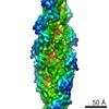
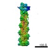
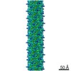
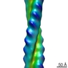
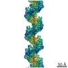
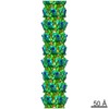
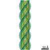
| 登録情報 | データベース: PDB / ID: 3j0s | ||||||
|---|---|---|---|---|---|---|---|
| タイトル | Remodeling of actin filaments by ADF cofilin proteins | ||||||
 要素 要素 |
| ||||||
 キーワード キーワード |  CONTRACTILE PROTEIN/PROTEIN BINDING / helical polymer / CONTRACTILE PROTEIN-ACTIN BINDING PROTEIN complex / CONTRACTILE PROTEIN-PROTEIN BINDING complex CONTRACTILE PROTEIN/PROTEIN BINDING / helical polymer / CONTRACTILE PROTEIN-ACTIN BINDING PROTEIN complex / CONTRACTILE PROTEIN-PROTEIN BINDING complex | ||||||
| 機能・相同性 |  機能・相同性情報 機能・相同性情報Gap junction degradation / Formation of annular gap junctions / RHO GTPases activate IQGAPs / UCH proteinases / DNA Damage Recognition in GG-NER / Adherens junctions interactions / Clathrin-mediated endocytosis / RHO GTPases Activate Formins / actin filament fragmentation / positive regulation of embryonic development ...Gap junction degradation / Formation of annular gap junctions / RHO GTPases activate IQGAPs / UCH proteinases / DNA Damage Recognition in GG-NER / Adherens junctions interactions / Clathrin-mediated endocytosis / RHO GTPases Activate Formins / actin filament fragmentation / positive regulation of embryonic development / establishment of spindle localization / structural constituent of postsynaptic actin cytoskeleton / EPH-ephrin mediated repulsion of cells / dense body / actin filament severing / EPHB-mediated forward signaling / VEGFA-VEGFR2 Pathway / regulation of dendritic spine morphogenesis / positive regulation by host of viral process / actin filament depolymerization / RHO GTPases Activate ROCKs / regulation of cell morphogenesis / NuA4 histone acetyltransferase complex / lamellipodium membrane / mitotic cytokinesis / Rho protein signal transduction / Sema3A PAK dependent Axon repulsion / cytoskeleton organization / EPHB-mediated forward signaling / Gene and protein expression by JAK-STAT signaling after Interleukin-12 stimulation / 軸索誘導 / 運動性 / マイクロフィラメント / response to virus / Regulation of actin dynamics for phagocytic cup formation / ruffle membrane / nuclear matrix / actin filament binding / マイクロフィラメント / Platelet degranulation / lamellipodium / 成長円錐 / actin cytoskeleton organization / vesicle / 細胞骨格 / 神経繊維 / focal adhesion / シナプス / negative regulation of apoptotic process / protein kinase binding / extracellular space / extracellular exosome / ATP binding / 生体膜 / 細胞核 / 細胞膜 / 細胞質基質 / 細胞質類似検索 - 分子機能 | ||||||
| 生物種 |  Homo sapiens (ヒト) Homo sapiens (ヒト) Gallus gallus (ニワトリ) Gallus gallus (ニワトリ) | ||||||
| 手法 | 電子顕微鏡法 / らせん対称体再構成法 / クライオ電子顕微鏡法 / 解像度: 9 Å | ||||||
 データ登録者 データ登録者 | Galkin, V.E. / Orlova, A. / Kudryashov, D.S. / Solodukhin, A. / Reisler, E. / Schroeder, G.F. / Egelman, E.H. | ||||||
 引用 引用 | ジャーナル: Proc Natl Acad Sci U S A / 年: 2011 タイトル: Remodeling of actin filaments by ADF/cofilin proteins. 著者: Vitold E Galkin / Albina Orlova / Dmitri S Kudryashov / Alexander Solodukhin / Emil Reisler / Gunnar F Schröder / Edward H Egelman /  要旨: Cofilin/ADF proteins play key roles in the dynamics of actin, one of the most abundant and highly conserved eukaryotic proteins. We used cryoelectron microscopy to generate a 9-Å resolution three- ...Cofilin/ADF proteins play key roles in the dynamics of actin, one of the most abundant and highly conserved eukaryotic proteins. We used cryoelectron microscopy to generate a 9-Å resolution three-dimensional reconstruction of cofilin-decorated actin filaments, the highest resolution achieved for a complex of F-actin with an actin-binding protein. We show that the cofilin-induced change in the filament twist is due to a unique conformation of the actin molecule unrelated to any previously observed state. The changes between the actin protomer in naked F-actin and in the actin-cofilin filament are greater than the conformational changes between G- and F-actin. Our results show the structural plasticity of actin, suggest that other actin-binding proteins may also induce large but different conformational changes, and show that F-actin cannot be described by a single molecular model. | ||||||
| 履歴 |
|
- 構造の表示
構造の表示
| ムービー |
ムービービューア |
|---|---|
| 構造ビューア | 分子: MolmilJmol/JSmol |
- ダウンロードとリンク
-ダウンロード
| PDBx/mmCIF形式 | 3j0s.cif.gz | 1 MB | 表示 | PDBx/mmCIF形式 |
|---|---|---|---|---|
| PDB形式 | pdb3j0s.ent.gz | 852.8 KB | 表示 | PDB形式 |
| PDBx/mmJSON形式 | 3j0s.json.gz | ツリー表示 | PDBx/mmJSON形式 | |
| その他 |  その他のダウンロード その他のダウンロード |
-検証レポート
| アーカイブディレクトリ | https://data.pdbj.org/pub/pdb/validation_reports/j0/3j0sftp://data.pdbj.org/pub/pdb/validation_reports/j0/3j0s | HTTPS FTP |
|---|
-関連構造データ
-リンク
 PDBj
PDBj


- 集合体
集合体
| 登録構造単位 | 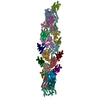
|
|---|---|
| 1 |
|
| 詳細 | THE ASSEMBLY REPRESENTED IN THIS ENTRY HAS REGULAR HELICAL SYMMETRY WITH THE FOLLOWING PARAMETERS: ROTATION PER SUBUNIT (TWIST) = -162.1 DEGREES; RISE PER SUBUNIT (HEIGHT) = 27.6 ANGSTROM |
-要素
| #1: タンパク質 | アクチン / Beta-actin 分子量: 41651.465 Da / 分子数: 12 / 由来タイプ: 天然 / 由来: (天然) Gallus gallus (ニワトリ) / 組織: muscle骨格筋 / 参照: UniProt: P60706#2: タンパク質 | / Cofilin / muscle isoform分子量: 18532.531 Da / 分子数: 12 / Fragment: SEE REMARK 999 / 由来タイプ: 組換発現 / 由来: (組換発現) Homo sapiens (ヒト) / 遺伝子: CFL1, CFL / 発現宿主:  Escherichia coli (大腸菌) / 参照: UniProt: P23528*PLUS Escherichia coli (大腸菌) / 参照: UniProt: P23528*PLUS配列の詳細 | THE AUTHORS STATE THAT HUMAN COFILIN-2 WAS USED IN THE EXPERIMENT, BUT HUMAN COFILIN-1 (UNP P23528) ...THE AUTHORS STATE THAT HUMAN COFILIN-2 WAS USED IN THE EXPERIMENT | |
|---|
-実験情報
-実験
| 実験 | 手法: 電子顕微鏡法 |
|---|---|
| EM実験 | 試料の集合状態: FILAMENT / 3次元再構成法: らせん対称体再構成法 |
- 試料調製
試料調製
| 構成要素 | 名称: actin decorated with cofilin / タイプ: COMPLEX / 詳細: filament containing one cofilin to one actin |
|---|---|
| 試料 | 包埋: NO / シャドウイング: NO / 染色: NO / 凍結: YES |
| 急速凍結 | 装置: FEI VITROBOT MARK I / 凍結剤: ETHANE / 湿度: 100 % |
- 電子顕微鏡撮影
| 実験機器 |  モデル: Tecnai F20 / 画像提供: FEI Company |
|---|---|
| 顕微鏡 | モデル: FEI TECNAI F20 / 日付: 2010年1月1日 |
| 電子銃 | 電子線源: FIELD EMISSION GUN / 加速電圧: 200 kV / 照射モード: FLOOD BEAM |
| 電子レンズ | モード: BRIGHT FIELDBright-field microscopy / 倍率(公称値): 50000 X / 最大 デフォーカス(公称値): 5300 nm / 最小 デフォーカス(公称値): 1100 nm / Cs: 2 mm |
| 試料ホルダ | 試料ホルダーモデル: GATAN LIQUID NITROGEN / 資料ホルダタイプ: SIDE ENTRY |
| 撮影 | フィルム・検出器のモデル: KODAK SO-163 FILM |
| 画像スキャン | デジタル画像の数: 125 |
- 解析
解析
| ソフトウェア |
| ||||||||||||
|---|---|---|---|---|---|---|---|---|---|---|---|---|---|
| EMソフトウェア |
| ||||||||||||
| CTF補正 | 詳細: each EM | ||||||||||||
| らせん対称 | 回転角度/サブユニット: 162.1 ° / 軸方向距離/サブユニット: 27.6 Å / らせん対称軸の対称性: C1 | ||||||||||||
| 3次元再構成 | 手法: IHRSR / 解像度: 9 Å / 解像度の算出法: FSC / 対称性のタイプ: HELICAL | ||||||||||||
| 原子モデル構築 | 空間: REAL | ||||||||||||
| 精密化ステップ | サイクル: LAST
|
