 ムービー
ムービー コントローラー


+ データを開く

- 基本情報
基本情報
| 登録情報 | データベース: PDB / ID: 1lu3 | ||||||
|---|---|---|---|---|---|---|---|
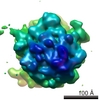
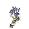
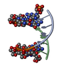
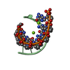
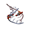
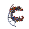
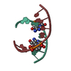
| タイトル | Separate Fitting of the Anticodon Loop Region of tRNA (nucleotide 26-42) in the Low Resolution Cryo-EM Map of an EF-Tu Ternary Complex (GDP and Kirromycin) Bound to E. coli 70S Ribosome | ||||||
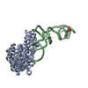
 要素 要素 | PHENYLALANINE TRANSFER RNA | ||||||
 キーワード キーワード |  RNA (リボ核酸) / tRNA (転移RNA) / ternary complex / cryo-EM (低温電子顕微鏡法) / 70S E.coli ribosome / conformational change (コンフォメーション変化) RNA (リボ核酸) / tRNA (転移RNA) / ternary complex / cryo-EM (低温電子顕微鏡法) / 70S E.coli ribosome / conformational change (コンフォメーション変化) | ||||||
| 機能・相同性 | リボ核酸 / RNA (> 10) 機能・相同性情報 機能・相同性情報 | ||||||
| 生物種 |  Saccharomyces cerevisiae (パン酵母) Saccharomyces cerevisiae (パン酵母) | ||||||
| 手法 | 電子顕微鏡法 / 単粒子再構成法 / クライオ電子顕微鏡法 / 解像度: 16.8 Å | ||||||
 データ登録者 データ登録者 | Valle, M. / Sengupta, J. / Swami, N.K. / Grassucci, R.A. / Burkhardt, N. / Nierhaus, K.H. / Agrawal, R.K. / Frank, J. | ||||||
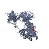
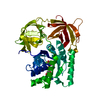
 引用 引用 | ジャーナル: EMBO J / 年: 2002 タイトル: Cryo-EM reveals an active role for aminoacyl-tRNA in the accommodation process. 著者: Mikel Valle / Jayati Sengupta / Neil K Swami / Robert A Grassucci / Nils Burkhardt / Knud H Nierhaus / Rajendra K Agrawal / Joachim Frank /  要旨: During the elongation cycle of protein biosynthesis, the specific amino acid coded for by the mRNA is delivered by a complex that is comprised of the cognate aminoacyl-tRNA, elongation factor Tu and ...During the elongation cycle of protein biosynthesis, the specific amino acid coded for by the mRNA is delivered by a complex that is comprised of the cognate aminoacyl-tRNA, elongation factor Tu and GTP. As this ternary complex binds to the ribosome, the anticodon end of the tRNA reaches the decoding center in the 30S subunit. Here we present the cryo- electron microscopy (EM) study of an Escherichia coli 70S ribosome-bound ternary complex stalled with an antibiotic, kirromycin. In the cryo-EM map the anticodon arm of the tRNA presents a new conformation that appears to facilitate the initial codon-anticodon interaction. Furthermore, the elbow region of the tRNA is seen to contact the GTPase-associated center on the 50S subunit of the ribosome, suggesting an active role of the tRNA in the transmission of the signal prompting the GTP hydrolysis upon codon recognition. #1: ジャーナル: J.Mol.Biol. / 年: 1999タイトル: Crystal structure of intact elongation factor EF-Tu from E.coli in GDP conformation at 2.05A resolution 著者: Song, H. / Parsons, M.R. / Rowsell, S. / Leonard, G. / Phillips, S.E. #2: ジャーナル: Science / 年: 1995タイトル: Crystal structure of the ternary complex of the Phe-tRNAPhe, EF-Tu, and a GTP analog 著者: Nissen, P. / Kjeldgaard, M. / Thirup, S. / Polekhina, G. / Reshetnikova, L. / Clark, B.F.C. / Nyborg, J. #3: ジャーナル: Nature / 年: 1997タイトル: Visualization of elongation factor Tu on E.coli ribosome 著者: Stark, H. / Rodnina, M.V. / Rinke-Appel, J. / Brimacombe, R. / Wintermeyer, W. / van Heel, M. #4: ジャーナル: Structure / 年: 1993タイトル: The crystal structure of elongation factor EF-Tu from Thermus aquaticus in the GTP conformation 著者: Kjeldgaard, M. / Nissen, P. / Thirup, S. / Nyborg, J. #5: ジャーナル: Structure / 年: 1996タイトル: Helix unwinding in the effector region of elongation factor EF-Tu-GDP 著者: Polekhina, G. / Thirup, S. / Kjeldgaard, M. / Nissen, P. / Lippmann, C. / Nyborg, J. | ||||||
| 履歴 |
|
- 構造の表示
構造の表示
| ムービー |
ムービービューア |
|---|---|
| 構造ビューア | 分子: MolmilJmol/JSmol |
- ダウンロードとリンク
-ダウンロード
| PDBx/mmCIF形式 | 1lu3.cif.gz | 20.8 KB | 表示 | PDBx/mmCIF形式 |
|---|---|---|---|---|
| PDB形式 | pdb1lu3.ent.gz | 10.9 KB | 表示 | PDB形式 |
| PDBx/mmJSON形式 | 1lu3.json.gz | ツリー表示 | PDBx/mmJSON形式 | |
| その他 |  その他のダウンロード その他のダウンロード |
-検証レポート
| アーカイブディレクトリ | https://data.pdbj.org/pub/pdb/validation_reports/lu/1lu3ftp://data.pdbj.org/pub/pdb/validation_reports/lu/1lu3 | HTTPS FTP |
|---|
-関連構造データ


-リンク
 PDBj
PDBj


- 集合体
集合体
| 登録構造単位 | 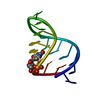
|
|---|---|
| 1 |
|
-要素
| #1: RNA鎖 | 分子量: 5466.326 Da / 分子数: 1 / 断片: anticodon loop region / 由来タイプ: 天然 / 由来: (天然) Saccharomyces cerevisiae (パン酵母) |
|---|
-実験情報
-実験
| 実験 | 手法: 電子顕微鏡法 |
|---|---|
| EM実験 | 試料の集合状態: PARTICLE / 3次元再構成法: 単粒子再構成法 |
- 試料調製
試料調製
| 構成要素 | 名称: E.coli 70S ribosome-Ternary complex-GDP-Kirromycin / タイプ: RIBOSOME / 詳細: E.coli 70S ribosome-Ternary complex-GDP-Kirromycin |
|---|---|
| 緩衝液 | 名称: Hepes-KOH buffer at pH 7.5 / pH: 7.5 / 詳細: Hepes-KOH buffer at pH 7.5 |
| 試料 | 包埋: NO / シャドウイング: NO / 染色: NO / 凍結: YES |
| 急速凍結 | 装置: HOMEMADE PLUNGER / 凍結剤: ETHANE / 詳細: Rapid-freezing in liquid ethane |
- 電子顕微鏡撮影
| 顕微鏡 | モデル: FEI/PHILIPS EM420 / 日付: 2001年3月1日 |
|---|---|
| 電子銃 | 電子線源: LAB6 / 加速電圧: 100 kV / 照射モード: FLOOD BEAM |
| 電子レンズ | モード: BRIGHT FIELDBright-field microscopy / 倍率(公称値): 50000 X / 倍率(補正後): 52000 X / 最大 デフォーカス(公称値): 3500 nm / 最小 デフォーカス(公称値): 1000 nm / Cs: 2 mm |
| 試料ホルダ | 温度: 93 K / 傾斜角・最大: 0 ° / 傾斜角・最小: 0 ° |
| 撮影 | 電子線照射量: 10 e/Å2 / フィルム・検出器のモデル: KODAK SO-163 FILM |
- 解析
解析
| EMソフトウェア |
| ||||||||||||
|---|---|---|---|---|---|---|---|---|---|---|---|---|---|
| CTF補正 | 詳細: CTF correction of 3D-maps | ||||||||||||
| 対称性 | 点対称性: C1 (非対称) | ||||||||||||
| 3次元再構成 | 手法: reference based alignment / 解像度: 16.8 Å / 粒子像の数: 7985 / ピクセルサイズ(実測値): 2.69 Å / 倍率補正: TMV / 詳細: SPIDER package / 対称性のタイプ: POINT | ||||||||||||
| 原子モデル構築 | プロトコル: OTHER / 詳細: METHOD--Manual | ||||||||||||
| 原子モデル構築 | PDB-ID: 1TTT Accession code: 1TTT / Source name: PDB / タイプ: experimental model | ||||||||||||
| 精密化 | 最高解像度: 16.8 Å | ||||||||||||
| 精密化ステップ | サイクル: LAST / 最高解像度: 16.8 Å
|
