National Institutes of Health/National Institute Of Allergy and Infectious Diseases (NIH/NIAID)
AI114902
米国
French National Research Agency
ANR14CE14001002
フランス
Canadian Institutes of Health Research (CIHR)
MOP125959
カナダ
Fondation pour la Recherche Medicale
フランス
引用
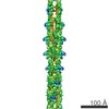
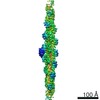
ジャーナル: Structure / 年: 2017 タイトル: Cryoelectron Microscopy Reconstructions of the Pseudomonas aeruginosa and Neisseria gonorrhoeae Type IV Pili at Sub-nanometer Resolution. 著者: Fengbin Wang / Mathieu Coureuil / Tomasz Osinski / Albina Orlova / Tuba Altindal / Gaël Gesbert / Xavier Nassif / Edward H Egelman / Lisa Craig / 要旨: We report here cryoelectron microscopy reconstructions of type IV pili (T4P) from two important human pathogens, Pseudomonas aeruginosa and Neisseria gonorrhoeae, at ∼ 8 and 5 Å resolution, ...We report here cryoelectron microscopy reconstructions of type IV pili (T4P) from two important human pathogens, Pseudomonas aeruginosa and Neisseria gonorrhoeae, at ∼ 8 and 5 Å resolution, respectively. The two structures reveal distinct arrangements of the pilin globular domains on the pilus surfaces, which impart different helical parameters, but similar packing of the conserved N-terminal α helices, α1, in the filament core. In contrast to the continuous α helix seen in the X-ray crystal structures of the P. aeruginosa and N. gonorrhoeae pilin subunits, α1 in the pilus filaments has a melted segment located between conserved helix-breaking residues Gly14 and Pro22, as seen for the Neisseria meningitidis T4P. Using mutagenesis we show that Pro22 is critical for pilus assembly, as are Thr2 and Glu5, which are positioned to interact in the hydrophobic filament core. These structures provide a framework for understanding T4P assembly, function, and biophysical properties.
フィルム・検出器のモデル: FEI FALCON II (4k x 4k) 検出モード: INTEGRATING / 平均露光時間: 2.0 sec. / 平均電子線量: 20.0 e/Å2 詳細: Images were stored containing seven parts, where each part represented a set of frames corresponding to a dose of ~20 electrons per Angstrom^2. The full dose image stack was used for the ...詳細: Images were stored containing seven parts, where each part represented a set of frames corresponding to a dose of ~20 electrons per Angstrom^2. The full dose image stack was used for the estimation of the CTF as well as for boxing filaments. Only the first two parts were used for the reconstruction (~5 electrons per Angstrom^2)
 ムービー
ムービー コントローラー


 データを開く
 基本情報
基本情報 マップデータ
 試料
試料 機能・相同性情報
 Pseudomonas aeruginosa PAK (緑膿菌)
 データ登録者
 米国,
米国,  フランス,
フランス,  カナダ, 4件
  引用
引用 構造の表示
構造の表示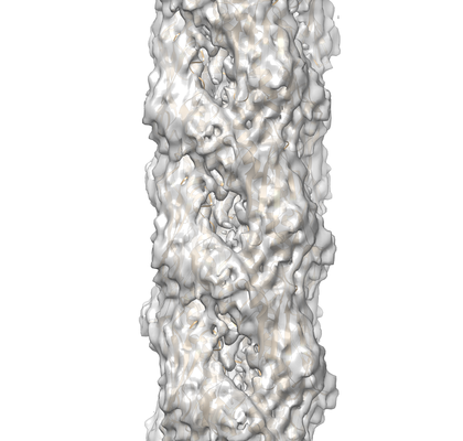
 ダウンロードとリンク
 emd_8740.png
 http://ftp.pdbj.org/pub/emdb/structures/EMD-8740


 試料の構成要素
 解析
解析 電子顕微鏡法

