 Movie
Movie Controller


+ Open data

- Basic information
| Entry | Database: EMDB / ID: EMD-7134 | ||||||||||||
|---|---|---|---|---|---|---|---|---|---|---|---|---|---|
| Title | AlfA Filament bound to AMPPNP | ||||||||||||
 Map data Map data | AlfA Filament | ||||||||||||
 Sample Sample |
| ||||||||||||
 Keywords Keywords |  actin / plasmid segregation / filament / CYTOSOLIC PROTEIN actin / plasmid segregation / filament / CYTOSOLIC PROTEIN | ||||||||||||
| Function / homology | Actin-like protein, N-terminal / Actin like proteins N terminal domain / ATPase, nucleotide binding domain / Actin-like protein N-terminal domain-containing protein Function and homology information Function and homology information | ||||||||||||
| Biological species |  Bacillus subtilis (bacteria) Bacillus subtilis (bacteria) | ||||||||||||
| Method | helical reconstruction / cryo EM / Resolution: 4.2 Å | ||||||||||||
 Authors Authors | Usluer GD / Kollman JM | ||||||||||||
| Funding support |  United States, United States,  Canada, Canada,  Turkey, 3 items Turkey, 3 items
| ||||||||||||
 Citation Citation | Journal: Proc Natl Acad Sci U S A / Year: 2018 Title: Cryo-EM structure of the bacterial actin AlfA reveals unique assembly and ATP-binding interactions and the absence of a conserved subdomain. Authors: Gülsima D Usluer / Frank DiMaio / Shun Kai Yang / Jesse M Hansen / Jessica K Polka / R Dyche Mullins / Justin M Kollman / Abstract: Bacterial actins are an evolutionarily diverse family of ATP-dependent filaments built from protomers with a conserved structural fold. Actin-based segregation systems are encoded on many bacterial ...Bacterial actins are an evolutionarily diverse family of ATP-dependent filaments built from protomers with a conserved structural fold. Actin-based segregation systems are encoded on many bacterial plasmids and function to partition plasmids into daughter cells. The bacterial actin AlfA segregates plasmids by a mechanism distinct from other partition systems, dependent on its unique dynamic properties. Here, we report the near-atomic resolution electron cryo-microscopy structure of the AlfA filament, which reveals a strikingly divergent filament architecture resulting from the loss of a subdomain conserved in all other actins and a mode of ATP binding. Its unusual assembly interfaces and nucleotide interactions provide insight into AlfA dynamics, and expand the range of evolutionary variation accessible to actin quaternary structure. | ||||||||||||
| History |
|
- Structure visualization
| Movie |
Movie viewer |
|---|---|
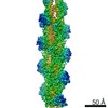
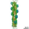
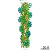
| Structure viewer | EM map: SurfViewMolmilJmol/JSmol |
| Supplemental images |

- Downloads & links
-EMDB archive
| Map data | emd_7134.map.gz | 135 MB | EMDB map data format | |
|---|---|---|---|---|
| Header (meta data) | emd-7134-v30.xmlemd-7134.xml | 11.2 KB 11.2 KB | Display Display | EMDB header |
| FSC (resolution estimation) | emd_7134_fsc.xml | 12 KB | Display | FSC data file |
| Images | 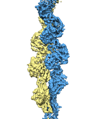 emd_7134.png emd_7134.png | 93.8 KB | ||
| Filedesc metadata | emd-7134.cif.gz | 5.3 KB | ||
| Archive directory |  http://ftp.pdbj.org/pub/emdb/structures/EMD-7134ftp://ftp.pdbj.org/pub/emdb/structures/EMD-7134 http://ftp.pdbj.org/pub/emdb/structures/EMD-7134ftp://ftp.pdbj.org/pub/emdb/structures/EMD-7134 | HTTPS FTP |
-Related structure data
| Related structure data |  6bqwMC M: atomic model generated by this map C: citing same article ( |
|---|---|
| Similar structure data |


-Links
| EMDB pages | EMDB (EBI/PDBe) / EMDataResource |
|---|
-Map
| File | Download / File: emd_7134.map.gz / Format: CCP4 / Size: 144.7 MB / Type: IMAGE STORED AS FLOATING POINT NUMBER (4 BYTES) | ||||||||||||||||||||||||||||||||||||||||||||||||||||||||||||
|---|---|---|---|---|---|---|---|---|---|---|---|---|---|---|---|---|---|---|---|---|---|---|---|---|---|---|---|---|---|---|---|---|---|---|---|---|---|---|---|---|---|---|---|---|---|---|---|---|---|---|---|---|---|---|---|---|---|---|---|---|---|
| Annotation | AlfA Filament | ||||||||||||||||||||||||||||||||||||||||||||||||||||||||||||
| Voxel size | X=Y=Z: 1 Å | ||||||||||||||||||||||||||||||||||||||||||||||||||||||||||||
| Density |
| ||||||||||||||||||||||||||||||||||||||||||||||||||||||||||||
| Symmetry | Space group: 1 | ||||||||||||||||||||||||||||||||||||||||||||||||||||||||||||
| Details | EMDB XML:
CCP4 map header:
| ||||||||||||||||||||||||||||||||||||||||||||||||||||||||||||
-Supplemental data
- Sample components
-Entire : AlfA filament
| Entire | Name: AlfA filament |
|---|---|
| Components |
|
-Supramolecule #1: AlfA filament
| Supramolecule | Name: AlfA filament / type: complex / ID: 1 / Parent: 0 / Macromolecule list: #1 Details: Each protomer is bound to the non-hydrolyzable nucleotide analog AMPPNP |
|---|---|
| Source (natural) | Organism: Bacillus subtilis (bacteria) |
| Molecular weight | Theoretical: 12750 kDa/nm |
-Macromolecule #1: Bacterial actin AlfA
| Macromolecule | Name: Bacterial actin AlfA / type: protein_or_peptide / ID: 1 / Number of copies: 9 / Enantiomer: LEVO |
|---|---|
| Source (natural) | Organism: Bacillus subtilis (bacteria) |
| Molecular weight | Theoretical: 31.150414 KDa |
| Recombinant expression | Organism: Escherichia coli (E. coli) |
| Sequence | String: MTLTTVIDIG NFSTKYAYKD KKQIKVGSFP SILHSYKPLE DYEGMERVEY NGLDYYVGET VKNFYFGREE QMYFGNTRKG HMEGQIRLV YALYTIFKET GKKEFNLILT CPYESMVTDK KYFVQHFEGE REVIVEGKSF KFTVHNIVMA AEGLGALNFS D SLNCVIVD ...String: MTLTTVIDIG NFSTKYAYKD KKQIKVGSFP SILHSYKPLE DYEGMERVEY NGLDYYVGET VKNFYFGREE QMYFGNTRKG HMEGQIRLV YALYTIFKET GKKEFNLILT CPYESMVTDK KYFVQHFEGE REVIVEGKSF KFTVHNIVMA AEGLGALNFS D SLNCVIVD AGSKTLNVLY LINGSISKMD SHTINGGTID NSIMDLAKTF AKTCSNIDYD YPIVCTGGKA EEMKECLENV GY STVSSAE LGEDKPSYYV NSVGLLLKYG RKFEEMFA UniProtKB: Actin-like protein N-terminal domain-containing protein |
-Macromolecule #2: PHOSPHOAMINOPHOSPHONIC ACID-ADENYLATE ESTER
| Macromolecule | Name: PHOSPHOAMINOPHOSPHONIC ACID-ADENYLATE ESTER / type: ligand / ID: 2 / Number of copies: 9 / Formula: ANP |
|---|---|
| Molecular weight | Theoretical: 506.196 Da |
| Chemical component information |  ChemComp-ANP: |
-Experimental details
-Structure determination
| Method | cryo EM |
|---|---|
 Processing Processing | helical reconstruction |
| Aggregation state | filament |
-Sample preparation
| Concentration | 0.16 mg/mL |
|---|---|
| Buffer | pH: 7.5 |
| Vitrification | Cryogen name: ETHANE |
| Details | Filaments were assembled in 5 mM AMPPNP for 15 minutes at room temperature |
- Electron microscopy
| Microscope | FEI TITAN KRIOS |
|---|---|
| Electron beam | Acceleration voltage: 300 kV / Electron source: FIELD EMISSION GUN |
| Electron optics | Illumination mode: FLOOD BEAM / Imaging mode: BRIGHT FIELDBright-field microscopy |
| Sample stage | Specimen holder model: FEI TITAN KRIOS AUTOGRID HOLDER / Cooling holder cryogen: NITROGEN |
| Image recording | Film or detector model: GATAN K2 SUMMIT (4k x 4k) / Detector mode: SUPER-RESOLUTION / Average electron dose: 72.0 e/Å2 |
| Experimental equipment |  Model: Titan Krios / Image courtesy: FEI Company |
-Image processing
| Startup model | Type of model: INSILICO MODEL / In silico model: cylinder |
|---|---|
| Final angle assignment | Type: NOT APPLICABLE |
| Final reconstruction | Applied symmetry - Helical parameters - Δz: 24.4 Å Applied symmetry - Helical parameters - Δ&Phi: 157.7 ° Applied symmetry - Helical parameters - Axial symmetry: C1 (asymmetric) Resolution.type: BY AUTHOR / Resolution: 4.2 Å / Resolution method: FSC 0.143 CUT-OFF / Software - Name: RELION (ver. 2.0) / Number images used: 113222 |
| FSC plot (resolution estimation) |  |
-Atomic model buiding 1
| Refinement | Space: REAL / Protocol: FLEXIBLE FIT |
|---|---|
| Output model | PDB-6bqw: |
