 ムービー
ムービー コントローラー


+ データを開く

- 基本情報
基本情報
| 登録情報 | データベース: EMDB / ID: EMD-6461 | |||||||||
|---|---|---|---|---|---|---|---|---|---|---|
| タイトル | Electron cryo-microscopy of the IST1-CHMP1B ESCRT-III copolymer | |||||||||
 マップデータ マップデータ | Reconstruction of IST1NTD-CHMP1B copolymer | |||||||||
 試料 試料 |
| |||||||||
 キーワード キーワード | ESCRT-III / IST1 / CHMP1B / membrane tubulation / helical filament | |||||||||
| 機能・相同性 |  機能・相同性情報 機能・相同性情報MIT domain binding / multivesicular body-lysosome fusion / amphisome membrane / vesicle fusion with vacuole / ESCRT III complex disassembly / late endosome to lysosome transport / ESCRT III complex / kinetochore microtubule / endosome transport via multivesicular body sorting pathway / cytoskeleton-dependent cytokinesis ...MIT domain binding / multivesicular body-lysosome fusion / amphisome membrane / vesicle fusion with vacuole / ESCRT III complex disassembly / late endosome to lysosome transport / ESCRT III complex / kinetochore microtubule / endosome transport via multivesicular body sorting pathway / cytoskeleton-dependent cytokinesis / collateral sprouting / membrane coat / regulation of centrosome duplication / nuclear membrane reassembly / multivesicular body sorting pathway / Sealing of the nuclear envelope (NE) by ESCRT-III / positive regulation of collateral sprouting / midbody abscission / membrane fission / plasma membrane repair / late endosome to vacuole transport / ubiquitin-dependent protein catabolic process via the multivesicular body sorting pathway / multivesicular body assembly / multivesicular body membrane / Flemming body / regulation of mitotic spindle assembly / mitotic metaphase chromosome alignment / nucleus organization / viral budding via host ESCRT complex / endoplasmic reticulum-Golgi intermediate compartment / positive regulation of proteolysis / autophagosome membrane / autophagosome maturation / nuclear pore / multivesicular body / viral budding from plasma membrane / establishment of protein localization / kinetochore / autophagy / azurophil granule lumen / intracellular protein localization / nuclear envelope / protein transport / midbody / endosome membrane / cadherin binding / protein domain specific binding / lysosomal membrane / cell division / intracellular membrane-bounded organelle / centrosome / Neutrophil degranulation / chromatin / protein-containing complex binding / extracellular exosome / extracellular region / nucleoplasm / identical protein binding / plasma membrane / cytosol 類似検索 - 分子機能 | |||||||||
| 生物種 |  Homo sapiens (ヒト) Homo sapiens (ヒト) | |||||||||
| 手法 | らせん対称体再構成法 / クライオ電子顕微鏡法 / 解像度: 4.0 Å | |||||||||
 データ登録者 データ登録者 | McCullough J / Clippinger AK / Talledge N / Skowyra ML / Saunders MG / Naismith TV / Colf LA / Afonine P / Arthur C / Sundquist WI ...McCullough J / Clippinger AK / Talledge N / Skowyra ML / Saunders MG / Naismith TV / Colf LA / Afonine P / Arthur C / Sundquist WI / Hanson PI / Frost A | |||||||||
 引用 引用 | ジャーナル: Science / 年: 2015 タイトル: Structure and membrane remodeling activity of ESCRT-III helical polymers. 著者: John McCullough / Amy K Clippinger / Nathaniel Talledge / Michael L Skowyra / Marissa G Saunders / Teresa V Naismith / Leremy A Colf / Pavel Afonine / Christopher Arthur / Wesley I Sundquist ...著者: John McCullough / Amy K Clippinger / Nathaniel Talledge / Michael L Skowyra / Marissa G Saunders / Teresa V Naismith / Leremy A Colf / Pavel Afonine / Christopher Arthur / Wesley I Sundquist / Phyllis I Hanson / Adam Frost /  要旨: The endosomal sorting complexes required for transport (ESCRT) proteins mediate fundamental membrane remodeling events that require stabilizing negative membrane curvature. These include endosomal ...The endosomal sorting complexes required for transport (ESCRT) proteins mediate fundamental membrane remodeling events that require stabilizing negative membrane curvature. These include endosomal intralumenal vesicle formation, HIV budding, nuclear envelope closure, and cytokinetic abscission. ESCRT-III subunits perform key roles in these processes by changing conformation and polymerizing into membrane-remodeling filaments. Here, we report the 4 angstrom resolution cryogenic electron microscopy reconstruction of a one-start, double-stranded helical copolymer composed of two different human ESCRT-III subunits, charged multivesicular body protein 1B (CHMP1B) and increased sodium tolerance 1 (IST1). The inner strand comprises "open" CHMP1B subunits that interlock in an elaborate domain-swapped architecture and is encircled by an outer strand of "closed" IST1 subunits. Unlike other ESCRT-III proteins, CHMP1B and IST1 polymers form external coats on positively curved membranes in vitro and in vivo. Our analysis suggests how common ESCRT-III filament architectures could stabilize different degrees and directions of membrane curvature. | |||||||||
| 履歴 |
|
- 構造の表示
構造の表示
| ムービー |
ムービービューア |
|---|---|
| 構造ビューア | EMマップ: SurfViewMolmilJmol/JSmol |
| 添付画像 |

- ダウンロードとリンク
-EMDBアーカイブ
| マップデータ | emd_6461.map.gz | 121.1 MB | EMDBマップデータ形式 | |
|---|---|---|---|---|
| ヘッダ (付随情報) | emd-6461-v30.xmlemd-6461.xml | 18 KB 18 KB | 表示 表示 | EMDBヘッダ |
| 画像 | 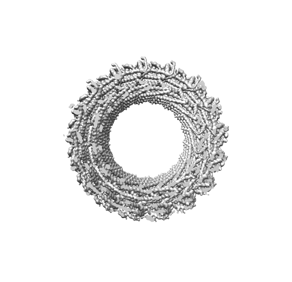 emd_6461.png emd_6461.png | 269.1 KB | ||
| アーカイブディレクトリ |  http://ftp.pdbj.org/pub/emdb/structures/EMD-6461ftp://ftp.pdbj.org/pub/emdb/structures/EMD-6461 http://ftp.pdbj.org/pub/emdb/structures/EMD-6461ftp://ftp.pdbj.org/pub/emdb/structures/EMD-6461 | HTTPS FTP |
-検証レポート
| 文書・要旨 | emd_6461_validation.pdf.gz | 455.5 KB | 表示 | EMDB検証レポート |
|---|---|---|---|---|
| 文書・詳細版 | emd_6461_full_validation.pdf.gz | 455 KB | 表示 | |
| XML形式データ | emd_6461_validation.xml.gz | 7 KB | 表示 | |
| アーカイブディレクトリ | https://ftp.pdbj.org/pub/emdb/validation_reports/EMD-6461ftp://ftp.pdbj.org/pub/emdb/validation_reports/EMD-6461 | HTTPS FTP |
-関連構造データ


-リンク
| EMDBのページ | EMDB (EBI/PDBe) / EMDataResource |
|---|
-マップ
| ファイル | ダウンロード / ファイル: emd_6461.map.gz / 形式: CCP4 / 大きさ: 126.7 MB / タイプ: IMAGE STORED AS FLOATING POINT NUMBER (4 BYTES) | ||||||||||||||||||||||||||||||||||||||||||||||||||||||||||||
|---|---|---|---|---|---|---|---|---|---|---|---|---|---|---|---|---|---|---|---|---|---|---|---|---|---|---|---|---|---|---|---|---|---|---|---|---|---|---|---|---|---|---|---|---|---|---|---|---|---|---|---|---|---|---|---|---|---|---|---|---|---|
| 注釈 | Reconstruction of IST1NTD-CHMP1B copolymer | ||||||||||||||||||||||||||||||||||||||||||||||||||||||||||||
| 投影像・断面図 | 画像のコントロール
画像は Spider により作成 | ||||||||||||||||||||||||||||||||||||||||||||||||||||||||||||
| ボクセルのサイズ | X=Y=Z: 1.2 Å | ||||||||||||||||||||||||||||||||||||||||||||||||||||||||||||
| 密度 |
| ||||||||||||||||||||||||||||||||||||||||||||||||||||||||||||
| 対称性 | 空間群: 1 | ||||||||||||||||||||||||||||||||||||||||||||||||||||||||||||
| 詳細 | EMDB XML:
CCP4マップ ヘッダ情報:
| ||||||||||||||||||||||||||||||||||||||||||||||||||||||||||||
 Z (Sec.)
 Y (Row.)
 X (Col.)


-添付データ
- 試料の構成要素
-全体 : ESCRT-III copolymer of IST1 and CHMP1B
| 全体 | 名称: ESCRT-III copolymer of IST1 and CHMP1B |
|---|---|
| 要素 |
|
-超分子 #1000: ESCRT-III copolymer of IST1 and CHMP1B
| 超分子 | 名称: ESCRT-III copolymer of IST1 and CHMP1B / タイプ: sample / ID: 1000 集合状態: 2-stranded helical filament composed of one strand of IST1 subunits and one strand of CHMP1B subunits Number unique components: 2 |
|---|
-分子 #1: IST1
| 分子 | 名称: IST1 / タイプ: protein_or_peptide / ID: 1 Name.synonym: Increased Sodium Tolerance 1, hIST1, Putatuve MAPK-activating protein PM28 詳細: IST1 N-terminal domain, residues 1-189 / 集合状態: Polymer / 組換発現: Yes |
|---|---|
| 由来(天然) | 生物種: Homo sapiens (ヒト) / 別称: Human |
| 分子量 | 実験値: 22 KDa / 理論値: 22 KDa |
| 組換発現 | 生物種:  |
| 配列 | UniProtKB: IST1 homolog |
-分子 #2: Charged multivesicular body protein 1b
| 分子 | 名称: Charged multivesicular body protein 1b / タイプ: protein_or_peptide / ID: 2 Name.synonym: CHMP1B, Chromatin-modifying protein 1b (CHMP1.5), Vacuolar protein sorting-associated protein 46-2 (hVps46-2) 詳細: full-length CHMP1B / 集合状態: Polymer / 組換発現: Yes |
|---|---|
| 由来(天然) | 生物種: Homo sapiens (ヒト) / 別称: Human |
| 分子量 | 実験値: 22 KDa / 理論値: 22 KDa |
| 組換発現 | 生物種: |
| 配列 | UniProtKB: Charged multivesicular body protein 1b |
-実験情報
-構造解析
| 手法 | クライオ電子顕微鏡法 |
|---|---|
 解析 解析 | らせん対称体再構成法 |
| 試料の集合状態 | filament |
-試料調製
| 濃度 | 0.7 mg/mL |
|---|---|
| 緩衝液 | pH: 8 / 詳細: 25 mM Tris, 25 mM NaCl |
| グリッド | 詳細: 3.5 uL of pelleted and resuspended liposome-nucleated IST1NTD-CHMP1B copolymers were applied to glow-discharged Quantifoil holey carbon grids (2 micron hole size, 2-4 micron spacing, 200 mesh). |
| 凍結 | 凍結剤: ETHANE / チャンバー内湿度: 100 % / 装置: FEI VITROBOT MARK I 手法: Blotted 7-9 seconds (-2 mm offset) and plunge-frozen |
- 電子顕微鏡法 #1
| Microscopy ID | 1 |
|---|---|
| 顕微鏡 | FEI TECNAI F20 |
| 日付 | 2013年6月1日 |
| 撮影 | カテゴリ: FILM / フィルム・検出器のモデル: KODAK SO-163 FILM デジタル化 - スキャナー: NIKON SUPER COOLSCAN 9000 実像数: 2493 / 平均電子線量: 10 e/Å2 詳細: 118,467 particles were selected from 2454 micrographs collected on a Titan Krios microscope with a Falcon I direct electron detector; 12,142 particles were selected from 39 micrographs ...詳細: 118,467 particles were selected from 2454 micrographs collected on a Titan Krios microscope with a Falcon I direct electron detector; 12,142 particles were selected from 39 micrographs collected on a JEM3200FSC microscope with a DE-12 direct electron detector. |
| 電子線 | 加速電圧: 200 kV / 電子線源:  FIELD EMISSION GUN FIELD EMISSION GUN |
| 電子光学系 | 照射モード: FLOOD BEAM / 撮影モード: BRIGHT FIELD / Cs: 2.0 mm / 最大 デフォーカス(公称値): 3.0 µm / 最小 デフォーカス(公称値): 0.6 µm / 倍率(公称値): 50000 |
| 試料ステージ | 試料ホルダーモデル: GATAN LIQUID NITROGEN |
| 実験機器 |  モデル: Tecnai F20 / 画像提供: FEI Company |
-電子顕微鏡法 #2
| Microscopy ID | 2 |
|---|---|
| 顕微鏡 | FEI TITAN KRIOS |
| 日付 | 2013年7月1日 |
| 撮影 | カテゴリ: FILM フィルム・検出器のモデル: FEI FALCON I (4k x 4k) デジタル化 - スキャナー: NIKON SUPER COOLSCAN 9000 実像数: 2493 / 平均電子線量: 15 e/Å2 詳細: 118,467 particles were selected from 2454 micrographs collected on a Titan Krios microscope with a Falcon I direct electron detector; 12,142 particles were selected from 39 micrographs ...詳細: 118,467 particles were selected from 2454 micrographs collected on a Titan Krios microscope with a Falcon I direct electron detector; 12,142 particles were selected from 39 micrographs collected on a JEM3200FSC microscope with a DE-12 direct electron detector. |
| 電子線 | 加速電圧: 300 kV / 電子線源: FIELD EMISSION GUN |
| 電子光学系 | 照射モード: FLOOD BEAM / 撮影モード: BRIGHT FIELD / 最大 デフォーカス(公称値): 3.0 µm / 最小 デフォーカス(公称値): 0.6 µm / 倍率(公称値): 59000 |
| 試料ステージ | 試料ホルダーモデル: FEI TITAN KRIOS AUTOGRID HOLDER |
| 実験機器 |  モデル: Titan Krios / 画像提供: FEI Company |
-電子顕微鏡法 #3
| Microscopy ID | 3 |
|---|---|
| 顕微鏡 | JEOL 3200FSC |
| 日付 | 2013年8月1日 |
| 撮影 | カテゴリ: FILM フィルム・検出器のモデル: DIRECT ELECTRON DE-12 (4k x 3k) デジタル化 - スキャナー: NIKON SUPER COOLSCAN 9000 実像数: 2493 / 平均電子線量: 20 e/Å2 詳細: 118,467 particles were selected from 2454 micrographs collected on a Titan Krios microscope with a Falcon I direct electron detector; 12,142 particles were selected from 39 micrographs ...詳細: 118,467 particles were selected from 2454 micrographs collected on a Titan Krios microscope with a Falcon I direct electron detector; 12,142 particles were selected from 39 micrographs collected on a JEM3200FSC microscope with a DE-12 direct electron detector. |
| Tilt angle min | 0 |
| Tilt angle max | 0 |
| 電子線 | 加速電圧: 300 kV / 電子線源: FIELD EMISSION GUN |
| 電子光学系 | 照射モード: FLOOD BEAM / 撮影モード: BRIGHT FIELD / Cs: 3.40 mm / 最大 デフォーカス(公称値): 3.0 µm / 最小 デフォーカス(公称値): 0.6 µm / 倍率(公称値): 59000 |
| 試料ステージ | 試料ホルダーモデル: JEOL 3200FSC CRYOHOLDER |
-画像解析
| 詳細 | Particles were picked manually using the e2helixboxer.py function of EMAN2. Particles were aligned with IHRSR initially, followed by high resolution asymmetric refinement in RELION, followed by helical averaging in real space. |
|---|---|
| 最終 再構成 | 想定した対称性 - らせんパラメータ - Δz: 2.96 Å 想定した対称性 - らせんパラメータ - ΔΦ: 21.060 ° 想定した対称性 - らせんパラメータ - 軸対称性: C1 (非対称) 解像度のタイプ: BY AUTHOR / 解像度: 4.0 Å / 解像度の算出法: OTHER ソフトウェア - 名称: CTFFIND3, EMAN2, IHRSR, SPIDER, RELION 詳細: 51 copies (3 complete turns) of the asymmetric RELION reconstruction were transformed according to the helical symmetry, resampled on the original grid, and summed together, with Iterative ...詳細: 51 copies (3 complete turns) of the asymmetric RELION reconstruction were transformed according to the helical symmetry, resampled on the original grid, and summed together, with Iterative Helical Real Space Reconstruction (IHRSR) single-particle algorithm as implemented in SPIDER and high-resolution refinement in RELION. |
| CTF補正 | 詳細: Each particle as implemented in RELION |
-原子モデル構築 1
| 初期モデル | PDB ID: Chain - Chain ID: A |
|---|---|
| ソフトウェア | 名称: Coot, Chimera, NAMD MDFF, Rosetta |
| 詳細 | 3FRR was docked manually into the segmented density using Chimera. Regions with poor fit to density were identified using the Rosetta loops-from-density algorithm and iteratively fitted using alternating cycles of Rosetta's rebuild and refine protocol and manual refinement in Coot. The full ring of IST1 structures was then refined using Rosetta's symmetry constraints. Finally, backbone hydrogen bonds in the helical regions were constrained and two cycles of loop rebuilding with constraints were performed. The IST1 model was then combined with the CHMP1B model to form a heterodimer and this structure was refined by iterating between manual rebuilding and refinement using MDFF |
| 精密化 | 空間: REAL / プロトコル: FLEXIBLE FIT 当てはまり具合の基準: Molprobity validation, cross-correlation |
| 得られたモデル | 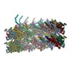 PDB-3jc1: |

