protein insertion into membrane from inner side / cell envelope Sec protein transport complex / protein transport by the Sec complex / intracellular protein transmembrane transport / protein-transporting ATPase activity / SRP-dependent cotranslational protein targeting to membrane, translocation / signal sequence binding / protein secretion / protein transmembrane transporter activity / intracellular protein transport ...protein insertion into membrane from inner side / cell envelope Sec protein transport complex / protein transport by the Sec complex / intracellular protein transmembrane transport / protein-transporting ATPase activity / SRP-dependent cotranslational protein targeting to membrane, translocation / signal sequence binding / protein secretion / protein transmembrane transporter activity / intracellular protein transport / ribosomal large subunit assembly / large ribosomal subunit rRNA binding / cytosolic large ribosomal subunit / cytoplasmic translation / rRNA binding / structural constituent of ribosome / 翻訳 (生物学) / 生体膜 / 細胞膜 / 細胞質基質 / 細胞質 類似検索 - 分子機能
Preprotein translocase SecG subunit / Preprotein translocase SecG subunit / SecE subunit of protein translocation complex, bacterial-like / SecE superfamily / Protein translocase subunit SecY / Protein secE/sec61-gamma signature. / Protein secY signature 1. / Protein secY signature 2. / SecE/Sec61-gamma subunits of protein translocation complex / Protein translocase complex, SecE/Sec61-gamma subunit ...Preprotein translocase SecG subunit / Preprotein translocase SecG subunit / SecE subunit of protein translocation complex, bacterial-like / SecE superfamily / Protein translocase subunit SecY / Protein secE/sec61-gamma signature. / Protein secY signature 1. / Protein secY signature 2. / SecE/Sec61-gamma subunits of protein translocation complex / Protein translocase complex, SecE/Sec61-gamma subunit / SecY/SEC61-alpha family / SecY domain superfamily / SecY conserved site / SecY / Ribosomal proteins 50S L24/mitochondrial 39S L24 / Ribosomal protein L24 / Ribosomal protein L23/L25, conserved site / Ribosomal protein L23 signature. / Ribosomal protein L29, conserved site / Ribosomal protein L29 signature. / Ribosomal protein L25/L23 / Ribosomal L29 protein / Ribosomal protein L29/L35 / Ribosomal protein L29/L35 superfamily / Ribosomal protein L23 / Ribosomal protein L24 signature. / Ribosomal protein L24/L26, conserved site / KOW (Kyprides, Ouzounis, Woese) motif. / Ribosomal protein L26/L24, KOW domain / Translation protein SH3-like domain superfamily / Ribosomal protein L23/L15e core domain superfamily / KOW motif / KOW / Ribosomal protein L2, domain 2 / Nucleotide-binding alpha-beta plait domain superfamily 類似検索 - ドメイン・相同性
Large ribosomal subunit protein uL29 / Large ribosomal subunit protein uL23 / Protein translocase subunit SecE / Protein-export membrane protein SecG / Protein translocase subunit SecY / Large ribosomal subunit protein uL24 類似検索 - 構成要素
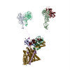
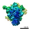
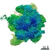
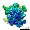
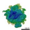
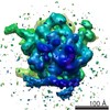
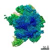
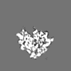
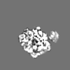
ジャーナル: Nature / 年: 2014 タイトル: Structure of the SecY channel during initiation of protein translocation. 著者: Eunyong Park / Jean-François Ménétret / James C Gumbart / Steven J Ludtke / Weikai Li / Andrew Whynot / Tom A Rapoport / Christopher W Akey / 要旨: Many secretory proteins are targeted by signal sequences to a protein-conducting channel, formed by prokaryotic SecY or eukaryotic Sec61 complexes, and are translocated across the membrane during ...Many secretory proteins are targeted by signal sequences to a protein-conducting channel, formed by prokaryotic SecY or eukaryotic Sec61 complexes, and are translocated across the membrane during their synthesis. Crystal structures of the inactive channel show that the SecY subunit of the heterotrimeric complex consists of two halves that form an hourglass-shaped pore with a constriction in the middle of the membrane and a lateral gate that faces the lipid phase. The closed channel has an empty cytoplasmic funnel and an extracellular funnel that is filled with a small helical domain, called the plug. During initiation of translocation, a ribosome-nascent chain complex binds to the SecY (or Sec61) complex, resulting in insertion of the nascent chain. However, the mechanism of channel opening during translocation is unclear. Here we have addressed this question by determining structures of inactive and active ribosome-channel complexes with cryo-electron microscopy. Non-translating ribosome-SecY channel complexes derived from Methanocaldococcus jannaschii or Escherichia coli show the channel in its closed state, and indicate that ribosome binding per se causes only minor changes. The structure of an active E. coli ribosome-channel complex demonstrates that the nascent chain opens the channel, causing mostly rigid body movements of the amino- and carboxy-terminal halves of SecY. In this early translocation intermediate, the polypeptide inserts as a loop into the SecY channel with the hydrophobic signal sequence intercalated into the open lateral gate. The nascent chain also forms a loop on the cytoplasmic surface of SecY rather than entering the channel directly.
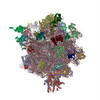
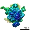
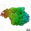
全体 : E. coli 70S ribosome with recombinant E. coli SecYEG
全体
名称: E. coli 70S ribosome with recombinant E. coli SecYEG
要素
試料: E. coli 70S ribosome with recombinant E. coli SecYEG
複合体: non-translating 70S ribosome
タンパク質・ペプチド: SecYEGSec61
-
超分子 #1000: E. coli 70S ribosome with recombinant E. coli SecYEG
超分子
名称: E. coli 70S ribosome with recombinant E. coli SecYEG タイプ: sample / ID: 1000 詳細: Ribosome-SecY complexes were prepared by mixing ribosomes at 4 uM with SecY (32 uM) and incubating them on ice for 30 min before freezing. 集合状態: one ribosome and one SecYEG / Number unique components: 2
分子量
理論値: 2.5 MDa
-
超分子 #1: non-translating 70S ribosome
超分子
名称: non-translating 70S ribosome / タイプ: complex / ID: 1 / 組換発現: No / データベース: NCBI / Ribosome-details: ribosome-prokaryote: ALL
Legacy - 非点収差: imaging of carbon film at 175,000 times magnification
詳細
low dose imaging with manual data collection
日付
2006年4月10日
撮影
カテゴリ: FILM / フィルム・検出器のモデル: KODAK SO-163 FILM / デジタル化 - スキャナー: OTHER / デジタル化 - サンプリング間隔: 4.5 µm / 実像数: 360 / 平均電子線量: 20 e/Å2 / Od range: 1 / ビット/ピクセル: 16
Tilt angle min
0
-
画像解析
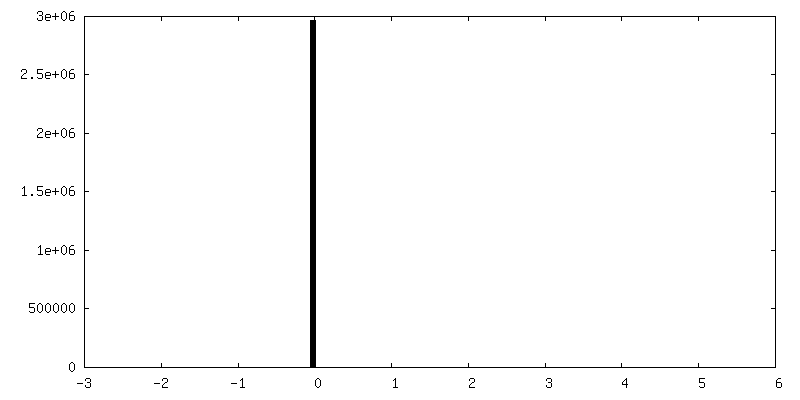
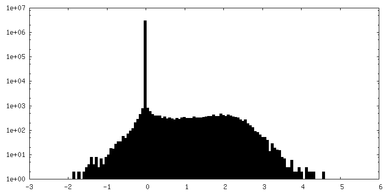
CTF補正
詳細: per micrograph
最終 2次元分類
クラス数: 1900
最終 再構成
アルゴリズム: OTHER / 解像度のタイプ: BY AUTHOR / 解像度: 9.5 Å / 解像度の算出法: OTHER / ソフトウェア - 名称: EMAN1 詳細: CTF correction was done on untilted and 30 degree tilted images. 使用した粒子像数: 39000
詳細
Particles were picked with boxer and CTF-corrected with EMAN1.
 ムービー
ムービー コントローラー


 データを開く
 基本情報
基本情報 マップデータ
 試料
試料 キーワード
キーワード SecYEG (Sec61)
 機能・相同性情報
 データ登録者
 引用
引用
 構造の表示
構造の表示
 ダウンロードとリンク
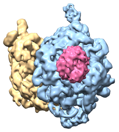 emd_5692.jpg
 http://ftp.pdbj.org/pub/emdb/structures/EMD-5692


 Z
Z Y
Y X
X


 試料の構成要素
 解析
解析 電子顕微鏡法