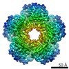
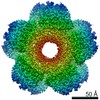
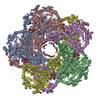
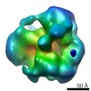
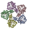
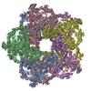
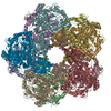
Journal: Structure / Year: 2019 Title: Structure, Function, and Evolution of the Pseudomonas aeruginosa Lysine Decarboxylase LdcA. Authors: Eaazhisai Kandiah / Diego Carriel / Pierre Simon Garcia / Jan Felix / Manuel Banzhaf / George Kritikos / Maria Bacia-Verloop / Céline Brochier-Armanet / Sylvie Elsen / Irina Gutsche / Abstract: The only enzyme responsible for cadaverine production in the major multidrug-resistant human pathogen Pseudomonas aeruginosa is the lysine decarboxylase LdcA. This enzyme modulates the general ...The only enzyme responsible for cadaverine production in the major multidrug-resistant human pathogen Pseudomonas aeruginosa is the lysine decarboxylase LdcA. This enzyme modulates the general polyamine homeostasis, promotes growth, and reduces bacterial persistence during carbenicillin treatment. Here we present a 3.7-Å resolution cryoelectron microscopy structure of LdcA. We introduce an original approach correlating phylogenetic signal with structural information and reveal possible recombination among LdcA and arginine decarboxylase subfamilies within structural domain boundaries. We show that LdcA is involved in full virulence in an insect pathogenesis model. Furthermore, unlike its enterobacterial counterparts, LdcA is regulated neither by the stringent response alarmone ppGpp nor by the AAA+ ATPase RavA. Instead, the P. aeruginosa ravA gene seems to play a defensive role. Altogether, our study identifies LdcA as an important player in P. aeruginosa physiology and virulence and as a potential drug target.
History
Deposition
Dec 11, 2018
-
Header (metadata) release
Sep 25, 2019
-
Map release
Sep 25, 2019
-
Update
Oct 23, 2019
-
Current status
Oct 23, 2019
Processing site: PDBe / Status: Released
-
Structure visualization
Movie
Surface view with section colored by density value
In the structure databanks used in Yorodumi, some data are registered as the other names, "COVID-19 virus" and "2019-nCoV". Here are the details of the virus and the list of structure data.
Jan 31, 2019. EMDB accession codes are about to change! (news from PDBe EMDB page)
EMDB accession codes are about to change! (news from PDBe EMDB page)
The allocation of 4 digits for EMDB accession codes will soon come to an end. Whilst these codes will remain in use, new EMDB accession codes will include an additional digit and will expand incrementally as the available range of codes is exhausted. The current 4-digit format prefixed with “EMD-” (i.e. EMD-XXXX) will advance to a 5-digit format (i.e. EMD-XXXXX), and so on. It is currently estimated that the 4-digit codes will be depleted around Spring 2019, at which point the 5-digit format will come into force.
The EM Navigator/Yorodumi systems omit the EMD- prefix.
Related info.:Q: What is EMD? / ID/Accession-code notation in Yorodumi/EM Navigator
Yorodumi is a browser for structure data from EMDB, PDB, SASBDB, etc.
This page is also the successor to EM Navigator detail page, and also detail information page/front-end page for Omokage search.
The word "yorodu" (or yorozu) is an old Japanese word meaning "ten thousand". "mi" (miru) is to see.
Related info.:EMDB / PDB / SASBDB / Comparison of 3 databanks / Yorodumi Search / Aug 31, 2016. New EM Navigator & Yorodumi / Yorodumi Papers / Jmol/JSmol / Function and homology information / Changes in new EM Navigator and Yorodumi
 Movie
Movie Controller


 Open data
 Basic information
 Map data
 Sample
 Function and homology information
 arginine decarboxylase /

 Authors
 France, 2 items
  Citation
 Structure visualization
 Downloads & links
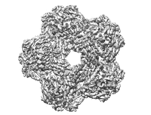 emd_4468.png
 http://ftp.pdbj.org/pub/emdb/structures/EMD-4468
 Sample components
 Processing
 Electron microscopy