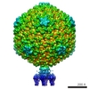
 Movie
Movie Controller


+ Open data

- Basic information
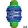
| Entry | Database: EMDB / ID: EMD-4459 | ||||||||||||
|---|---|---|---|---|---|---|---|---|---|---|---|---|---|
| Title | Structure of native bacteriophage P68 | ||||||||||||
 Map data Map data | None | ||||||||||||
 Sample Sample |
| ||||||||||||
 Keywords Keywords |  structural protein / bacteriophage / receptor binding protein / complex / VIRUS structural protein / bacteriophage / receptor binding protein / complex / VIRUS | ||||||||||||
| Function / homology | : / Phage 5-bladed beta propeller receptor binding platform domain / Uncharacterized protein / Uncharacterized protein / Major head protein / Lower collar protein / Minor structural protein Function and homology information Function and homology information | ||||||||||||
| Biological species |  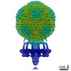 Staphylococcus phage P68 (virus) Staphylococcus phage P68 (virus) | ||||||||||||
| Method | single particle reconstruction / cryo EM / Resolution: 3.8 Å | ||||||||||||
 Authors Authors | Dominik H / Karel S / Fuzik T / Plevka P | ||||||||||||
| Funding support |  Czech Republic, 3 items Czech Republic, 3 items
| ||||||||||||
 Citation Citation | Journal: Sci Adv / Year: 2019 Title: Structure and genome ejection mechanism of phage P68. Authors: Dominik Hrebík / Dana Štveráková / Karel Škubník / Tibor Füzik / Roman Pantůček / Pavel Plevka / Abstract: Phages infecting can be used as therapeutics against antibiotic-resistant bacterial infections. However, there is limited information about the mechanism of genome delivery of phages that infect ...Phages infecting can be used as therapeutics against antibiotic-resistant bacterial infections. However, there is limited information about the mechanism of genome delivery of phages that infect Gram-positive bacteria. Here, we present the structures of native phage P68, genome ejection intermediate, and empty particle. The P68 head contains 72 subunits of inner core protein, 15 of which bind to and alter the structure of adjacent major capsid proteins and thus specify attachment sites for head fibers. Unlike in the previously studied phages, the head fibers of P68 enable its virion to position itself at the cell surface for genome delivery. The unique interaction of one end of P68 DNA with one of the 12 portal protein subunits is disrupted before the genome ejection. The inner core proteins are released together with the DNA and enable the translocation of phage genome across the bacterial membrane into the cytoplasm. | ||||||||||||
| History |
|
- Structure visualization
| Movie |
Movie viewer |
|---|---|
| Structure viewer | EM map: SurfViewMolmilJmol/JSmol |
| Supplemental images |

- Downloads & links
-EMDB archive
| Map data | emd_4459.map.gz | 214.8 MB | EMDB map data format | |
|---|---|---|---|---|
| Header (meta data) | emd-4459-v30.xmlemd-4459.xml | 29.7 KB 29.7 KB | Display Display | EMDB header |
| Images | 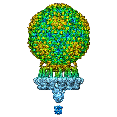 emd_4459.png emd_4459.png | 153.4 KB | ||
| Filedesc metadata | emd-4459.cif.gz | 8.1 KB | ||
| Others | emd_4459_additional.map.gz | 609.1 MB | ||
| Archive directory |  http://ftp.pdbj.org/pub/emdb/structures/EMD-4459ftp://ftp.pdbj.org/pub/emdb/structures/EMD-4459 http://ftp.pdbj.org/pub/emdb/structures/EMD-4459ftp://ftp.pdbj.org/pub/emdb/structures/EMD-4459 | HTTPS FTP |
-Related structure data
| Related structure data |  6q3gMC  4435C 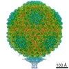 4436C  4437C  4438C  4440C 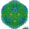 4442C  4449C  4450C 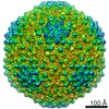 4451C  4453C  4454C  4455C 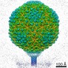 4456C  4457C  4458C  6iabC 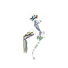 6iacC  6iatC  6iawC  6ib1C C: citing same article ( M: atomic model generated by this map |
|---|---|
| Similar structure data |
-Links
| EMDB pages | EMDB (EBI/PDBe) / EMDataResource |
|---|
-Map
| File | Download / File: emd_4459.map.gz / Format: CCP4 / Size: 3.7 GB / Type: IMAGE STORED AS FLOATING POINT NUMBER (4 BYTES) | ||||||||||||||||||||||||||||||||||||||||||||||||||||||||||||
|---|---|---|---|---|---|---|---|---|---|---|---|---|---|---|---|---|---|---|---|---|---|---|---|---|---|---|---|---|---|---|---|---|---|---|---|---|---|---|---|---|---|---|---|---|---|---|---|---|---|---|---|---|---|---|---|---|---|---|---|---|---|
| Annotation | None | ||||||||||||||||||||||||||||||||||||||||||||||||||||||||||||
| Voxel size | X=Y=Z: 1.063 Å | ||||||||||||||||||||||||||||||||||||||||||||||||||||||||||||
| Density |
| ||||||||||||||||||||||||||||||||||||||||||||||||||||||||||||
| Symmetry | Space group: 1 | ||||||||||||||||||||||||||||||||||||||||||||||||||||||||||||
| Details | EMDB XML:
CCP4 map header:
| ||||||||||||||||||||||||||||||||||||||||||||||||||||||||||||
-Supplemental data
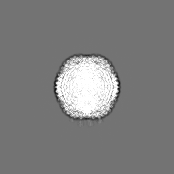
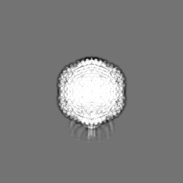
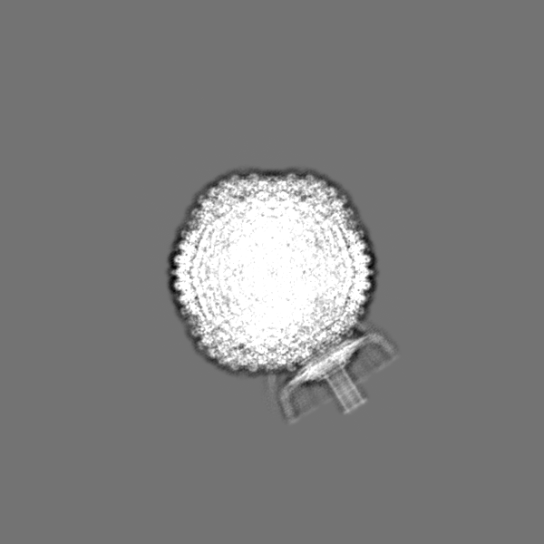
-Additional map: Masked asymmetric reconstruction of native bacteriophage P68
| File | emd_4459_additional.map | ||||||||||||
|---|---|---|---|---|---|---|---|---|---|---|---|---|---|
| Annotation | Masked asymmetric reconstruction of native bacteriophage P68 | ||||||||||||
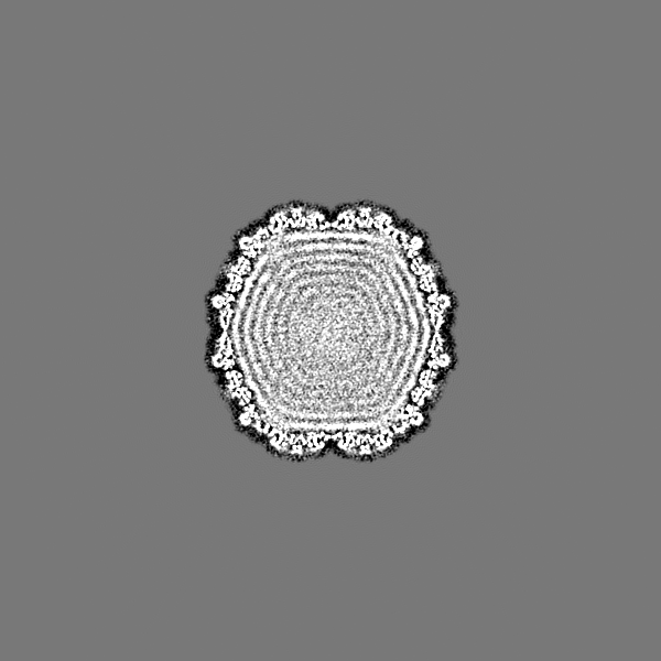
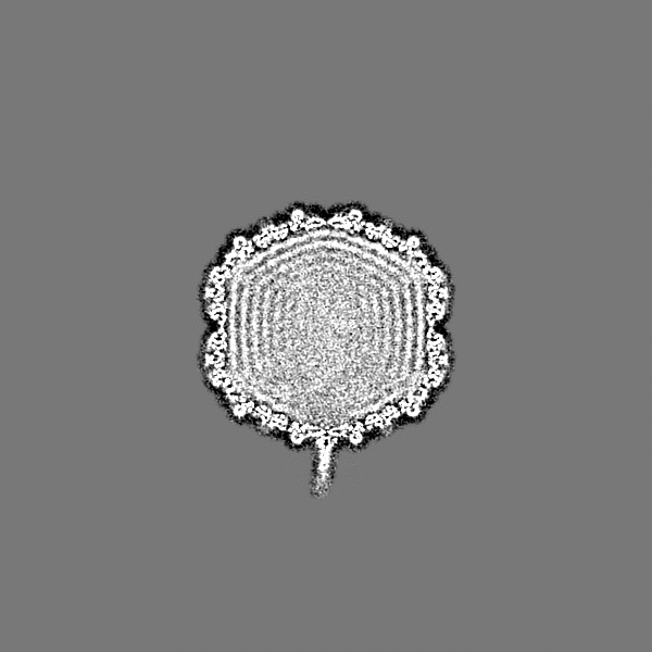
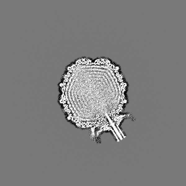
| Projections & Slices |
| ||||||||||||
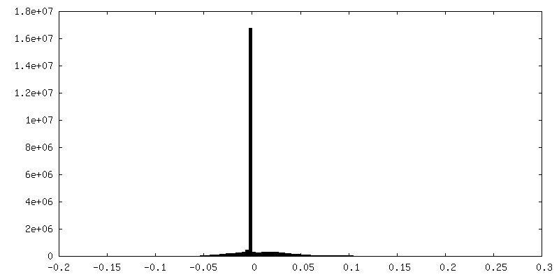
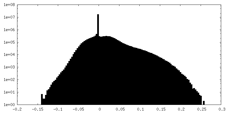
| Density Histograms |
 Z
Z Y
Y X
X

- Sample components
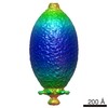
+Entire : Staphylococcus phage P68
+Supramolecule #1: Staphylococcus phage P68


+Supramolecule #2: Capsid of native phage P68
+Supramolecule #3: Tail complex
+Supramolecule #4: Portal protein
+Supramolecule #5: Inner core protein
+Macromolecule #1: Major head protein
+Macromolecule #2: Arstotzka protein
+Macromolecule #3: Portal protein
+Macromolecule #4: Lower collar protein
+Macromolecule #5: Minor structural protein
+Macromolecule #6: Tail fibre protein
+Macromolecule #7: Head fiber protein
+Macromolecule #8: Inner core protein
-Experimental details
-Structure determination
| Method | cryo EM |
|---|---|
 Processing Processing | single particle reconstruction |
| Aggregation state | particle |
-Sample preparation
| Concentration | 2 mg/mL | ||||||||||||
|---|---|---|---|---|---|---|---|---|---|---|---|---|---|
| Buffer | pH: 8 Component:
| ||||||||||||
| Grid | Model: Quantifoil R2/1 / Material: COPPER / Mesh: 200 / Support film - Material: CARBON / Support film - topology: HOLEY / Pretreatment - Type: GLOW DISCHARGE / Pretreatment - Time: 30 sec. / Pretreatment - Atmosphere: NITROGEN | ||||||||||||
| Vitrification | Cryogen name: ETHANE / Chamber humidity: 100 % / Chamber temperature: 293 K / Instrument: FEI VITROBOT MARK IV / Details: blot time 2s; blot force -2; 3.6 ul of sample. |
- Electron microscopy
| Microscope | FEI TITAN KRIOS |
|---|---|
| Electron beam | Acceleration voltage: 300 kV / Electron source: FIELD EMISSION GUN |
| Electron optics | C2 aperture diameter: 70.0 µm / Illumination mode: FLOOD BEAM / Imaging mode: BRIGHT FIELDBright-field microscopy / Cs: 2.7 mm / Nominal defocus max: 0.003 µm / Nominal defocus min: 0.001 µm / Nominal magnification: 75000 |
| Sample stage | Specimen holder model: FEI TITAN KRIOS AUTOGRID HOLDER / Cooling holder cryogen: NITROGEN |
| Image recording | Film or detector model: FEI FALCON II (4k x 4k) / Detector mode: INTEGRATING / Digitization - Dimensions - Width: 4096 pixel / Digitization - Dimensions - Height: 4096 pixel / Digitization - Frames/image: 1-7 / Number grids imaged: 2 / Number real images: 2891 / Average exposure time: 1.0 sec. / Average electron dose: 21.0 e/Å2 |
| Experimental equipment |  Model: Titan Krios / Image courtesy: FEI Company |
-Image processing
| Particle selection | Number selected: 37218 |
|---|---|
| Startup model | Type of model: EMDB MAP EMDB ID: Details: The initial model was scaled and clipped in EMAN2 to match the dimensions of phage P68. command: e2proc3d.py --clip=600 --scale=0.73 |
| Initial angle assignment | Type: MAXIMUM LIKELIHOOD / Software - Name: RELION (ver. 2.1) |
| Final angle assignment | Type: MAXIMUM LIKELIHOOD / Software - Name: RELION (ver. 2.1) |
| Final reconstruction | Applied symmetry - Point group: C1 (asymmetric) / Algorithm: BACK PROJECTION / Resolution.type: BY AUTHOR / Resolution: 3.8 Å / Resolution method: FSC 0.143 CUT-OFF / Software - Name: RELION (ver. 2.1) Details: The final map is combination of maps of individual parts of bacteriophage P68 as described in the assembly section. Number images used: 33612 |
-Atomic model buiding 1
| Initial model |
| ||||||||||
|---|---|---|---|---|---|---|---|---|---|---|---|
| Refinement | Space: REAL / Protocol: RIGID BODY FIT / Target criteria: Cross-correlation coefficient | ||||||||||
| Output model | PDB-6q3g: |
