Movie
Movie Controller


+ Open data

- Basic information
| Entry |  | |||||||||
|---|---|---|---|---|---|---|---|---|---|---|
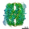
| Title | yeast TRiC-plp2-substrate complex at S2 ATP binding state | |||||||||
 Map data Map data | ||||||||||
 Sample Sample |
| |||||||||
| Function / homology |  Function and homology information Function and homology informationnegative regulation of chaperone-mediated protein folding / Association of TriC/CCT with target proteins during biosynthesis / Cooperation of PDCL (PhLP1) and TRiC/CCT in G-protein beta folding / response to pheromone / chaperone mediated protein folding independent of cofactor / chaperonin-containing T-complex /  vascular endothelial growth factor receptor 2 binding / negative regulation of signal transduction / protein folding chaperone / Neutrophil degranulation ...negative regulation of chaperone-mediated protein folding / Association of TriC/CCT with target proteins during biosynthesis / Cooperation of PDCL (PhLP1) and TRiC/CCT in G-protein beta folding / response to pheromone / chaperone mediated protein folding independent of cofactor / chaperonin-containing T-complex / vascular endothelial growth factor receptor 2 binding / negative regulation of signal transduction / protein folding chaperone / Neutrophil degranulation / ATP-dependent protein folding chaperone / G-protein beta/gamma-subunit complex binding / unfolded protein binding / protein folding / actin binding / actin cytoskeleton organization / regulation of cell cycle / positive regulation of gene expression / ATP hydrolysis activity / positive regulation of transcription by RNA polymerase II / ATP binding / plasma membrane / cytoplasm vascular endothelial growth factor receptor 2 binding / negative regulation of signal transduction / protein folding chaperone / Neutrophil degranulation ...negative regulation of chaperone-mediated protein folding / Association of TriC/CCT with target proteins during biosynthesis / Cooperation of PDCL (PhLP1) and TRiC/CCT in G-protein beta folding / response to pheromone / chaperone mediated protein folding independent of cofactor / chaperonin-containing T-complex / vascular endothelial growth factor receptor 2 binding / negative regulation of signal transduction / protein folding chaperone / Neutrophil degranulation / ATP-dependent protein folding chaperone / G-protein beta/gamma-subunit complex binding / unfolded protein binding / protein folding / actin binding / actin cytoskeleton organization / regulation of cell cycle / positive regulation of gene expression / ATP hydrolysis activity / positive regulation of transcription by RNA polymerase II / ATP binding / plasma membrane / cytoplasmSimilarity search - Function | |||||||||
| Biological species |  Saccharomyces cerevisiae (brewer's yeast) Saccharomyces cerevisiae (brewer's yeast) | |||||||||
| Method | single particle reconstruction / cryo EM / Resolution: 3.91 Å | |||||||||
 Authors Authors | Han WY | |||||||||
| Funding support |  China, 1 items China, 1 items
| |||||||||
 Citation Citation | Journal: Sci Adv / Year: 2023 Title: Structural basis of plp2-mediated cytoskeletal protein folding by TRiC/CCT. Authors: Wenyu Han / Mingliang Jin / Caixuan Liu / Qiaoyu Zhao / Shutian Wang / Yifan Wang / Yue Yin / Chao Peng / Yanxing Wang / Yao Cong / Abstract: The cytoskeletal proteins tubulin and actin are the obligate substrates of TCP-1 ring complex/Chaperonin containing TCP-1 (TRiC/CCT), and their folding involves co-chaperone. Through cryo-electron ...The cytoskeletal proteins tubulin and actin are the obligate substrates of TCP-1 ring complex/Chaperonin containing TCP-1 (TRiC/CCT), and their folding involves co-chaperone. Through cryo-electron microscopy analysis, we present a more complete picture of TRiC-assisted tubulin/actin folding along TRiC adenosine triphosphatase cycle, under the coordination of co-chaperone plp2. In the open S1/S2 states, plp2 and tubulin/actin engaged within opposite TRiC chambers. Notably, we captured an unprecedented TRiC-plp2-tubulin complex in the closed S3 state, engaged with a folded full-length -tubulin and loaded with a guanosine triphosphate, and a plp2 occupying opposite rings. Another closed S4 state revealed an actin in the intermediate folding state and a plp2. Accompanying TRiC ring closure, plp2 translocation could coordinate substrate translocation on the CCT6 hemisphere, facilitating substrate stabilization and folding. Our findings reveal the folding mechanism of the major cytoskeletal proteins tubulin/actin under the coordination of the biogenesis machinery TRiC and plp2 and extend our understanding of the links between cytoskeletal proteostasis and related human diseases. | |||||||||
| History |
|
- Structure visualization
| Supplemental images |
|---|

- Downloads & links
-EMDB archive
| Map data | emd_33918.map.gz | 60 MB | EMDB map data format | |
|---|---|---|---|---|
| Header (meta data) | emd-33918-v30.xmlemd-33918.xml | 27.8 KB 27.8 KB | Display Display | EMDB header |
| Images | 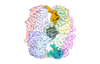 emd_33918.png emd_33918.png | 85.8 KB | ||
| Others | emd_33918_half_map_1.map.gzemd_33918_half_map_2.map.gz | 49.6 MB 49.6 MB | ||
| Archive directory |  http://ftp.pdbj.org/pub/emdb/structures/EMD-33918ftp://ftp.pdbj.org/pub/emdb/structures/EMD-33918 http://ftp.pdbj.org/pub/emdb/structures/EMD-33918ftp://ftp.pdbj.org/pub/emdb/structures/EMD-33918 | HTTPS FTP |
-Related structure data
| Related structure data |  7ylvMC  7yluC  7ylwC 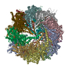 7ylxC  7ylyC M: atomic model generated by this map C: citing same article ( |
|---|---|
| Similar structure data |


-Links
| EMDB pages | EMDB (EBI/PDBe) / EMDataResource |
|---|---|
| Related items in Molecule of the Month |


-Map
| File | Download / File: emd_33918.map.gz / Format: CCP4 / Size: 64 MB / Type: IMAGE STORED AS FLOATING POINT NUMBER (4 BYTES) | ||||||||||||||||||||
|---|---|---|---|---|---|---|---|---|---|---|---|---|---|---|---|---|---|---|---|---|---|
| Voxel size | X=Y=Z: 1.318 Å | ||||||||||||||||||||
| Density |
| ||||||||||||||||||||
| Symmetry | Space group: 1 | ||||||||||||||||||||
| Details | EMDB XML:
|
-Supplemental data
-Half map: #1
| File | emd_33918_half_map_1.map | ||||||||||||
|---|---|---|---|---|---|---|---|---|---|---|---|---|---|
| Projections & Slices |
| ||||||||||||
| Density Histograms |
 Z
Z Y
Y X
X


-Half map: #2
| File | emd_33918_half_map_2.map | ||||||||||||
|---|---|---|---|---|---|---|---|---|---|---|---|---|---|
| Projections & Slices |
| ||||||||||||
| Density Histograms |


- Sample components
+Entire : yeast TRiC-plp2-substrate complex at C2 ATP binding state
+Supramolecule #1: yeast TRiC-plp2-substrate complex at C2 ATP binding state
+Supramolecule #2: TRiC
+Supramolecule #3: plp2
+Macromolecule #1: T-complex protein 1 subunit alpha
+Macromolecule #2: T-complex protein 1 subunit beta
+Macromolecule #3: T-complex protein 1 subunit gamma
+Macromolecule #4: T-complex protein 1 subunit delta
+Macromolecule #5: T-complex protein 1 subunit epsilon
+Macromolecule #6: T-complex protein 1 subunit zeta
+Macromolecule #7: T-complex protein 1 subunit eta
+Macromolecule #8: T-complex protein 1 subunit theta
+Macromolecule #9: Phosducin-like protein 2

-Experimental details
-Structure determination
| Method | cryo EM |
|---|---|
 Processing Processing | single particle reconstruction |
| Aggregation state | particle |
-Sample preparation
| Buffer | pH: 7.5 |
|---|---|
| Vitrification | Cryogen name: ETHANE |
- Electron microscopy
| Microscope | FEI TITAN KRIOS |
|---|---|
| Electron beam | Acceleration voltage: 300 kV / Electron source: FIELD EMISSION GUN |
| Electron optics | Illumination mode: FLOOD BEAM / Imaging mode: BRIGHT FIELDBright-field microscopy / Nominal defocus max: 2.5 µm / Nominal defocus min: 0.8 µm |
| Image recording | Film or detector model: GATAN K2 SUMMIT (4k x 4k) / Average electron dose: 38.0 e/Å2 |
| Experimental equipment |  Model: Titan Krios / Image courtesy: FEI Company |
-Image processing
| Startup model | Type of model: EMDB MAP EMDB ID: |
|---|---|
| Initial angle assignment | Type: MAXIMUM LIKELIHOOD |
| Final angle assignment | Type: MAXIMUM LIKELIHOOD |
| Final reconstruction | Resolution.type: BY AUTHOR / Resolution: 3.91 Å / Resolution method: FSC 0.143 CUT-OFF / Number images used: 33866 |