 Movie
Movie Controller


+ Open data

- Basic information
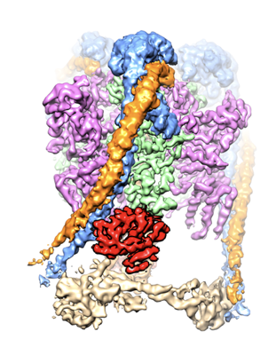
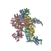
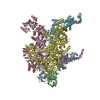
| Entry | Database: EMDB / ID: EMD-31541 | ||||||||||||
|---|---|---|---|---|---|---|---|---|---|---|---|---|---|
| Title | CryoEM Structures of Reconstituted V-ATPase, Oxr1 bound V1 | ||||||||||||
 Map data Map data | |||||||||||||
 Sample Sample |
| ||||||||||||
| Function / homology |  Function and homology information Function and homology informationvacuole-mitochondrion membrane contact site / Insulin receptor recycling / Transferrin endocytosis and recycling / ROS and RNS production in phagocytes / Amino acids regulate mTORC1 / Golgi lumen acidification / proteasome storage granule assembly / vacuolar proton-transporting V-type ATPase, V1 domain / endosomal lumen acidification / pexophagy ...vacuole-mitochondrion membrane contact site / Insulin receptor recycling / Transferrin endocytosis and recycling / ROS and RNS production in phagocytes / Amino acids regulate mTORC1 / Golgi lumen acidification / proteasome storage granule assembly / vacuolar proton-transporting V-type ATPase, V1 domain / endosomal lumen acidification / pexophagy / proton-transporting V-type ATPase complex / vacuolar proton-transporting V-type ATPase complex / vacuolar acidification / fungal-type vacuole membrane / ATP metabolic process / Neutrophil degranulation / proton-transporting ATPase activity, rotational mechanism / proton transmembrane transport / transmembrane transport / intracellular calcium ion homeostasis / cytoplasmic stress granule / cellular response to oxidative stress / response to oxidative stress /  Golgi membrane / mitochondrion / ATP binding / membrane / nucleus / cytoplasm Golgi membrane / mitochondrion / ATP binding / membrane / nucleus / cytoplasmSimilarity search - Function | ||||||||||||
| Biological species |  Saccharomyces cerevisiae S288c (yeast) / Saccharomyces cerevisiae S288C (yeast) Saccharomyces cerevisiae S288c (yeast) / Saccharomyces cerevisiae S288C (yeast) | ||||||||||||
| Method | single particle reconstruction / cryo EM / Resolution: 3.8 Å | ||||||||||||
 Authors Authors | Khan MM / Lee S / Oot RA / Couoh-Cardel S / KIm H / Wilkens S / Roh SH | ||||||||||||
| Funding support |  United States, 3 items United States, 3 items
| ||||||||||||
 Citation Citation | Journal: EMBO J / Year: 2022 Title: Oxidative stress protein Oxr1 promotes V-ATPase holoenzyme disassembly in catalytic activity-independent manner. Authors: Md Murad Khan / Seowon Lee / Sergio Couoh-Cardel / Rebecca A Oot / Hyunmin Kim / Stephan Wilkens / Soung-Hun Roh /  Abstract: The vacuolar ATPase (V-ATPase) is a rotary motor proton pump that is regulated by an assembly equilibrium between active holoenzyme and autoinhibited V -ATPase and V proton channel subcomplexes. ...The vacuolar ATPase (V-ATPase) is a rotary motor proton pump that is regulated by an assembly equilibrium between active holoenzyme and autoinhibited V -ATPase and V proton channel subcomplexes. Here, we report cryo-EM structures of yeast V-ATPase assembled in vitro from lipid nanodisc reconstituted V and mutant V . Our analysis identified holoenzymes in three active rotary states, indicating that binding of V to V provides sufficient free energy to overcome V autoinhibition. Moreover, the structures suggest that the unequal spacing of V 's proton-carrying glutamic acid residues serves to alleviate the symmetry mismatch between V and V motors, a notion that is supported by mutagenesis experiments. We also uncover a structure of free V bound to Oxr1, a conserved but poorly characterized factor involved in the oxidative stress response. Biochemical experiments show that Oxr1 inhibits V -ATPase and causes disassembly of the holoenzyme, suggesting that Oxr1 plays a direct role in V-ATPase regulation. | ||||||||||||
| History |
|
- Structure visualization
| Movie |
Movie viewer |
|---|---|
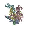
| Structure viewer | EM map: SurfViewMolmilJmol/JSmol |
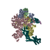
| Supplemental images |
- Downloads & links
-EMDB archive
| Map data | emd_31541.map.gz | 185.5 MB | EMDB map data format | |
|---|---|---|---|---|
| Header (meta data) | emd-31541-v30.xmlemd-31541.xml | 21.5 KB 21.5 KB | Display Display | EMDB header |
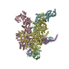
| Images |  emd_31541.png emd_31541.png | 175.6 KB | ||
| Archive directory |  http://ftp.pdbj.org/pub/emdb/structures/EMD-31541ftp://ftp.pdbj.org/pub/emdb/structures/EMD-31541 http://ftp.pdbj.org/pub/emdb/structures/EMD-31541ftp://ftp.pdbj.org/pub/emdb/structures/EMD-31541 | HTTPS FTP |
-Related structure data
| Related structure data |  7fdeMC  7fdaC  7fdbC  7fdcC M: atomic model generated by this map C: citing same article ( |
|---|---|
| Similar structure data |


-Links
| EMDB pages | EMDB (EBI/PDBe) / EMDataResource |
|---|---|
| Related items in Molecule of the Month |


-Map
| File | Download / File: emd_31541.map.gz / Format: CCP4 / Size: 244.1 MB / Type: IMAGE STORED AS FLOATING POINT NUMBER (4 BYTES) | ||||||||||||||||||||||||||||||||||||||||||||||||||||||||||||||||||||
|---|---|---|---|---|---|---|---|---|---|---|---|---|---|---|---|---|---|---|---|---|---|---|---|---|---|---|---|---|---|---|---|---|---|---|---|---|---|---|---|---|---|---|---|---|---|---|---|---|---|---|---|---|---|---|---|---|---|---|---|---|---|---|---|---|---|---|---|---|---|
| Voxel size | X=Y=Z: 1.08 Å | ||||||||||||||||||||||||||||||||||||||||||||||||||||||||||||||||||||
| Density |
| ||||||||||||||||||||||||||||||||||||||||||||||||||||||||||||||||||||
| Symmetry | Space group: 1 | ||||||||||||||||||||||||||||||||||||||||||||||||||||||||||||||||||||
| Details | EMDB XML:
CCP4 map header:
| ||||||||||||||||||||||||||||||||||||||||||||||||||||||||||||||||||||
-Supplemental data
- Sample components
+Entire : Oxr1 bound V1 subcomplex of Yeast Vacuolar ATPase
+Supramolecule #1: Oxr1 bound V1 subcomplex of Yeast Vacuolar ATPase
+Supramolecule #2: Yeast Vacuolar ATPase subunit C
+Supramolecule #3: Yeast V-type proton ATPase subunits
+Macromolecule #1: V-type proton ATPase subunit C

+Macromolecule #2: V-type proton ATPase subunit E
+Macromolecule #3: V-type proton ATPase subunit G
+Macromolecule #4: Yeast Vacuolar ATPase A subunit
+Macromolecule #5: V-type proton ATPase subunit B
+Macromolecule #6: V-type proton ATPase subunit D
+Macromolecule #7: V-type proton ATPase subunit F
+Macromolecule #8: Oxidation resistance protein 1
-Experimental details
-Structure determination
| Method | cryo EM |
|---|---|
 Processing Processing | single particle reconstruction |
| Aggregation state | particle |
-Sample preparation
| Concentration | 1 mg/mL |
|---|---|
| Buffer | pH: 7.4 |
| Grid | Model: UltrAuFoil / Material: GOLD / Pretreatment - Type: GLOW DISCHARGE / Pretreatment - Atmosphere: AIR |
| Vitrification | Cryogen name: ETHANE / Chamber humidity: 90 % / Chamber temperature: 277 K / Instrument: HOMEMADE PLUNGER |
- Electron microscopy
| Microscope | TFS KRIOS |
|---|---|
| Electron beam | Acceleration voltage: 300 kV / Electron source: FIELD EMISSION GUN |
| Electron optics | Illumination mode: FLOOD BEAM / Imaging mode: BRIGHT FIELDBright-field microscopy / Nominal defocus max: 2.5 µm / Nominal defocus min: 1.0 µm |
| Specialist optics | Energy filter - Name: GIF Bioquantum / Energy filter - Slit width: 20 eV |
| Image recording | Film or detector model: GATAN K2 SUMMIT (4k x 4k) / Detector mode: COUNTING / Number real images: 20447 / Average exposure time: 10.0 sec. / Average electron dose: 50.0 e/Å2 |
| Experimental equipment |  Model: Titan Krios / Image courtesy: FEI Company |
-Image processing
| Initial angle assignment | Type: MAXIMUM LIKELIHOOD |
|---|---|
| Final angle assignment | Type: MAXIMUM LIKELIHOOD / Software - Name: cryoSPARC |
| Final reconstruction | Algorithm: BACK PROJECTION / Resolution.type: BY AUTHOR / Resolution: 3.8 Å / Resolution method: FSC 0.143 CUT-OFF / Software - Name: cryoSPARC / Number images used: 20447 |
-Atomic model buiding 1
| Refinement | Space: REAL / Protocol: FLEXIBLE FIT |
|---|---|
| Output model | PDB-7fde: |
