National Natural Science Foundation of China (NSFC)
31901054
China
Howard Hughes Medical Institute (HHMI)
United States
Citation
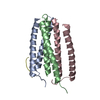
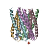
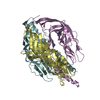
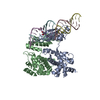
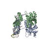
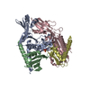
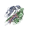
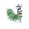
Journal: Nature / Year: 2020 Title: Computational design of transmembrane pores. Authors: Chunfu Xu / Peilong Lu / Tamer M Gamal El-Din / Xue Y Pei / Matthew C Johnson / Atsuko Uyeda / Matthew J Bick / Qi Xu / Daohua Jiang / Hua Bai / Gabriella Reggiano / Yang Hsia / T J Brunette ...Authors: Chunfu Xu / Peilong Lu / Tamer M Gamal El-Din / Xue Y Pei / Matthew C Johnson / Atsuko Uyeda / Matthew J Bick / Qi Xu / Daohua Jiang / Hua Bai / Gabriella Reggiano / Yang Hsia / T J Brunette / Jiayi Dou / Dan Ma / Eric M Lynch / Scott E Boyken / Po-Ssu Huang / Lance Stewart / Frank DiMaio / Justin M Kollman / Ben F Luisi / Tomoaki Matsuura / William A Catterall / David Baker / Abstract: Transmembrane channels and pores have key roles in fundamental biological processes and in biotechnological applications such as DNA nanopore sequencing, resulting in considerable interest in the ...Transmembrane channels and pores have key roles in fundamental biological processes and in biotechnological applications such as DNA nanopore sequencing, resulting in considerable interest in the design of pore-containing proteins. Synthetic amphiphilic peptides have been found to form ion channels, and there have been recent advances in de novo membrane protein design and in redesigning naturally occurring channel-containing proteins. However, the de novo design of stable, well-defined transmembrane protein pores that are capable of conducting ions selectively or are large enough to enable the passage of small-molecule fluorophores remains an outstanding challenge. Here we report the computational design of protein pores formed by two concentric rings of α-helices that are stable and monodisperse in both their water-soluble and their transmembrane forms. Crystal structures of the water-soluble forms of a 12-helical pore and a 16-helical pore closely match the computational design models. Patch-clamp electrophysiology experiments show that, when expressed in insect cells, the transmembrane form of the 12-helix pore enables the passage of ions across the membrane with high selectivity for potassium over sodium; ion passage is blocked by specific chemical modification at the pore entrance. When incorporated into liposomes using in vitro protein synthesis, the transmembrane form of the 16-helix pore-but not the 12-helix pore-enables the passage of biotinylated Alexa Fluor 488. A cryo-electron microscopy structure of the 16-helix transmembrane pore closely matches the design model. The ability to produce structurally and functionally well-defined transmembrane pores opens the door to the creation of designer channels and pores for a wide variety of applications.
History
Deposition
Mar 16, 2020
-
Header (metadata) release
Jun 24, 2020
-
Map release
Jun 24, 2020
-
Update
Mar 27, 2024
-
Current status
Mar 27, 2024
Processing site: PDBj / Status: Released
-
Structure visualization
Movie
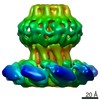
Surface view with section colored by density value
In the structure databanks used in Yorodumi, some data are registered as the other names, "COVID-19 virus" and "2019-nCoV". Here are the details of the virus and the list of structure data.
Jan 31, 2019. EMDB accession codes are about to change! (news from PDBe EMDB page)
EMDB accession codes are about to change! (news from PDBe EMDB page)
The allocation of 4 digits for EMDB accession codes will soon come to an end. Whilst these codes will remain in use, new EMDB accession codes will include an additional digit and will expand incrementally as the available range of codes is exhausted. The current 4-digit format prefixed with “EMD-” (i.e. EMD-XXXX) will advance to a 5-digit format (i.e. EMD-XXXXX), and so on. It is currently estimated that the 4-digit codes will be depleted around Spring 2019, at which point the 5-digit format will come into force.
The EM Navigator/Yorodumi systems omit the EMD- prefix.
Related info.:Q: What is EMD? / ID/Accession-code notation in Yorodumi/EM Navigator
Yorodumi is a browser for structure data from EMDB, PDB, SASBDB, etc.
This page is also the successor to EM Navigator detail page, and also detail information page/front-end page for Omokage search.
The word "yorodu" (or yorozu) is an old Japanese word meaning "ten thousand". "mi" (miru) is to see.
Related info.:EMDB / PDB / SASBDB / Comparison of 3 databanks / Yorodumi Search / Aug 31, 2016. New EM Navigator & Yorodumi / Yorodumi Papers / Jmol/JSmol / Function and homology information / Changes in new EM Navigator and Yorodumi
 Movie
Movie Controller


 Open data
 Basic information
 Map data
 Sample
 Keywords
 MEMBRANE PROTEIN /
 Authors
 China,
  United States, 2 items
  Citation

 Structure visualization
 Movie viewer
 Downloads & links
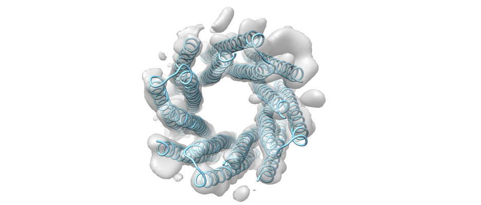 emd_30126.png
 http://ftp.pdbj.org/pub/emdb/structures/EMD-30126

 Z
Z Y
Y X
X


 Sample components
 Processing
 Electron microscopy

