 Movie
Movie Controller

[English] 日本語
 Yorodumi
- EMDB-23084: Outer dynein arm docking complex bound to doublet microtubules fr... -
+ Open data

- Basic information
| Entry | Database: EMDB / ID: EMD-23084 | |||||||||
|---|---|---|---|---|---|---|---|---|---|---|
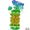
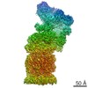
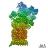
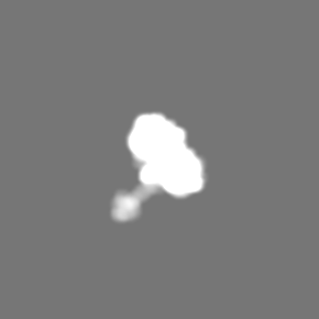
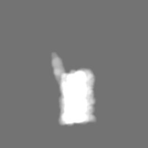
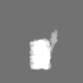
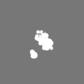
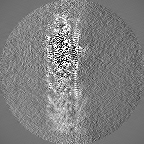
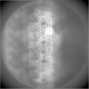
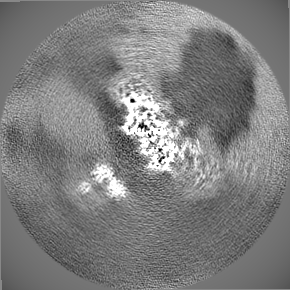
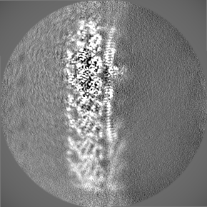
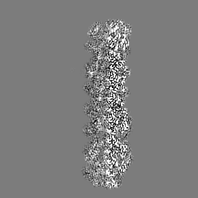
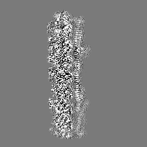
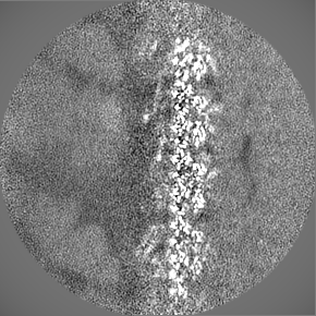
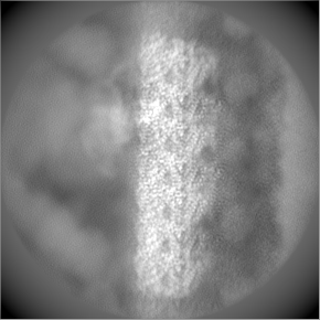
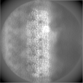
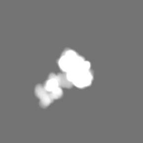
| Title | Outer dynein arm docking complex bound to doublet microtubules from C. reinhardtii | |||||||||
 Map data Map data | composite map of ODA-DC bound to doublet microtubules | |||||||||
 Sample Sample |
| |||||||||
 Keywords Keywords | dynein / microtubule / cilia / MOTOR PROTEIN | |||||||||
| Function / homology |  Function and homology information Function and homology informationouter dynein arm / axonemal dynein complex / outer dynein arm assembly / cilium movement involved in cell motility / minus-end-directed microtubule motor activity / dynein light intermediate chain binding / motile cilium / dynein intermediate chain binding / Hydrolases; Acting on acid anhydrides; Acting on GTP to facilitate cellular and subcellular movement / structural constituent of cytoskeleton ...outer dynein arm / axonemal dynein complex / outer dynein arm assembly / cilium movement involved in cell motility / minus-end-directed microtubule motor activity / dynein light intermediate chain binding / motile cilium / dynein intermediate chain binding / Hydrolases; Acting on acid anhydrides; Acting on GTP to facilitate cellular and subcellular movement / structural constituent of cytoskeleton / microtubule cytoskeleton organization / mitotic cell cycle / microtubule / hydrolase activity / GTPase activity / calcium ion binding / GTP binding / ATP hydrolysis activity / ATP binding / metal ion binding / cytoplasm Similarity search - Function | |||||||||
| Biological species |   Chlamydomonas reinhardtii (plant) Chlamydomonas reinhardtii (plant) | |||||||||
| Method | helical reconstruction / cryo EM / Resolution: 3.3 Å | |||||||||
 Authors Authors | Walton T / Wu H | |||||||||
 Citation Citation | Journal: Nat Commun / Year: 2021 Title: Structure of a microtubule-bound axonemal dynein. Authors: Travis Walton / Hao Wu / Alan Brown /  Abstract: Axonemal dyneins are tethered to doublet microtubules inside cilia to drive ciliary beating, a process critical for cellular motility and extracellular fluid flow. Axonemal dyneins are evolutionarily ...Axonemal dyneins are tethered to doublet microtubules inside cilia to drive ciliary beating, a process critical for cellular motility and extracellular fluid flow. Axonemal dyneins are evolutionarily and biochemically distinct from cytoplasmic dyneins that transport cargo, and the mechanisms regulating their localization and function are poorly understood. Here, we report a single-particle cryo-EM reconstruction of a three-headed axonemal dynein natively bound to doublet microtubules isolated from cilia. The slanted conformation of the axonemal dynein causes interaction of its motor domains with the neighboring dynein complex. Our structure shows how a heterotrimeric docking complex specifically localizes the linear array of axonemal dyneins to the doublet microtubule by directly interacting with the heavy chains. Our structural analysis establishes the arrangement of conserved heavy, intermediate and light chain subunits, and provides a framework to understand the roles of individual subunits and the interactions between dyneins during ciliary waveform generation. | |||||||||
| History |
|
- Structure visualization
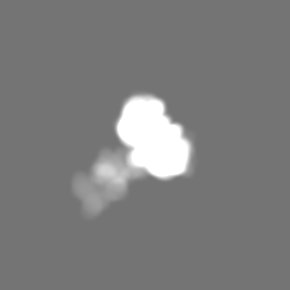
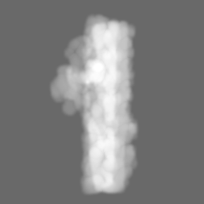
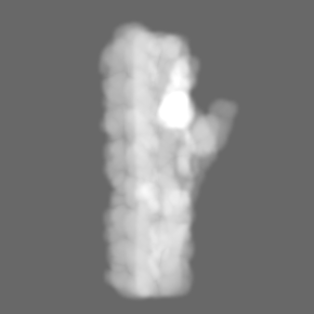
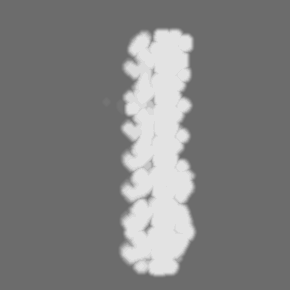
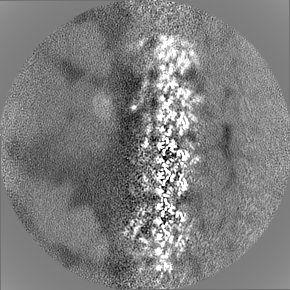
| Movie |
Movie viewer |
|---|---|
| Structure viewer | EM map: SurfViewMolmilJmol/JSmol |
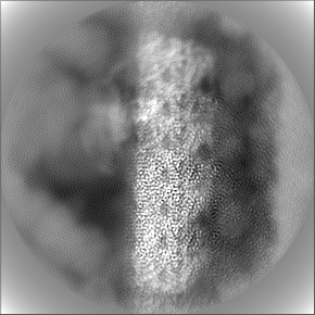
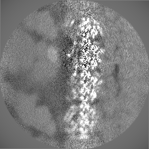
| Supplemental images |

- Downloads & links
-EMDB archive
| Map data | emd_23084.map.gz | 3.7 MB | EMDB map data format | |
|---|---|---|---|---|
| Header (meta data) | emd-23084-v30.xmlemd-23084.xml | 56.3 KB 56.3 KB | Display Display | EMDB header |
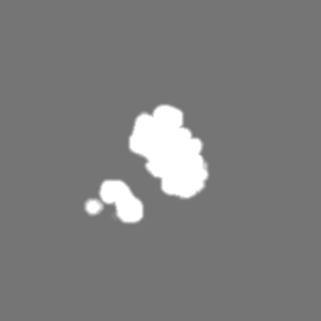
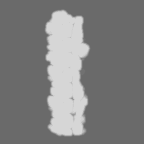
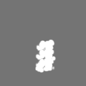
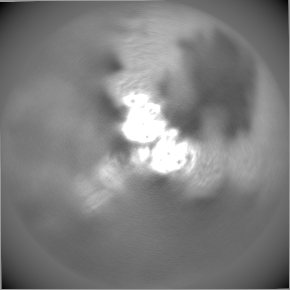
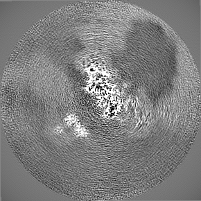
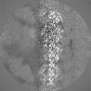
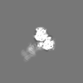
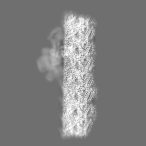
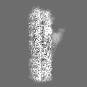
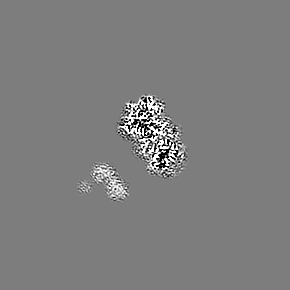
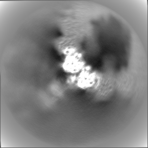
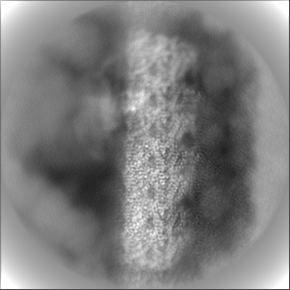
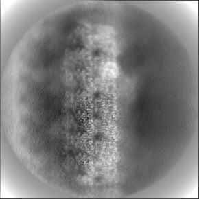
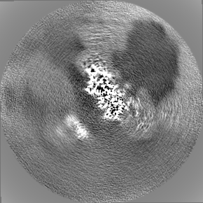
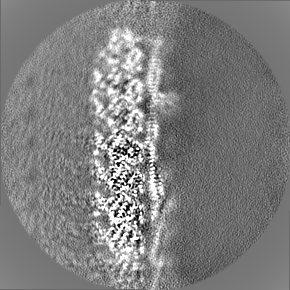
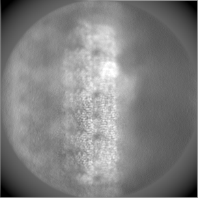
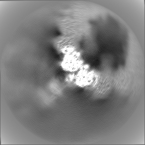
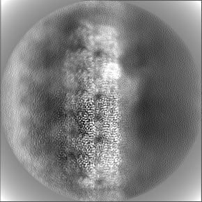
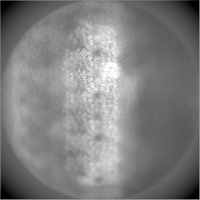
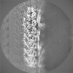
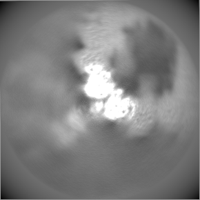
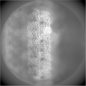
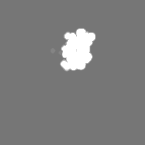
| Images | 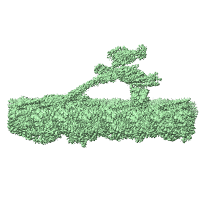 emd_23084.png emd_23084.png | 112.2 KB | ||
| Masks | emd_23084_msk_1.map | 93 MB | Mask map | |
| Filedesc metadata | emd-23084.cif.gz | 11.2 KB | ||
| Others | emd_23084_additional_1.map.gzemd_23084_additional_10.map.gzemd_23084_additional_11.map.gzemd_23084_additional_12.map.gzemd_23084_additional_13.map.gzemd_23084_additional_14.map.gzemd_23084_additional_15.map.gzemd_23084_additional_2.map.gzemd_23084_additional_3.map.gzemd_23084_additional_4.map.gzemd_23084_additional_5.map.gzemd_23084_additional_6.map.gzemd_23084_additional_7.map.gzemd_23084_additional_8.map.gzemd_23084_additional_9.map.gz | 305.5 KB 83.4 MB 84 MB 83.4 MB 8.1 MB 83.7 MB 83.6 MB 83.7 MB 83.5 MB 84.2 MB 83.5 MB 236.3 KB 83.5 MB 84.1 MB 305.1 KB | ||
| Archive directory |  http://ftp.pdbj.org/pub/emdb/structures/EMD-23084ftp://ftp.pdbj.org/pub/emdb/structures/EMD-23084 http://ftp.pdbj.org/pub/emdb/structures/EMD-23084ftp://ftp.pdbj.org/pub/emdb/structures/EMD-23084 | HTTPS FTP |
-Validation report
| Summary document | emd_23084_validation.pdf.gz | 437.5 KB | Display | EMDB validaton report |
|---|---|---|---|---|
| Full document | emd_23084_full_validation.pdf.gz | 437 KB | Display | |
| Data in XML | emd_23084_validation.xml.gz | 6.2 KB | Display | |
| Data in CIF | emd_23084_validation.cif.gz | 7 KB | Display | |
| Arichive directory | https://ftp.pdbj.org/pub/emdb/validation_reports/EMD-23084ftp://ftp.pdbj.org/pub/emdb/validation_reports/EMD-23084 | HTTPS FTP |
-Related structure data
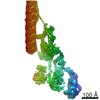
| Related structure data |  7kzoMC  7kzmC  7kznC M: atomic model generated by this map C: citing same article ( |
|---|---|
| Similar structure data |


-Links
| EMDB pages | EMDB (EBI/PDBe) / EMDataResource |
|---|---|
| Related items in Molecule of the Month |


-Map
| File | Download / File: emd_23084.map.gz / Format: CCP4 / Size: 93 MB / Type: IMAGE STORED AS FLOATING POINT NUMBER (4 BYTES) | ||||||||||||||||||||||||||||||||||||||||||||||||||||||||||||
|---|---|---|---|---|---|---|---|---|---|---|---|---|---|---|---|---|---|---|---|---|---|---|---|---|---|---|---|---|---|---|---|---|---|---|---|---|---|---|---|---|---|---|---|---|---|---|---|---|---|---|---|---|---|---|---|---|---|---|---|---|---|
| Annotation | composite map of ODA-DC bound to doublet microtubules | ||||||||||||||||||||||||||||||||||||||||||||||||||||||||||||
| Voxel size | X=Y=Z: 1.36 Å | ||||||||||||||||||||||||||||||||||||||||||||||||||||||||||||
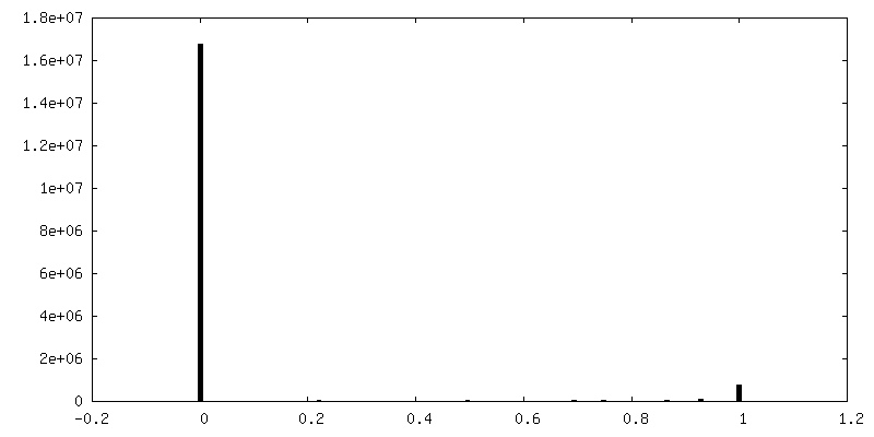
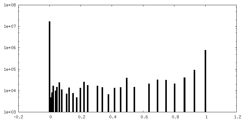
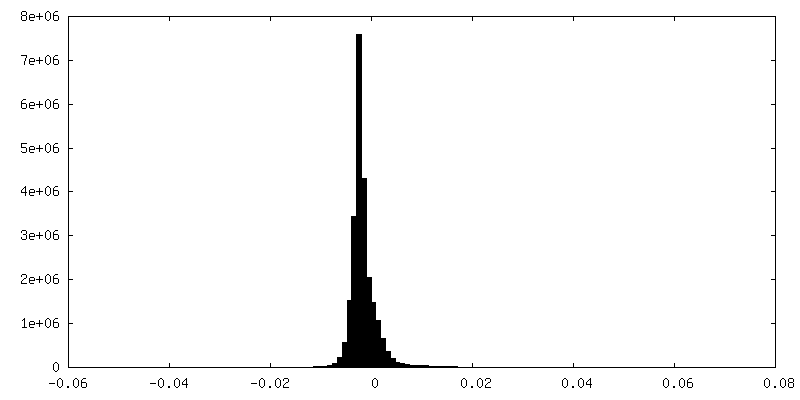
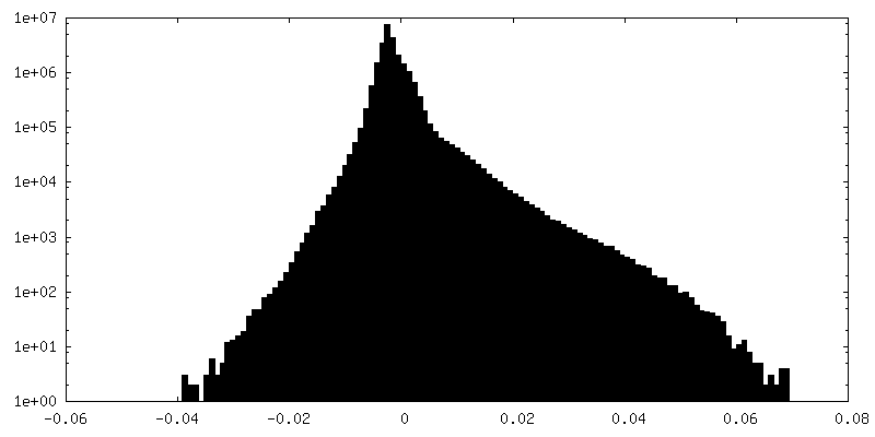
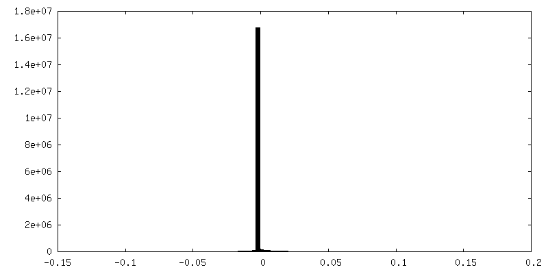
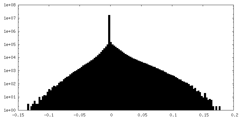
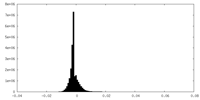
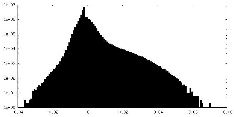
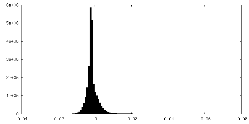
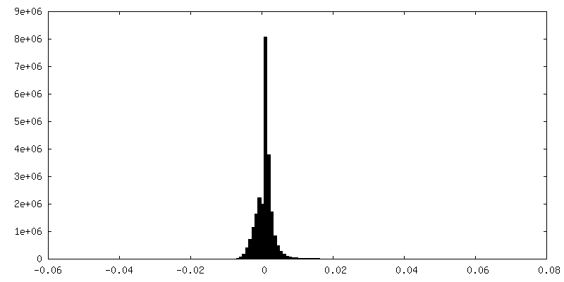
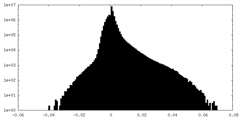
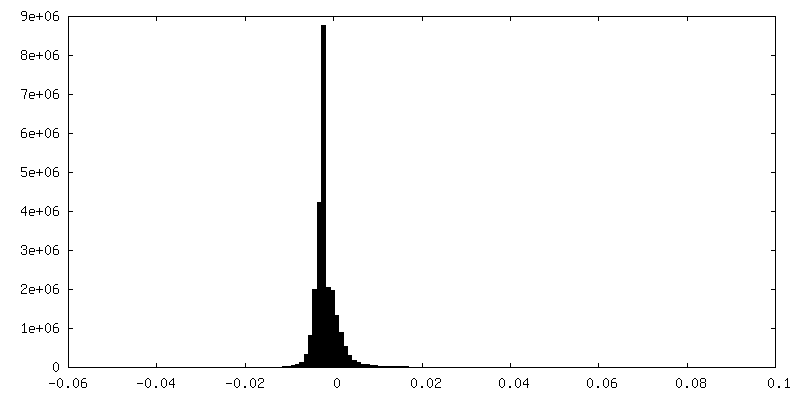
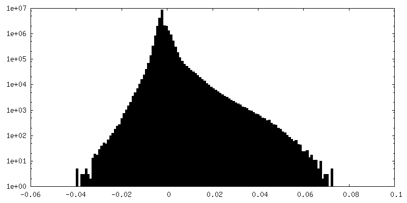
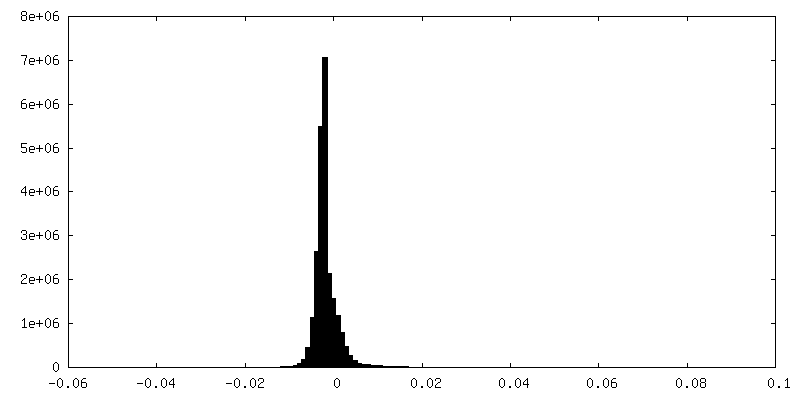
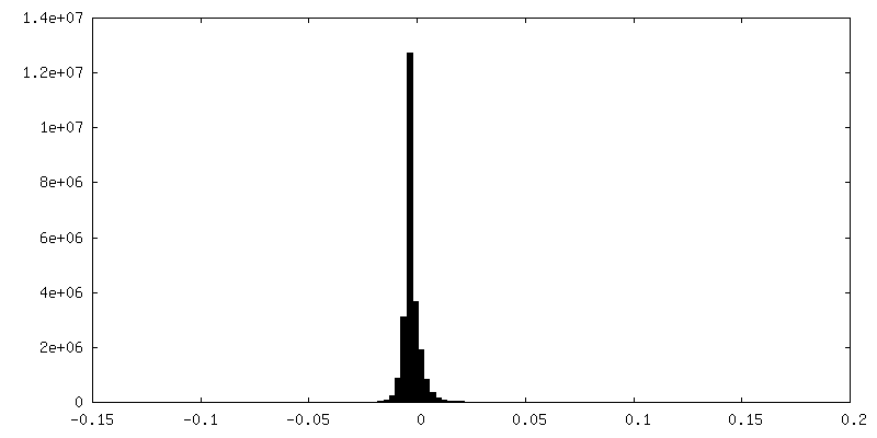
| Density |
| ||||||||||||||||||||||||||||||||||||||||||||||||||||||||||||
| Symmetry | Space group: 1 | ||||||||||||||||||||||||||||||||||||||||||||||||||||||||||||
| Details | EMDB XML:
CCP4 map header:
| ||||||||||||||||||||||||||||||||||||||||||||||||||||||||||||
-Supplemental data
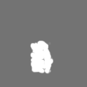
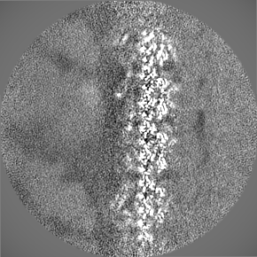
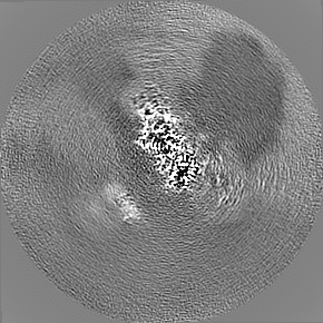
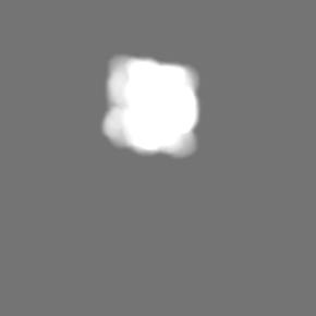
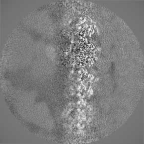
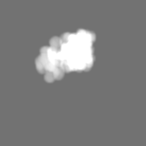
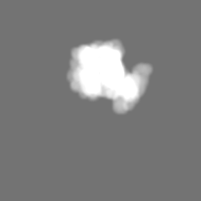
+Mask #1
 Z
Z Y
Y X
X

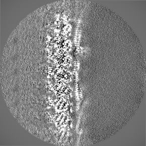
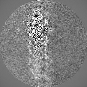
+Additional map: mask for focused refinement of DC1/2
+Additional map: half map 1 for focused refinement of gamma HC N-terminal tail


+Additional map: focused refinement of gamma HC N-terminal tail


+Additional map: half map 2 for focused refinement of gamma HC N-terminal tail


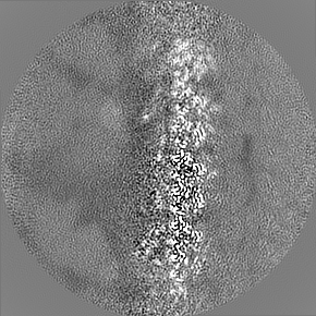
+Additional map: refinement of entire ODA-DC
+Additional map: half map 2 for refinement of entire ODA-DC


+Additional map: half map 1 for refinement of entire ODA-DC


+Additional map: half map 1 for focused refinement of DC1/2
+Additional map: half map 2 for focused refinement of DC1/2


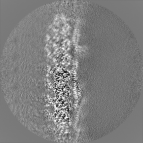
+Additional map: focused refinement of DC1/2
+Additional map: half map 1 for focused refinement of DC3


+Additional map: mask for focused refinement of DC3


+Additional map: half map 2 for focused refinement of DC3


+Additional map: focused refinement of DC3


+Additional map: mask for focused refinement of gamma HC N-terminal tail


- Sample components
+Entire : ODA docking complex
+Supramolecule #1: ODA docking complex
+Macromolecule #1: Tubulin beta
+Macromolecule #2: Tubulin alpha
+Macromolecule #3: Dynein gamma chain, flagellar outer arm
+Macromolecule #4: Outer dynein arm-docking complex subunit 1
+Macromolecule #5: Outer dynein arm protein 1
+Macromolecule #6: Outer dynein arm-docking complex protein DC3
+Macromolecule #7: GUANOSINE-5'-TRIPHOSPHATE

+Macromolecule #8: MAGNESIUM ION
+Macromolecule #9: GUANOSINE-5'-DIPHOSPHATE

-Experimental details
-Structure determination
| Method | cryo EM |
|---|---|
 Processing Processing | helical reconstruction |
| Aggregation state | filament |
-Sample preparation
| Concentration | 20 mg/mL | |||||||||||||||||||||
|---|---|---|---|---|---|---|---|---|---|---|---|---|---|---|---|---|---|---|---|---|---|---|
| Buffer | pH: 7.4 Component:
Details: Buffer also contained 1x Protease Arrest (G-Biosciences) | |||||||||||||||||||||
| Grid | Model: C-flat-1.2/1.3 / Material: COPPER / Mesh: 400 / Pretreatment - Type: GLOW DISCHARGE / Pretreatment - Time: 30 sec. / Details: 15 mA | |||||||||||||||||||||
| Vitrification | Cryogen name: ETHANE / Chamber humidity: 100 % / Chamber temperature: 298 K / Instrument: FEI VITROBOT MARK IV Details: 2.5 ul of splayed axoneme solution was then dispensed onto glow-discharged C-Flat 1.2/1.3-4Cu grids inside a Vitrobot Mark IV under 100% humidity. After a 10 s delay time, cryo-EM samples ...Details: 2.5 ul of splayed axoneme solution was then dispensed onto glow-discharged C-Flat 1.2/1.3-4Cu grids inside a Vitrobot Mark IV under 100% humidity. After a 10 s delay time, cryo-EM samples were prepared by first blotting for 10 s with blot force set to 16 and immediately plunged into liquid ethane.. | |||||||||||||||||||||
| Details | Splayed axonemes isolated from Chlamydomonas reinhardtii flagella. |
- Electron microscopy
| Microscope | TFS KRIOS |
|---|---|
| Specialist optics | Energy filter - Name: GIF Bioquantum / Energy filter - Slit width: 25 eV |
| Image recording | Film or detector model: GATAN K3 BIOQUANTUM (6k x 4k) / Number grids imaged: 2 / Number real images: 20524 / Average exposure time: 3.7 sec. / Average electron dose: 61.48 e/Å2 |
| Electron beam | Acceleration voltage: 300 kV / Electron source:  FIELD EMISSION GUN FIELD EMISSION GUN |
| Electron optics | C2 aperture diameter: 50.0 µm / Illumination mode: FLOOD BEAM / Imaging mode: BRIGHT FIELD / Cs: 2.7 mm |
| Sample stage | Specimen holder model: FEI TITAN KRIOS AUTOGRID HOLDER / Cooling holder cryogen: NITROGEN |
| Experimental equipment |  Model: Titan Krios / Image courtesy: FEI Company |
-Image processing
| Final reconstruction | Applied symmetry - Helical parameters - Δz: 82.0 Å Applied symmetry - Helical parameters - Δ&Phi: 0 ° Applied symmetry - Helical parameters - Axial symmetry: C1 (asymmetric) Resolution.type: BY AUTHOR / Resolution: 3.3 Å / Resolution method: FSC 0.143 CUT-OFF / Software - Name: RELION (ver. 3.1) Details: The composite map was generated from three focused refinements of the full ODA-DC map. The three focused refinements centered on DC1/2 (3.1 A), DC3 (3.2 A), and heavy chain gamma (3.2 A). Number images used: 485694 |
|---|---|
| Segment selection | Number selected: 5584147 / Software - Name: RELION (ver. 3.1) |
| Startup model | Type of model: PDB ENTRY PDB model - PDB ID: |
| Final angle assignment | Type: NOT APPLICABLE / Software - Name: RELION (ver. 3.1) |
-Atomic model buiding 1
| Refinement | Space: REAL / Protocol: OTHER / Target criteria: Correlation coefficient |
|---|---|
| Output model | PDB-7kzo: |
