 Movie
Movie Controller


+ Open data

- Basic information
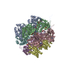
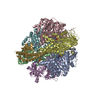
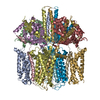
| Entry | Database: EMDB / ID: EMD-21016 | |||||||||
|---|---|---|---|---|---|---|---|---|---|---|
| Title | Structure of the Clostridioides difficile transferase toxin | |||||||||
 Map data Map data | Cryo-EM Reconstruction of CDT | |||||||||
 Sample Sample |
| |||||||||
 Keywords Keywords |  Clostridium / Clostridioides / Binary / CDT / Iota / Toxin / TRANSLOCASE Clostridium / Clostridioides / Binary / CDT / Iota / Toxin / TRANSLOCASE | |||||||||
| Function / homology |  Function and homology information Function and homology informationprotein homooligomerization / transferase activity / nucleotide binding / extracellular region / identical protein bindingSimilarity search - Function | |||||||||
| Biological species |  Clostridioides difficile (bacteria) Clostridioides difficile (bacteria) | |||||||||
| Method | single particle reconstruction / cryo EM / Resolution: 3.8 Å | |||||||||
 Authors Authors | Sheedlo MJ / Anderson DM | |||||||||
| Funding support |  United States, 2 items United States, 2 items
| |||||||||
 Citation Citation | Journal: Proc Natl Acad Sci U S A / Year: 2020 Title: Structural elucidation of the transferase toxin reveals a single-site binding mode for the enzyme. Authors: Michael J Sheedlo / David M Anderson / Audrey K Thomas / D Borden Lacy / Abstract: is a Gram-positive, pathogenic bacterium and a prominent cause of hospital-acquired diarrhea in the United States. The symptoms of infection are caused by the activity of three large toxins known ... is a Gram-positive, pathogenic bacterium and a prominent cause of hospital-acquired diarrhea in the United States. The symptoms of infection are caused by the activity of three large toxins known as toxin A (TcdA), toxin B (TcdB), and the transferase toxin (CDT). Reported here is a 3.8-Å cryo-electron microscopy (cryo-EM) structure of CDT, a bipartite toxin comprised of the proteins CDTa and CDTb. We observe a single molecule of CDTa bound to a CDTb heptamer. The formation of the CDT complex relies on the interaction of an N-terminal adaptor and pseudoenzyme domain of CDTa with six subunits of the CDTb heptamer. CDTb is observed in a preinsertion state, a conformation observed in the transition of prepore to β-barrel pore, although we also observe a single bound CDTa in the prepore and β-barrel conformations of CDTb. The binding interaction appears to prime CDTa for translocation as the adaptor subdomain enters the lumen of the preinsertion state channel. These structural observations advance the understanding of how a single protein, CDTb, can mediate the delivery of a large enzyme, CDTa, into the cytosol of mammalian cells. | |||||||||
| History |
|
- Structure visualization
| Movie |
Movie viewer |
|---|---|
| Structure viewer | EM map: SurfViewMolmilJmol/JSmol |
| Supplemental images |

- Downloads & links
-EMDB archive
| Map data | emd_21016.map.gz | 38 MB | EMDB map data format | |
|---|---|---|---|---|
| Header (meta data) | emd-21016-v30.xmlemd-21016.xml | 12.9 KB 12.9 KB | Display Display | EMDB header |
| Images |  emd_21016.png emd_21016.png | 119.9 KB | ||
| Filedesc metadata | emd-21016.cif.gz | 6.2 KB | ||
| Archive directory |  http://ftp.pdbj.org/pub/emdb/structures/EMD-21016ftp://ftp.pdbj.org/pub/emdb/structures/EMD-21016 http://ftp.pdbj.org/pub/emdb/structures/EMD-21016ftp://ftp.pdbj.org/pub/emdb/structures/EMD-21016 | HTTPS FTP |
-Related structure data
| Related structure data |  6v1sMC M: atomic model generated by this map C: citing same article ( |
|---|---|
| Similar structure data |


-Links
| EMDB pages | EMDB (EBI/PDBe) / EMDataResource |
|---|---|
| Related items in Molecule of the Month |

-Map
| File | Download / File: emd_21016.map.gz / Format: CCP4 / Size: 40.6 MB / Type: IMAGE STORED AS FLOATING POINT NUMBER (4 BYTES) | ||||||||||||||||||||||||||||||||||||||||||||||||||||||||||||||||||||
|---|---|---|---|---|---|---|---|---|---|---|---|---|---|---|---|---|---|---|---|---|---|---|---|---|---|---|---|---|---|---|---|---|---|---|---|---|---|---|---|---|---|---|---|---|---|---|---|---|---|---|---|---|---|---|---|---|---|---|---|---|---|---|---|---|---|---|---|---|---|
| Annotation | Cryo-EM Reconstruction of CDT | ||||||||||||||||||||||||||||||||||||||||||||||||||||||||||||||||||||
| Voxel size | X=Y=Z: 1.13 Å | ||||||||||||||||||||||||||||||||||||||||||||||||||||||||||||||||||||
| Density |
| ||||||||||||||||||||||||||||||||||||||||||||||||||||||||||||||||||||
| Symmetry | Space group: 1 | ||||||||||||||||||||||||||||||||||||||||||||||||||||||||||||||||||||
| Details | EMDB XML:
CCP4 map header:
| ||||||||||||||||||||||||||||||||||||||||||||||||||||||||||||||||||||
-Supplemental data
- Sample components
-Entire : C. difficile transferase toxin
| Entire | Name: C. difficile transferase toxin |
|---|---|
| Components |
|
-Supramolecule #1: C. difficile transferase toxin
| Supramolecule | Name: C. difficile transferase toxin / type: complex / ID: 1 / Parent: 0 / Macromolecule list: #1-#2 |
|---|---|
| Source (natural) | Organism: Clostridioides difficile (bacteria) |
| Molecular weight | Theoretical: 600 KDa |
-Macromolecule #1: ADP-ribosyltransferase binding component
| Macromolecule | Name: ADP-ribosyltransferase binding component / type: protein_or_peptide / ID: 1 / Number of copies: 7 / Enantiomer: LEVO |
|---|---|
| Source (natural) | Organism: Clostridioides difficile (bacteria) |
| Molecular weight | Theoretical: 98.916828 KDa |
| Recombinant expression | Organism: Escherichia coli (E. coli) |
| Sequence | String: MKIQMRNKKV LSFLTLTAIV SQALVYPVYA QTSTSNHSNK KKEIVNEDIL PNNGLMGYYF TDEHFKDLKL MAPIKDGNLK FEEKKVDKL LDKDKSDVKS IRWTGRIIPS KDGEYTLSTD RDDVLMQVNT ESTISNTLKV NMKKGKEYKV RIELQDKNLG S IDNLSSPN ...String: MKIQMRNKKV LSFLTLTAIV SQALVYPVYA QTSTSNHSNK KKEIVNEDIL PNNGLMGYYF TDEHFKDLKL MAPIKDGNLK FEEKKVDKL LDKDKSDVKS IRWTGRIIPS KDGEYTLSTD RDDVLMQVNT ESTISNTLKV NMKKGKEYKV RIELQDKNLG S IDNLSSPN LYWELDGMKK IIPEENLFLR DYSNIEKDDP FIPNNNFFDP KLMSDWEDED LDTDNDNIPD SYERNGYTIK DL IAVKWED SFAEQGYKKY VSNYLESNTA GDPYTDYEKA SGSFDKAIKT EARDPLVAAY PIVGVGMEKL IISTNEHAST DQG KTVSRA TTNSKTESNT AGVSVNVGYQ NGFTANVTTN YSHTTDNSTA VQDSNGESWN TGLSINKGES AYINANVRYY NTGT APMYK VTPTTNLVLD GDTLSTIKAQ ENQIGNNLSP GDTYPKKGLS PLALNTMDQF SSRLIPINYD QLKKLDAGKQ IKLET TQVS GNFGTKNSSG QIVTEGNSWS DYISQIDSIS ASIILDTENE SYERRVTAKN LQDPEDKTPE LTIGEAIEKA FGATKK DGL LYFNDIPIDE SCVELIFDDN TANKIKDSLK TLSDKKIYNV KLERGMNILI KTPTYFTNFD DYNNYPSTWS NVNTTNQ DG LQGSANKLNG ETKIKIPMSE LKPYKRYVFS GYSKDPLTSN SIIVKIKAKE EKTDYLVPEQ GYTKFSYEFE TTEKDSSN I EITLIGSGTT YLDNLSITEL NSTPEILDEP EVKIPTDQEI MDAHKIYFAD LNFNPSTGNT YINGMYFAPT QTNKEALDY IQKYRVEATL QYSGFKDIGT KDKEMRNYLG DPNQPKTNYV NLRSYFTGGE NIMTYKKLRI YAITPDDREL LVLSVD UniProtKB: ADP-ribosyltransferase binding component |
-Macromolecule #2: ADP-ribosylating binary toxin enzymatic subunit CdtA
| Macromolecule | Name: ADP-ribosylating binary toxin enzymatic subunit CdtA / type: protein_or_peptide / ID: 2 / Number of copies: 1 / Enantiomer: LEVO EC number: Transferases; Glycosyltransferases; Pentosyltransferases |
|---|---|
| Source (natural) | Organism: Clostridioides difficile (bacteria) |
| Molecular weight | Theoretical: 53.323336 KDa |
| Recombinant expression | Organism: Escherichia coli (E. coli) |
| Sequence | String: MKKFRKHKRI SNCISILLIL YLTLGGLLPN NIYAQDLQSY SEKVCNTTYK APIERPEDFL KDKEKAKEWE RKEAERIEQK LERSEKEAL ESYKKDSVEI SKYSQTRNYF YDYQIEANSR EKEYKELRNA ISKNKIDKPM YVYYFESPEK FAFNKVIRTE N QNEISLEK ...String: MKKFRKHKRI SNCISILLIL YLTLGGLLPN NIYAQDLQSY SEKVCNTTYK APIERPEDFL KDKEKAKEWE RKEAERIEQK LERSEKEAL ESYKKDSVEI SKYSQTRNYF YDYQIEANSR EKEYKELRNA ISKNKIDKPM YVYYFESPEK FAFNKVIRTE N QNEISLEK FNEFKETIQN KLFKQDGFKD ISLYEPGKGD EKPTPLLMHL KLPRNTGMLP YTNTNNVSTL IEQGYSIKID KI VRIVIDG KHYIKAEASV VSSLDFKDDV SKGDSWGKAN YNDWSNKLTP NELADVNDYM RGGYTAINNY LISNGPVNNP NPE LDSKIT NIENALKREP IPTNLTVYRR SGPQEFGLTL TSPEYDFNKL ENIDAFKSKW EGQALSYPNF ISTSIGSVNM SAFA KRKIV LRITIPKGSP GAYLSAIPGY AGEYEVLLNH GSKFKINKID SYKDGTITKL IVDATLIP UniProtKB: ADP-ribosyltransferase enzymatic component |
-Macromolecule #3: CALCIUM ION
| Macromolecule | Name: CALCIUM ION / type: ligand / ID: 3 / Number of copies: 14 / Formula: CA |
|---|---|
| Molecular weight | Theoretical: 40.078 Da |
-Experimental details
-Structure determination
| Method | cryo EM |
|---|---|
 Processing Processing | single particle reconstruction |
| Aggregation state | particle |
-Sample preparation
| Buffer | pH: 8 |
|---|---|
| Grid | Model: Quantifoil R2/1 / Material: COPPER / Mesh: 200 / Pretreatment - Type: GLOW DISCHARGE |
| Vitrification | Cryogen name: ETHANE / Chamber humidity: 100 % / Chamber temperature: 295 K |
- Electron microscopy
| Microscope | FEI TITAN KRIOS |
|---|---|
| Electron beam | Acceleration voltage: 300 kV / Electron source: FIELD EMISSION GUN |
| Electron optics | Illumination mode: FLOOD BEAM / Imaging mode: BRIGHT FIELDBright-field microscopy |
| Sample stage | Cooling holder cryogen: NITROGEN |
| Image recording | Film or detector model: GATAN K2 SUMMIT (4k x 4k) / Detector mode: COUNTING / Number grids imaged: 1 / Average electron dose: 61.34 e/Å2 |
| Experimental equipment |  Model: Titan Krios / Image courtesy: FEI Company |
-Image processing
| Startup model | Type of model: NONE |
|---|---|
| Initial angle assignment | Type: MAXIMUM LIKELIHOOD |
| Final angle assignment | Type: MAXIMUM LIKELIHOOD |
| Final reconstruction | Applied symmetry - Point group: C1 (asymmetric) / Resolution.type: BY AUTHOR / Resolution: 3.8 Å / Resolution method: FSC 0.143 CUT-OFF / Number images used: 31377 |
-Atomic model buiding 1
| Refinement | Space: REAL / Protocol: AB INITIO MODEL |
|---|---|
| Output model | PDB-6v1s: |
