 Movie
Movie Controller

[English] 日本語
 Yorodumi
- EMDB-17121: Cryo-EM structure of SH-SY5Y seeded with filaments from Alzheimer... -
+ Open data

- Basic information
| Entry |  | |||||||||
|---|---|---|---|---|---|---|---|---|---|---|
| Title | Cryo-EM structure of SH-SY5Y seeded with filaments from Alzheimer's Disease | |||||||||
 Map data Map data | Post processed map | |||||||||
 Sample Sample |
| |||||||||
 Keywords Keywords |  Amyloid / tau / PROTEIN FIBRIL Amyloid / tau / PROTEIN FIBRIL | |||||||||
| Function / homology |  Function and homology information Function and homology informationplus-end-directed organelle transport along microtubule / axonal transport / histone-dependent DNA binding / neurofibrillary tangle assembly / positive regulation of diacylglycerol kinase activity / negative regulation of establishment of protein localization to mitochondrion / neurofibrillary tangle / positive regulation of protein localization to synapse / microtubule lateral binding / tubulin complex ...plus-end-directed organelle transport along microtubule / axonal transport / histone-dependent DNA binding / neurofibrillary tangle assembly / positive regulation of diacylglycerol kinase activity / negative regulation of establishment of protein localization to mitochondrion / neurofibrillary tangle / positive regulation of protein localization to synapse / microtubule lateral binding / tubulin complex / phosphatidylinositol bisphosphate binding / main axon / regulation of long-term synaptic depression / negative regulation of kinase activity / negative regulation of tubulin deacetylation / generation of neurons / regulation of chromosome organization / positive regulation of protein localization / rRNA metabolic process / internal protein amino acid acetylation / regulation of mitochondrial fission / intracellular distribution of mitochondria / axonal transport of mitochondrion / axon development / central nervous system neuron development / regulation of microtubule polymerization / microtubule polymerization / minor groove of adenine-thymine-rich DNA binding / lipoprotein particle binding / dynactin binding / glial cell projection / negative regulation of mitochondrial membrane potential / apolipoprotein binding / protein polymerization / negative regulation of mitochondrial fission / axolemma / Caspase-mediated cleavage of cytoskeletal proteins / regulation of microtubule polymerization or depolymerization / positive regulation of axon extension / supramolecular fiber organization / Activation of AMPK downstream of NMDARs / regulation of microtubule cytoskeleton organization / cytoplasmic microtubule organization / stress granule assembly / regulation of cellular response to heat / axon cytoplasm / regulation of calcium-mediated signaling / positive regulation of microtubule polymerization / cellular response to brain-derived neurotrophic factor stimulus / somatodendritic compartment / synapse assembly / phosphatidylinositol binding / nuclear periphery / cellular response to nerve growth factor stimulus / positive regulation of superoxide anion generation / protein phosphatase 2A binding / regulation of autophagy / astrocyte activation / synapse organization / response to lead ion / microglial cell activation / regulation of synaptic plasticity / Hsp90 protein binding / PKR-mediated signaling / protein homooligomerization / cytoplasmic ribonucleoprotein granule / memory / microtubule cytoskeleton organization / cellular response to reactive oxygen species / SH3 domain binding / activation of cysteine-type endopeptidase activity involved in apoptotic process / neuron projection development / microtubule cytoskeleton / protein-macromolecule adaptor activity / single-stranded DNA binding / cell-cell signaling / cellular response to heat / cell body / actin binding / growth cone / protein-folding chaperone binding / double-stranded DNA binding / microtubule binding / microtubule / amyloid fibril formation / sequence-specific DNA binding / dendritic spine / learning or memory / neuron projection / nuclear speck / membrane raft / axon / negative regulation of gene expression / neuronal cell body / dendrite / DNA damage response / protein kinase binding / enzyme binding / mitochondrion / DNA bindingSimilarity search - Function | |||||||||
| Biological species |  Homo sapiens (human) Homo sapiens (human) | |||||||||
| Method | helical reconstruction / cryo EM / Resolution: 2.5 Å | |||||||||
 Authors Authors | Lovestam S / Scheres SHW / Goedert M | |||||||||
| Funding support |  United Kingdom, 1 items United Kingdom, 1 items
| |||||||||
 Citation Citation | Journal: FEBS Open Bio / Year: 2023 Title: Cryo-EM structures of tau filaments from SH-SY5Y cells seeded with brain extracts from cases of Alzheimer's disease and corticobasal degeneration. Authors: Airi Tarutani / Sofia Lövestam / Xianjun Zhang / Abhay Kotecha / Andrew C Robinson / David M A Mann / Yuko Saito / Shigeo Murayama / Taisuke Tomita / Michel Goedert / Sjors H W Scheres / Masato Hasegawa /   Abstract: The formation of amyloid filaments through templated seeding is believed to underlie the propagation of pathology in most human neurodegenerative diseases. A widely used model system to study this ...The formation of amyloid filaments through templated seeding is believed to underlie the propagation of pathology in most human neurodegenerative diseases. A widely used model system to study this process is to seed amyloid filament formation in cultured cells using human brain extracts. Here, we report the electron cryo-microscopy structures of tau filaments from undifferentiated seeded SH-SY5Y cells that transiently expressed N-terminally HA-tagged 1N3R or 1N4R human tau, using brain extracts from individuals with Alzheimer's disease or corticobasal degeneration. Although the resulting filament structures differed from those of the brain seeds, some degrees of structural templating were observed. Studying templated seeding in cultured cells, and determining the structures of the resulting filaments, can thus provide insights into the cellular aspects underlying neurodegenerative diseases. | |||||||||
| History |
|
- Structure visualization
| Supplemental images |
|---|

- Downloads & links
-EMDB archive
| Map data | emd_17121.map.gz | 17.9 MB | EMDB map data format | |
|---|---|---|---|---|
| Header (meta data) | emd-17121-v30.xmlemd-17121.xml | 16 KB 16 KB | Display Display | EMDB header |
| FSC (resolution estimation) | emd_17121_fsc.xml | 9.1 KB | Display | FSC data file |
| Images | 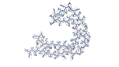 emd_17121.png emd_17121.png | 50.1 KB | ||
| Others | emd_17121_additional_1.map.gzemd_17121_half_map_1.map.gzemd_17121_half_map_2.map.gz | 49.8 MB 49.8 MB 49.8 MB | ||
| Archive directory |  http://ftp.pdbj.org/pub/emdb/structures/EMD-17121ftp://ftp.pdbj.org/pub/emdb/structures/EMD-17121 http://ftp.pdbj.org/pub/emdb/structures/EMD-17121ftp://ftp.pdbj.org/pub/emdb/structures/EMD-17121 | HTTPS FTP |
-Related structure data
| Related structure data |  8oreMC  8orfC  8orgC M: atomic model generated by this map C: citing same article ( |
|---|---|
| Similar structure data |


-Links
| EMDB pages | EMDB (EBI/PDBe) / EMDataResource |
|---|---|
| Related items in Molecule of the Month |


-Map
| File | Download / File: emd_17121.map.gz / Format: CCP4 / Size: 64 MB / Type: IMAGE STORED AS FLOATING POINT NUMBER (4 BYTES) | ||||||||||||||||||||||||||||||||||||
|---|---|---|---|---|---|---|---|---|---|---|---|---|---|---|---|---|---|---|---|---|---|---|---|---|---|---|---|---|---|---|---|---|---|---|---|---|---|
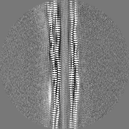
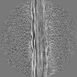
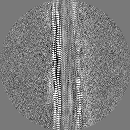
| Annotation | Post processed map | ||||||||||||||||||||||||||||||||||||
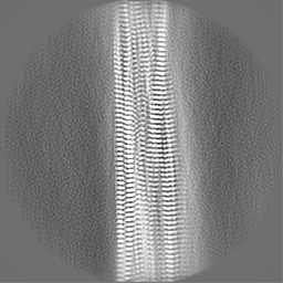
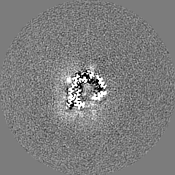
| Projections & slices | Image control
Images are generated by Spider. | ||||||||||||||||||||||||||||||||||||
| Voxel size | X=Y=Z: 1.09 Å | ||||||||||||||||||||||||||||||||||||
| Density |
| ||||||||||||||||||||||||||||||||||||
| Symmetry | Space group: 1 | ||||||||||||||||||||||||||||||||||||
| Details | EMDB XML:
|
 Z (Sec.)
 Y (Row.)
 X (Col.)


-Supplemental data
-Additional map: Unsharpened map
| File | emd_17121_additional_1.map | ||||||||||||
|---|---|---|---|---|---|---|---|---|---|---|---|---|---|
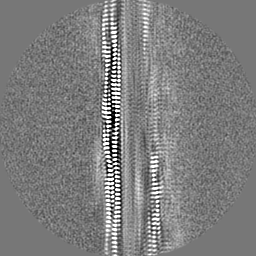
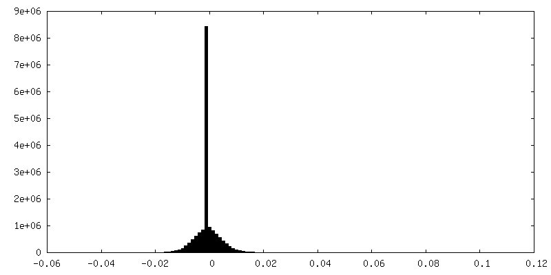
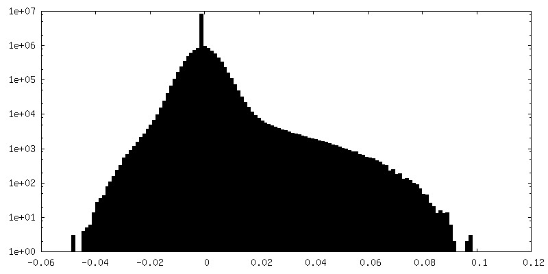
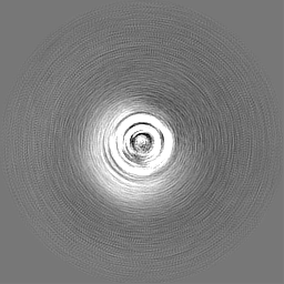
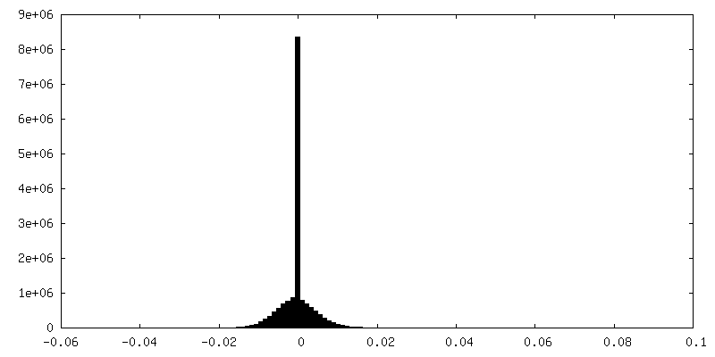
| Annotation | Unsharpened map | ||||||||||||
| Projections & Slices |
| ||||||||||||
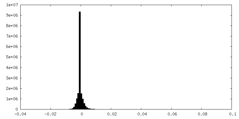
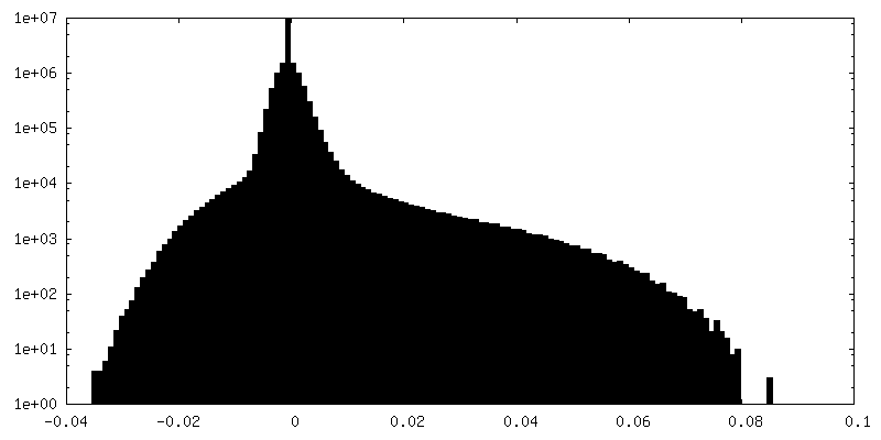
| Density Histograms |


-Half map: Half map 1
| File | emd_17121_half_map_1.map | ||||||||||||
|---|---|---|---|---|---|---|---|---|---|---|---|---|---|
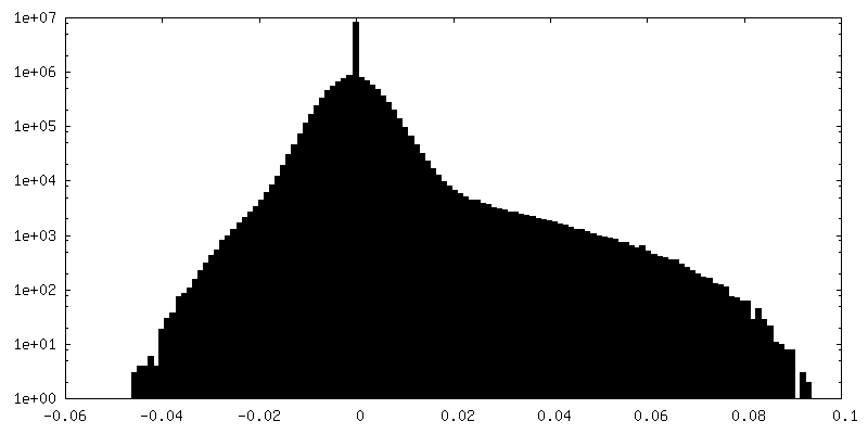
| Annotation | Half map 1 | ||||||||||||
| Projections & Slices |
| ||||||||||||
| Density Histograms |

-Half map: Half map 2
| File | emd_17121_half_map_2.map | ||||||||||||
|---|---|---|---|---|---|---|---|---|---|---|---|---|---|
| Annotation | Half map 2 | ||||||||||||
| Projections & Slices |
| ||||||||||||
| Density Histograms |
- Sample components
-Entire : Tau amyloid filaments
| Entire | Name: Tau amyloid filaments |
|---|---|
| Components |
|
-Supramolecule #1: Tau amyloid filaments
| Supramolecule | Name: Tau amyloid filaments / type: cell / ID: 1 / Parent: 0 / Macromolecule list: all |
|---|---|
| Source (natural) | Organism: Homo sapiens (human) |
-Macromolecule #1: Microtubule-associated protein tau
| Macromolecule | Name: Microtubule-associated protein tau / type: protein_or_peptide / ID: 1 / Number of copies: 3 / Enantiomer: LEVO |
|---|---|
| Source (natural) | Organism: Homo sapiens (human) / Organ: Brain |
| Molecular weight | Theoretical: 39.782066 KDa |
| Recombinant expression | Organism: Homo sapiens (human) |
| Sequence | String: MAEPRQEFEV MEDHAGTYGL GDRKDQGGYT MHQDQEGDTD AGLKESPLQT PTEDGSEEPG SETSDAKSTP TAEAEEAGIG DTPSLEDEA AGHVTQARMV SKSKDGTGSD DKKAKGADGK TKIATPRGAA PPGQKGQANA TRIPAKTPPA PKTPPSSGEP P KSGDRSGY ...String: MAEPRQEFEV MEDHAGTYGL GDRKDQGGYT MHQDQEGDTD AGLKESPLQT PTEDGSEEPG SETSDAKSTP TAEAEEAGIG DTPSLEDEA AGHVTQARMV SKSKDGTGSD DKKAKGADGK TKIATPRGAA PPGQKGQANA TRIPAKTPPA PKTPPSSGEP P KSGDRSGY SSPGSPGTPG SRSRTPSLPT PPTREPKKVA VVRTPPKSPS SAKSRLQTAP VPMPDLKNVK SKIGSTENLK HQ PGGGKVQ IVYKPVDLSK VTSKCGSLGN IHHKPGGGQV EVKSEKLDFK DRVQSKIGSL DNITHVPGGG NKKIETHKLT FRE NAKAKT DHGAEIVYKS PVVSGDTSPR HLSNVSSTGS IDMVDSPQLA TLADEVSASL AKQGL UniProtKB: Microtubule-associated protein tau |
-Experimental details
-Structure determination
| Method | cryo EM |
|---|---|
 Processing Processing | helical reconstruction |
| Aggregation state | filament |
-Sample preparation
| Buffer | pH: 7.4 |
|---|---|
| Vitrification | Cryogen name: ETHANE |
| Details | This sample was a filament |
- Electron microscopy
| Microscope | TFS KRIOS |
|---|---|
| Electron beam | Acceleration voltage: 300 kV / Electron source: FIELD EMISSION GUN |
| Electron optics | Illumination mode: FLOOD BEAM / Imaging mode: BRIGHT FIELDBright-field microscopy / Nominal defocus max: 3.0 µm / Nominal defocus min: 0.5 µm |
| Image recording | Film or detector model: FEI FALCON IV (4k x 4k) / Average electron dose: 40.0 e/Å2 |
| Experimental equipment |  Model: Titan Krios / Image courtesy: FEI Company |
-Image processing
| Startup model | Type of model: OTHER / Details: de novo |
|---|---|
| Final angle assignment | Type: NOT APPLICABLE |
| Final reconstruction | Applied symmetry - Helical parameters - Δz: 4.75 Å Applied symmetry - Helical parameters - Δ&Phi: -1.48 ° Applied symmetry - Helical parameters - Axial symmetry: C1 (asymmetric) Resolution.type: BY AUTHOR / Resolution: 2.5 Å / Resolution method: FSC 0.143 CUT-OFF / Software - Name: RELION / Software - details: Relion 4.1 ;) / Number images used: 16052 |
| FSC plot (resolution estimation) |  |
