 Movie
Movie Controller

[English] 日本語
 Yorodumi
- EMDB-10530: Structure of the chaperonin gp146 from the bacteriophage EL (Pseu... -
+ Open data

- Basic information
| Entry | Database: EMDB / ID: EMD-10530 | |||||||||
|---|---|---|---|---|---|---|---|---|---|---|
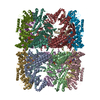
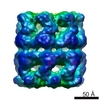
| Title | Structure of the chaperonin gp146 from the bacteriophage EL (Pseudomonas aeruginosa) in complex with ATPgammaS | |||||||||
 Map data Map data | Structure of the complex of gp146 with ATPgammaS | |||||||||
 Sample Sample |
| |||||||||
 Keywords Keywords |  molecular chaperone / ATPase / chaperonin / CHAPERONE molecular chaperone / ATPase / chaperonin / CHAPERONE | |||||||||
| Function / homology |  Function and homology information Function and homology informationATP-dependent protein folding chaperone / protein refolding / ATP binding / identical protein bindingSimilarity search - Function | |||||||||
| Biological species |  Pseudomonas phage EL (virus) Pseudomonas phage EL (virus) | |||||||||
| Method | single particle reconstruction / cryo EM / Resolution: 5.8 Å | |||||||||
 Authors Authors | Bracher A / Wang H | |||||||||
 Citation Citation | Journal: PLoS One / Year: 2020 Title: Structure and conformational cycle of a bacteriophage-encoded chaperonin. Authors: Andreas Bracher / Simanta S Paul / Huping Wang / Nadine Wischnewski / F Ulrich Hartl / Manajit Hayer-Hartl /  Abstract: Chaperonins are ubiquitous molecular chaperones found in all domains of life. They form ring-shaped complexes that assist in the folding of substrate proteins in an ATP-dependent reaction cycle. Key ...Chaperonins are ubiquitous molecular chaperones found in all domains of life. They form ring-shaped complexes that assist in the folding of substrate proteins in an ATP-dependent reaction cycle. Key to the folding cycle is the transient encapsulation of substrate proteins by the chaperonin. Here we present a structural and functional characterization of the chaperonin gp146 (ɸEL) from the phage EL of Pseudomonas aeruginosa. ɸEL, an evolutionarily distant homolog of bacterial GroEL, is active in ATP hydrolysis and prevents the aggregation of denatured protein in a nucleotide-dependent manner. However, ɸEL failed to refold the encapsulation-dependent model substrate rhodanese and did not interact with E. coli GroES, the lid-shaped co-chaperone of GroEL. ɸEL forms tetradecameric double-ring complexes, which dissociate into single rings in the presence of ATP. Crystal structures of ɸEL (at 3.54 and 4.03 Å) in presence of ATP•BeFx revealed two distinct single-ring conformational states, both with open access to the ring cavity. One state showed uniform ATP-bound subunit conformations (symmetric state), whereas the second combined distinct ATP- and ADP-bound subunit conformations (asymmetric state). Cryo-electron microscopy of apo-ɸEL revealed a double-ring structure composed of rings in the asymmetric state (3.45 Å resolution). We propose that the phage chaperonin undergoes nucleotide-dependent conformational switching between double- and single rings and functions in aggregation prevention without substrate protein encapsulation. Thus, ɸEL may represent an evolutionarily more ancient chaperonin prior to acquisition of the encapsulation mechanism. | |||||||||
| History |
|
- Structure visualization
| Movie |
Movie viewer |
|---|---|
| Structure viewer | EM map: SurfViewMolmilJmol/JSmol |
| Supplemental images |

- Downloads & links
-EMDB archive
| Map data | emd_10530.map.gz | 4.1 MB | EMDB map data format | |
|---|---|---|---|---|
| Header (meta data) | emd-10530-v30.xmlemd-10530.xml | 15.2 KB 15.2 KB | Display Display | EMDB header |
| FSC (resolution estimation) | emd_10530_fsc.xml | 5.8 KB | Display | FSC data file |
| Images | 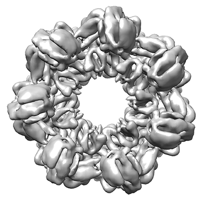 emd_10530.png emd_10530.png | 86.1 KB | ||
| Filedesc metadata | emd-10530.cif.gz | 6.1 KB | ||
| Archive directory |  http://ftp.pdbj.org/pub/emdb/structures/EMD-10530ftp://ftp.pdbj.org/pub/emdb/structures/EMD-10530 http://ftp.pdbj.org/pub/emdb/structures/EMD-10530ftp://ftp.pdbj.org/pub/emdb/structures/EMD-10530 | HTTPS FTP |
-Related structure data
| Related structure data |  6tmxMC  6tmtC  6tmuC  6tmvC  6tmwC C: citing same article ( M: atomic model generated by this map |
|---|---|
| Similar structure data |


-Links
| EMDB pages | EMDB (EBI/PDBe) / EMDataResource |
|---|---|
| Related items in Molecule of the Month |

-Map
| File | Download / File: emd_10530.map.gz / Format: CCP4 / Size: 15.6 MB / Type: IMAGE STORED AS FLOATING POINT NUMBER (4 BYTES) | ||||||||||||||||||||||||||||||||||||||||||||||||||||||||||||
|---|---|---|---|---|---|---|---|---|---|---|---|---|---|---|---|---|---|---|---|---|---|---|---|---|---|---|---|---|---|---|---|---|---|---|---|---|---|---|---|---|---|---|---|---|---|---|---|---|---|---|---|---|---|---|---|---|---|---|---|---|---|
| Annotation | Structure of the complex of gp146 with ATPgammaS | ||||||||||||||||||||||||||||||||||||||||||||||||||||||||||||
| Voxel size | X=Y=Z: 1.997 Å | ||||||||||||||||||||||||||||||||||||||||||||||||||||||||||||
| Density |
| ||||||||||||||||||||||||||||||||||||||||||||||||||||||||||||
| Symmetry | Space group: 1 | ||||||||||||||||||||||||||||||||||||||||||||||||||||||||||||
| Details | EMDB XML:
CCP4 map header:
| ||||||||||||||||||||||||||||||||||||||||||||||||||||||||||||
-Supplemental data
- Sample components
-Entire : The chaperonin gp146 from the bacteriophage EL (Pseudomonas aerug...
| Entire | Name: The chaperonin gp146 from the bacteriophage EL (Pseudomonas aeruginosa) in complex with ATPgammaS |
|---|---|
| Components |
|
-Supramolecule #1: The chaperonin gp146 from the bacteriophage EL (Pseudomonas aerug...
| Supramolecule | Name: The chaperonin gp146 from the bacteriophage EL (Pseudomonas aeruginosa) in complex with ATPgammaS type: complex / ID: 1 / Parent: 0 / Macromolecule list: #1 |
|---|---|
| Source (natural) | Organism: Pseudomonas phage EL (virus) |
| Molecular weight | Theoretical: 864 KDa |
-Macromolecule #1: Putative GroEL-like chaperonine protein
| Macromolecule | Name: Putative GroEL-like chaperonine protein / type: protein_or_peptide / ID: 1 / Number of copies: 14 / Enantiomer: LEVO |
|---|---|
| Source (natural) | Organism: Pseudomonas phage EL (virus) |
| Molecular weight | Theoretical: 61.760941 KDa |
| Recombinant expression | Organism:  Escherichia coli BL21(DE3) (bacteria) Escherichia coli BL21(DE3) (bacteria) |
| Sequence | String: MSQTLLVHGK DAQGIIKQVL SEVYDAVTST MGPNGQLVMI KNGVSTKTTK DGVTVARSIR FADEAHELVN RVITEPATKT DEECGDGTT TTIMLTHALY HLFKDFPGFQ HHRNIEDLVE RVIQRLESMA IRVEVDDPRL YQVALTSSNQ DEKLARLVSE L YANNKGSY ...String: MSQTLLVHGK DAQGIIKQVL SEVYDAVTST MGPNGQLVMI KNGVSTKTTK DGVTVARSIR FADEAHELVN RVITEPATKT DEECGDGTT TTIMLTHALY HLFKDFPGFQ HHRNIEDLVE RVIQRLESMA IRVEVDDPRL YQVALTSSNQ DEKLARLVSE L YANNKGSY PDIELKEGVN FEDQIEQTTG RTIRMFYANP WFAKGHQGGV TELTGFTAFV IDRRIDKEDT QKLIDGVNHL VK THKQHLA LPILLIARSF EEAANSTLMQ LNAAHPTLVE DGRPWLIPLS TPVGGAIGTS ELQDIAVMLN APMLSDVADL TKL DTHSIN GQHGQLELGG NRSILKSTTP KDEDRIEQHA RGIEELLEGF SLSDKFSVRA RYNERRIRTL RGKLITISVG GETY SEVKE RVDRYEDVVK AIRSALENGI LPGGGVSLVK AVFGTIKEGL EDKDQSAEFA KRYINSGIAN ELMRLSTIQH KLLFK DTAL YKENGSFHFN DDWLNTPTVM NLATGEIGTP EGLGIYDTAY ASITALKGGL QTAKILATTK TLILGEKLSA VKVR UniProtKB: Putative GroEL-like chaperonine protein |
-Macromolecule #2: MAGNESIUM ION
| Macromolecule | Name: MAGNESIUM ION / type: ligand / ID: 2 / Number of copies: 14 / Formula: MG |
|---|---|
| Molecular weight | Theoretical: 24.305 Da |
-Macromolecule #3: POTASSIUM ION
| Macromolecule | Name: POTASSIUM ION / type: ligand / ID: 3 / Number of copies: 14 / Formula: K |
|---|---|
| Molecular weight | Theoretical: 39.098 Da |
-Macromolecule #4: PHOSPHOTHIOPHOSPHORIC ACID-ADENYLATE ESTER
| Macromolecule | Name: PHOSPHOTHIOPHOSPHORIC ACID-ADENYLATE ESTER / type: ligand / ID: 4 / Number of copies: 14 / Formula: AGS |
|---|---|
| Molecular weight | Theoretical: 523.247 Da |
| Chemical component information |  ChemComp-AGS: |
-Experimental details
-Structure determination
| Method | cryo EM |
|---|---|
 Processing Processing | single particle reconstruction |
| Aggregation state | particle |
-Sample preparation
| Concentration | 1.125 mg/mL | ||||||||||||||||||
|---|---|---|---|---|---|---|---|---|---|---|---|---|---|---|---|---|---|---|---|
| Buffer | pH: 7.5 Component:
| ||||||||||||||||||
| Grid | Model: Quantifoil R2/1 / Material: COPPER / Mesh: 300 / Support film - Material: CARBON / Support film - topology: HOLEY / Pretreatment - Type: PLASMA CLEANING / Pretreatment - Time: 30 sec. / Pretreatment - Atmosphere: AIR | ||||||||||||||||||
| Vitrification | Cryogen name: ETHANE / Chamber humidity: 100 % / Chamber temperature: 298 K / Instrument: FEI VITROBOT MARK IV / Details: Blot time was 2 sec.. |
- Electron microscopy
| Microscope | FEI TALOS ARCTICA |
|---|---|
| Electron beam | Acceleration voltage: 200 kV / Electron source: FIELD EMISSION GUN |
| Electron optics | Illumination mode: FLOOD BEAM / Imaging mode: BRIGHT FIELDBright-field microscopy |
| Image recording | Film or detector model: FEI FALCON III (4k x 4k) / Detector mode: INTEGRATING / Number grids imaged: 1 / Number real images: 732 / Average exposure time: 0.074769 sec. / Average electron dose: 1.58 e/Å2 |
| Experimental equipment |  Model: Talos Arctica / Image courtesy: FEI Company |
-Image processing
| Particle selection | Number selected: 283285 |
|---|---|
| Startup model | Type of model: NONE |
| Initial angle assignment | Type: MAXIMUM LIKELIHOOD / Software - Name: RELION (ver. 3.0) |
| Final 3D classification | Number classes: 6 / Avg.num./class: 22877 / Software - Name: RELION (ver. 3.0) |
| Final angle assignment | Type: MAXIMUM LIKELIHOOD / Software - Name: RELION (ver. 3.0) |
| Final reconstruction | Number classes used: 1 / Applied symmetry - Point group: D7 (2x7 fold dihedral) / Resolution.type: BY AUTHOR / Resolution: 5.8 Å / Resolution method: FSC 0.143 CUT-OFF / Software - Name: RELION (ver. 3.0) / Number images used: 52885 |
| FSC plot (resolution estimation) | 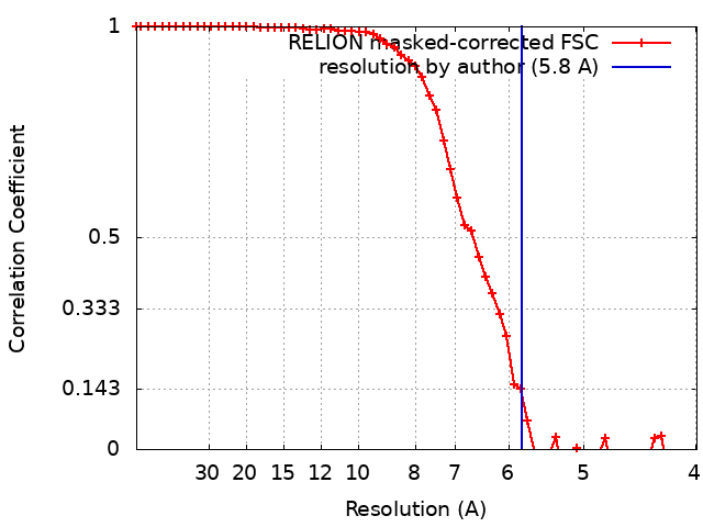 |
-Atomic model buiding 1
| Details | Jelly body refinement D7 symmetry NCS restraints |
|---|---|
| Refinement | Space: REAL / Protocol: RIGID BODY FIT |
| Output model | PDB-6tmx: |
