 ムービー
ムービー コントローラー


+ データを開く

- 基本情報
基本情報
| 登録情報 | データベース: EMDB / ID: EMD-0728 | |||||||||
|---|---|---|---|---|---|---|---|---|---|---|
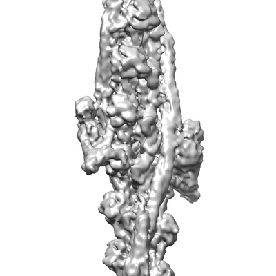
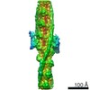
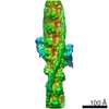
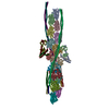
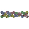
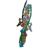
| タイトル | Structure of human cardiac thin filament in the calcium free state | |||||||||
 マップデータ マップデータ | ||||||||||
 試料 試料 |
| |||||||||
 キーワード キーワード |  Troponin (トロポニン) / Tropomyosin (トロポミオシン) / Actin (アクチン) / Thin filement / Muscle (骨格筋) / CONTRACTILE PROTEIN-ACTIN BINDING PROTEIN complex Troponin (トロポニン) / Tropomyosin (トロポミオシン) / Actin (アクチン) / Thin filement / Muscle (骨格筋) / CONTRACTILE PROTEIN-ACTIN BINDING PROTEIN complex | |||||||||
| 機能・相同性 |  機能・相同性情報 機能・相同性情報positive regulation of heart rate by epinephrine / muscle thin filament tropomyosin / regulation of systemic arterial blood pressure by ischemic conditions / troponin C binding / diaphragm contraction / regulation of ATP-dependent activity / regulation of muscle filament sliding speed / troponin T binding / cardiac Troponin complex / cardiac myofibril ...positive regulation of heart rate by epinephrine / muscle thin filament tropomyosin / regulation of systemic arterial blood pressure by ischemic conditions / troponin C binding / diaphragm contraction / regulation of ATP-dependent activity / regulation of muscle filament sliding speed / troponin T binding / cardiac Troponin complex / cardiac myofibril / regulation of smooth muscle contraction / トロポニン / bleb / negative regulation of vascular associated smooth muscle cell migration / regulation of muscle contraction / muscle filament sliding / transition between fast and slow fiber / negative regulation of ATP-dependent activity / ruffle organization / regulation of cardiac muscle contraction by calcium ion signaling / positive regulation of ATP-dependent activity / Striated Muscle Contraction / response to metal ion / regulation of heart contraction / sarcomere organization / structural constituent of muscle / cytoskeletal motor activator activity / ventricular cardiac muscle tissue morphogenesis / tropomyosin binding / myosin heavy chain binding / heart contraction / mesenchyme migration / troponin I binding / actin filament bundle / filamentous actin / negative regulation of vascular associated smooth muscle cell proliferation / skeletal muscle thin filament assembly / actin filament bundle assembly / striated muscle thin filament / skeletal muscle contraction / skeletal muscle myofibril / actin monomer binding / positive regulation of cell adhesion / Smooth Muscle Contraction / calcium channel inhibitor activity / 脈管形成 / skeletal muscle fiber development / stress fiber / titin binding / Ion homeostasis / cardiac muscle contraction / positive regulation of stress fiber assembly / cytoskeleton organization / actin filament polymerization / cytoskeletal protein binding / sarcomere / negative regulation of cell migration / filopodium / actin filament organization / マイクロフィラメント / 加水分解酵素; 酸無水物に作用; 酸無水物に作用・細胞または細胞小器官の運動に関与 / wound healing / structural constituent of cytoskeleton / intracellular calcium ion homeostasis / ruffle membrane / cellular response to reactive oxygen species / response to calcium ion / calcium-dependent protein binding / actin filament binding / マイクロフィラメント / lamellipodium / cell body / actin binding / heart development / regulation of cell shape / 細胞骨格 / hydrolase activity / protein heterodimerization activity / protein domain specific binding / calcium ion binding / positive regulation of gene expression / protein kinase binding / magnesium ion binding / protein homodimerization activity / ATP binding / identical protein binding / 細胞質基質 / 細胞質類似検索 - 分子機能 | |||||||||
| 生物種 |  Oryctolagus cuniculus (ウサギ) / Oryctolagus cuniculus (ウサギ) /  Homo sapiens (ヒト) Homo sapiens (ヒト) | |||||||||
| 手法 | 単粒子再構成法 / クライオ電子顕微鏡法 / 解像度: 6.6 Å | |||||||||
 データ登録者 データ登録者 | Fujii T / Yamada Y | |||||||||
 引用 引用 | ジャーナル: Nat Commun / 年: 2020 タイトル: Cardiac muscle thin filament structures reveal calcium regulatory mechanism. 著者: Yurika Yamada / Keiichi Namba / Takashi Fujii /  要旨: Contraction of striated muscles is driven by cyclic interactions of myosin head projecting from the thick filament with actin filament and is regulated by Ca released from sarcoplasmic reticulum. ...Contraction of striated muscles is driven by cyclic interactions of myosin head projecting from the thick filament with actin filament and is regulated by Ca released from sarcoplasmic reticulum. Muscle thin filament consists of actin, tropomyosin and troponin, and Ca binding to troponin triggers conformational changes of troponin and tropomyosin to allow actin-myosin interactions. However, the structural changes involved in this regulatory mechanism remain unknown. Here we report the structures of human cardiac muscle thin filament in the absence and presence of Ca by electron cryomicroscopy. Molecular models in the two states built based on available crystal structures reveal the structures of a C-terminal region of troponin I and an N-terminal region of troponin T in complex with the head-to-tail junction of tropomyosin together with the troponin core on actin filament. Structural changes of the thin filament upon Ca binding now reveal the mechanism of Ca regulation of muscle contraction. | |||||||||
| 履歴 |
|
- 構造の表示
構造の表示
| ムービー |
ムービービューア |
|---|---|
| 構造ビューア | EMマップ: SurfViewMolmilJmol/JSmol |
| 添付画像 |
- ダウンロードとリンク
-EMDBアーカイブ
| マップデータ | emd_0728.map.gz | 28 MB | EMDBマップデータ形式 | |
|---|---|---|---|---|
| ヘッダ (付随情報) | emd-0728-v30.xmlemd-0728.xml | 16.2 KB 16.2 KB | 表示 表示 | EMDBヘッダ |
| FSC (解像度算出) | emd_0728_fsc.xml | 7.2 KB | 表示 | FSCデータファイル |
| 画像 |  emd_0728.png emd_0728.png | 49.4 KB | ||
| Filedesc metadata | emd-0728.cif.gz | 6 KB | ||
| アーカイブディレクトリ |  http://ftp.pdbj.org/pub/emdb/structures/EMD-0728ftp://ftp.pdbj.org/pub/emdb/structures/EMD-0728 http://ftp.pdbj.org/pub/emdb/structures/EMD-0728ftp://ftp.pdbj.org/pub/emdb/structures/EMD-0728 | HTTPS FTP |
-関連構造データ


-リンク
| EMDBのページ | EMDB (EBI/PDBe) / EMDataResource |
|---|---|
| 「今月の分子」の関連する項目 |


-マップ
| ファイル | ダウンロード / ファイル: emd_0728.map.gz / 形式: CCP4 / 大きさ: 30.5 MB / タイプ: IMAGE STORED AS FLOATING POINT NUMBER (4 BYTES) | ||||||||||||||||||||||||||||||||||||||||||||||||||||||||||||
|---|---|---|---|---|---|---|---|---|---|---|---|---|---|---|---|---|---|---|---|---|---|---|---|---|---|---|---|---|---|---|---|---|---|---|---|---|---|---|---|---|---|---|---|---|---|---|---|---|---|---|---|---|---|---|---|---|---|---|---|---|---|
| ボクセルのサイズ | X=Y=Z: 2.22 Å | ||||||||||||||||||||||||||||||||||||||||||||||||||||||||||||
| 密度 |
| ||||||||||||||||||||||||||||||||||||||||||||||||||||||||||||
| 対称性 | 空間群: 1 | ||||||||||||||||||||||||||||||||||||||||||||||||||||||||||||
| 詳細 | EMDB XML:
CCP4マップ ヘッダ情報:
| ||||||||||||||||||||||||||||||||||||||||||||||||||||||||||||
-添付データ
- 試料の構成要素
+全体 : Cardiac muscle thin filament in the calcium free state
+超分子 #1: Cardiac muscle thin filament in the calcium free state
+超分子 #2: Actin, alpha skeletal muscle
+超分子 #3: Tropomyosin, Troponin
+分子 #1: Actin, alpha skeletal muscle
+分子 #2: Tropomyosin alpha-1 chain

+分子 #3: Tropomyosin alpha-1 chain
+分子 #4: Troponin T, cardiac muscle
+分子 #5: Troponin I, cardiac muscle
+分子 #6: Troponin C, slow skeletal and cardiac muscles
+分子 #7: ADENOSINE-5'-DIPHOSPHATE

-実験情報
-構造解析
| 手法 | クライオ電子顕微鏡法 |
|---|---|
 解析 解析 | 単粒子再構成法 |
| 試料の集合状態 | filament |
-試料調製
| 濃度 | 0.05 mg/mL |
|---|---|
| 緩衝液 | pH: 7.5 |
| 凍結 | 凍結剤: ETHANE |
- 電子顕微鏡法
| 顕微鏡 | JEOL CRYO ARM 200 |
|---|---|
| 電子線 | 加速電圧: 200 kV / 電子線源: FIELD EMISSION GUN |
| 電子光学系 | 照射モード: FLOOD BEAM / 撮影モード: BRIGHT FIELDBright-field microscopy |
| 撮影 | フィルム・検出器のモデル: GATAN K2 SUMMIT (4k x 4k) 平均電子線量: 65.0 e/Å2 |
-画像解析
| 初期モデル | モデルのタイプ: OTHER / 詳細: EM map of actin filament |
|---|---|
| 初期 角度割当 | タイプ: PROJECTION MATCHING |
| 最終 角度割当 | タイプ: PROJECTION MATCHING |
| 最終 再構成 | 解像度のタイプ: BY AUTHOR / 解像度: 6.6 Å / 解像度の算出法: FSC 0.143 CUT-OFF / 使用した粒子像数: 21588 |
| FSC曲線 (解像度の算出) | 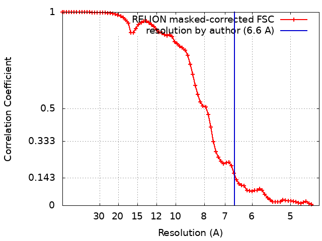 |
