[English] 日本語
Yorodumi
- EMDB-8508: Cryo-EM Structure of Immature Zika Virus -

+
Open data


ID or keywords:

Loading...

-
Basic information

Entry
Database: EMDB / ID: EMD-8508
TitleCryo-EM Structure of Immature Zika Virus
Map dataCryo-EM Reconstruction of Immature Zika Virus
Sample
  • Virus: Zika virus
    • Complex: Transmembrane domains
      • Protein or peptide: Protein E
      • Protein or peptide: M protein
    • Protein or peptide: E protein
    • Protein or peptide: pr domain
  • Ligand: 2-acetamido-2-deoxy-beta-D-glucopyranose
  • Ligand: beta-D-mannopyranose
Function / homology
Function and homology information


: / negative regulation of innate immune response / symbiont-mediated suppression of host JAK-STAT cascade via inhibition of host TYK2 activity / flavivirin / symbiont-mediated suppression of host JAK-STAT cascade via inhibition of STAT2 activity / symbiont-mediated suppression of host JAK-STAT cascade via inhibition of STAT1 activity / ribonucleoside triphosphate phosphatase activity / viral capsid / nucleoside-triphosphate phosphatase / double-stranded RNA binding ...: / negative regulation of innate immune response / symbiont-mediated suppression of host JAK-STAT cascade via inhibition of host TYK2 activity / flavivirin / symbiont-mediated suppression of host JAK-STAT cascade via inhibition of STAT2 activity / symbiont-mediated suppression of host JAK-STAT cascade via inhibition of STAT1 activity / ribonucleoside triphosphate phosphatase activity / viral capsid / nucleoside-triphosphate phosphatase / double-stranded RNA binding / 4 iron, 4 sulfur cluster binding / mRNA (guanine-N7)-methyltransferase / methyltransferase cap1 / clathrin-dependent endocytosis of virus by host cell / mRNA (nucleoside-2'-O-)-methyltransferase activity / mRNA 5'-cap (guanine-N7-)-methyltransferase activity / host cell surface / RNA helicase activity / host cell endoplasmic reticulum membrane / molecular adaptor activity / host cell perinuclear region of cytoplasm / membrane => GO:0016020 / protein dimerization activity / RNA helicase / induction by virus of host autophagy / RNA-directed RNA polymerase / symbiont entry into host cell / viral RNA genome replication / RNA-dependent RNA polymerase activity / serine-type endopeptidase activity / fusion of virus membrane with host endosome membrane / centrosome / viral envelope / lipid binding / symbiont-mediated suppression of host type I interferon-mediated signaling pathway / host cell nucleus / structural molecule activity / virion attachment to host cell / GTP binding / virion membrane / ATP hydrolysis activity / proteolysis / extracellular region / ATP binding / membrane / metal ion binding / cytoplasm
Similarity search - Function
Flavivirus envelope glycoprotein E, stem/anchor domain / : / RNA-directed RNA polymerase, thumb domain, Flavivirus / Flavivirus RNA-directed RNA polymerase, thumb domain / Flavivirus capsid protein C superfamily / Flavivirus NS3 helicase, C-terminal helical domain / Flavivirus non-structural protein NS2B / Genome polyprotein, Flavivirus / Flavivirus non-structural protein NS4A / Flavivirus non-structural protein NS2B ...Flavivirus envelope glycoprotein E, stem/anchor domain / : / RNA-directed RNA polymerase, thumb domain, Flavivirus / Flavivirus RNA-directed RNA polymerase, thumb domain / Flavivirus capsid protein C superfamily / Flavivirus NS3 helicase, C-terminal helical domain / Flavivirus non-structural protein NS2B / Genome polyprotein, Flavivirus / Flavivirus non-structural protein NS4A / Flavivirus non-structural protein NS2B / Flavivirus capsid protein C / Flavivirus non-structural protein NS4B / mRNA cap 0/1 methyltransferase / Flavivirus capsid protein C / Flavivirus non-structural protein NS4B / Flavivirus non-structural protein NS4A / Flavivirus NS2B domain profile. / mRNA cap 0 and cap 1 methyltransferase (EC 2.1.1.56 and EC 2.1.1.57) domain profile. / Flavivirus non-structural protein NS2A / Flavivirus non-structural protein NS2A / Flavivirus NS3, petidase S7 / Peptidase S7, Flavivirus NS3 serine protease / Flavivirus NS3 protease (NS3pro) domain profile. / Envelope glycoprotein M, flavivirus / Flavivirus envelope glycoprotein M / RNA-directed RNA polymerase, flavivirus / Flavivirus RNA-directed RNA polymerase, fingers and palm domains / Flavivirus non-structural Protein NS1 / Flavivirus non-structural protein NS1 / Envelope glycoprotein M superfamily, flavivirus / Flavivirus polyprotein propeptide / Flavivirus polyprotein propeptide superfamily / Flavivirus polyprotein propeptide / Flaviviral glycoprotein E, central domain, subdomain 1 / Flaviviral glycoprotein E, central domain, subdomain 2 / Flavivirus envelope glycoprotein E, Stem/Anchor domain / Flavivirus glycoprotein E, immunoglobulin-like domain / Flavivirus envelope glycoprotein E, Stem/Anchor domain superfamily / Flavivirus glycoprotein, immunoglobulin-like domain / Flavivirus glycoprotein central and dimerisation domain / Flavivirus glycoprotein, central and dimerisation domains / Ribosomal RNA methyltransferase, FtsJ domain / FtsJ-like methyltransferase / Flavivirus/Alphavirus glycoprotein, immunoglobulin-like domain superfamily / Flavivirus glycoprotein, central and dimerisation domain superfamily / Flaviviral glycoprotein E, dimerisation domain / DEAD box, Flavivirus / Flavivirus DEAD domain / helicase superfamily c-terminal domain / Immunoglobulin E-set / Superfamilies 1 and 2 helicase C-terminal domain profile. / Superfamilies 1 and 2 helicase ATP-binding type-1 domain profile. / DEAD-like helicases superfamily / Helicase, C-terminal / Helicase superfamily 1/2, ATP-binding domain / RNA-directed RNA polymerase, catalytic domain / RdRp of positive ssRNA viruses catalytic domain profile. / S-adenosyl-L-methionine-dependent methyltransferase superfamily / Peptidase S1, PA clan, chymotrypsin-like fold / Peptidase S1, PA clan / DNA/RNA polymerase superfamily / P-loop containing nucleoside triphosphate hydrolase
Similarity search - Domain/homology
Genome polyprotein / Core protein / Genome polyprotein / Genome polyprotein / Genome polyprotein
Similarity search - Component
Biological speciesZika virus / ZIKV (virus)
Methodsingle particle reconstruction / cryo EM / Resolution: 9.1 Å
AuthorsMangala Prasad V / Miller AS / Klose T / Sirohi D / Buda G / Jiang W / Kuhn RJ / Rossmann MG
Funding support United States, 2 items
OrganizationGrant numberCountry
National Institutes of Health/National Institute Of Allergy and Infectious Diseases (NIH/NIAID)RO1 AI076331 United States
National Institutes of Health/National Institute Of Allergy and Infectious Diseases (NIH/NIAID)RO1 AI073755 United States
Citation
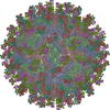
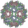
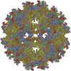
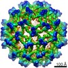
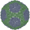
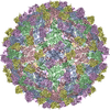
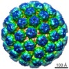
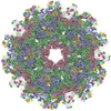
Journal: Nat Struct Mol Biol / Year: 2017
Title: Structure of the immature Zika virus at 9 Å resolution.
Authors: Vidya Mangala Prasad / Andrew S Miller / Thomas Klose / Devika Sirohi / Geeta Buda / Wen Jiang / Richard J Kuhn / Michael G Rossmann /
Abstract: The current Zika virus (ZIKV) epidemic is characterized by severe pathogenicity in both children and adults. Sequence changes in ZIKV since its first isolation are apparent when pre-epidemic strains ...The current Zika virus (ZIKV) epidemic is characterized by severe pathogenicity in both children and adults. Sequence changes in ZIKV since its first isolation are apparent when pre-epidemic strains are compared with those causing the current epidemic. However, the residues that are responsible for ZIKV pathogenicity are largely unknown. Here we report the cryo-electron microscopy (cryo-EM) structure of the immature ZIKV at 9-Å resolution. The cryo-EM map was fitted with the crystal structures of the precursor membrane and envelope glycoproteins and was shown to be similar to the structures of other known immature flaviviruses. However, the immature ZIKV contains a partially ordered capsid protein shell that is less prominent in other immature flaviviruses. Furthermore, six amino acids near the interface between pr domains at the top of the spikes were found to be different between the pre-epidemic and epidemic ZIKV, possibly influencing the composition and structure of the resulting viruses.
History
DepositionDec 6, 2016-
Header (metadata) releaseDec 21, 2016-
Map releaseJan 11, 2017-
UpdateJul 29, 2020-
Current statusJul 29, 2020Processing site: RCSB / Status: Released

-
Structure visualization
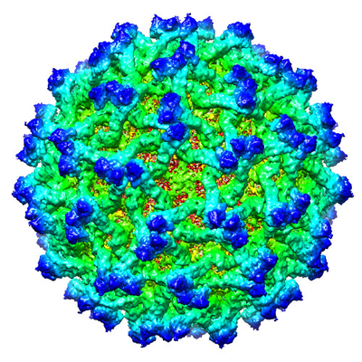
Movie
  • Surface view with section colored by density value
  • Surface level: 4.5
  • Imaged by UCSF Chimera
  • Download
  • Surface view colored by radius
  • Surface level: 4.5
  • Imaged by UCSF Chimera
  • Download
  • Surface view with fitted model
  • Atomic models: PDB-5u4w
  • Surface level: 4.5
  • Imaged by UCSF Chimera
  • Download
  • Simplified surface model + fitted atomic model
  • Atomic modelsPDB-5u4w
  • Imaged by Jmol
  • Download
Movie viewer
Structure viewerEM map:
SurfViewMolmilJmol/JSmol
Supplemental images

Downloads & links

-
Map

FileDownload / File: emd_8508.map.gz / Format: CCP4 / Size: 144.7 MB / Type: IMAGE STORED AS FLOATING POINT NUMBER (4 BYTES)
AnnotationCryo-EM Reconstruction of Immature Zika Virus
Voxel sizeX=Y=Z: 2.6 Å
Density
Contour LevelBy AUTHOR: 4.5 / Movie #1: 4.5
Minimum - Maximum-32.93714 - 22.248032
Average (Standard dev.)0.055202045 (±1.5859408)
SymmetrySpace group: 1
Details

EMDB XML:

Map geometry
Axis orderXYZ
Origin-168-168-168
Dimensions336336336
Spacing336336336
CellA=B=C: 873.6 Å
α=β=γ: 90.0 °

CCP4 map header:

modeImage stored as Reals
Å/pix. X/Y/Z2.62.62.6
M x/y/z336336336
origin x/y/z0.0000.0000.000
length x/y/z873.600873.600873.600
α/β/γ90.00090.00090.000
MAP C/R/S123
start NC/NR/NS-168-168-168
NC/NR/NS336336336
D min/max/mean-32.93722.2480.055

-
Supplemental data

-
Sample components

-
Entire : Zika virus

EntireName: Zika virus
Components
  • Virus: Zika virus
    • Complex: Transmembrane domains
      • Protein or peptide: Protein E
      • Protein or peptide: M protein
    • Protein or peptide: E protein
    • Protein or peptide: pr domain
  • Ligand: 2-acetamido-2-deoxy-beta-D-glucopyranose
  • Ligand: beta-D-mannopyranose

-
Supramolecule #1: Zika virus

SupramoleculeName: Zika virus / type: virus / ID: 1 / Parent: 0 / Macromolecule list: #1-#4 / NCBI-ID: 64320 / Sci species name: Zika virus / Virus type: VIRION / Virus isolate: STRAIN / Virus enveloped: Yes / Virus empty: No
Host (natural)Organism: Homo sapiens (human)
Virus shellShell ID: 1 / Name: prM-E glycoprotein / Diameter: 600.0 Å / T number (triangulation number): 1

-
Supramolecule #2: Transmembrane domains

SupramoleculeName: Transmembrane domains / type: complex / ID: 2 / Parent: 1 / Macromolecule list: #3-#4
Source (natural)Organism: Zika virus

-
Macromolecule #1: E protein

MacromoleculeName: E protein / type: protein_or_peptide / ID: 1 / Number of copies: 3 / Enantiomer: LEVO
Source (natural)Organism: Zika virus
Molecular weightTheoretical: 44.80141 KDa
Recombinant expressionOrganism: Drosophila melanogaster (fruit fly)
SequenceString: GENLYFQGMR CIGMSNRDFV EGVSGGSWVD IVLEHGSCVT TMAKNKPTLD FELIKTEAKQ PATLRKYCIE AKLTNTTTES RCPTQGEPS LNEEQDKRFV CKHSMVDRGW GNGCGLFGKG GIVTCAMFRC KKNMEGKVVQ PENLEYTIVI TPHSGEEHAV G NDTGKHGK ...String:
GENLYFQGMR CIGMSNRDFV EGVSGGSWVD IVLEHGSCVT TMAKNKPTLD FELIKTEAKQ PATLRKYCIE AKLTNTTTES RCPTQGEPS LNEEQDKRFV CKHSMVDRGW GNGCGLFGKG GIVTCAMFRC KKNMEGKVVQ PENLEYTIVI TPHSGEEHAV G NDTGKHGK EIKITPQSSI TEAELTGYGT VTMECSPRTG LDFNEMVLLQ MENKAWLVHR QWFLDLPLPW LPGADTQGSN WI QKETLVT FKNPHAKKQD VVVLGSQEGA MHTALTGATE IQMSSGNLLF TGHLKCRLRM DKLQLKGMSY SMCTGKFKVV KEI AETQHG TIVIRVQYEG DGSPCKIPFE IMDLEKRHVL GRLITVNPIV TEKDSPVNIE AEPPFGDSYI IIGVEPGQLK LNWF KK

-
Macromolecule #2: pr domain

MacromoleculeName: pr domain / type: protein_or_peptide / ID: 2 / Number of copies: 3 / Enantiomer: LEVO
Source (natural)Organism: Zika virus
Molecular weightTheoretical: 9.261531 KDa
Recombinant expressionOrganism: Drosophila melanogaster (fruit fly)
SequenceString:
FHLTTRNGEP HMIVSRQEKG KSLLFKTEDG VNMCTLMAMD LGELCEDTIT YKCPLLRQNE PEDIDCWCNS TSTWVTYGTC T

-
Macromolecule #3: Protein E

MacromoleculeName: Protein E / type: protein_or_peptide / ID: 3 / Number of copies: 3 / Enantiomer: LEVO
Source (natural)Organism: ZIKV (virus)
Molecular weightTheoretical: 6.892228 KDa
SequenceString:
GALNSLGKGI HQIFGAAFKS LFGGMSWFSQ ILIGTLLMWL GLNTKNGSIS LMCLALGGVL IFLSTA

-
Macromolecule #4: M protein

MacromoleculeName: M protein / type: protein_or_peptide / ID: 4 / Number of copies: 3 / Enantiomer: LEVO
Source (natural)Organism: ZIKV (virus)
Molecular weightTheoretical: 5.984065 KDa
SequenceString:
REYTKHLIRV ENWIFRNPGF ALAAAAIAWL LGSSTSQKVI YLVMILLIAP AYS

-
Macromolecule #5: 2-acetamido-2-deoxy-beta-D-glucopyranose

MacromoleculeName: 2-acetamido-2-deoxy-beta-D-glucopyranose / type: ligand / ID: 5 / Number of copies: 12 / Formula: NAG
Molecular weightTheoretical: 221.208 Da
Chemical component information

ChemComp-NAG:
2-acetamido-2-deoxy-beta-D-glucopyranose / N-Acetylglucosamine

-
Macromolecule #6: beta-D-mannopyranose

MacromoleculeName: beta-D-mannopyranose / type: ligand / ID: 6 / Number of copies: 6 / Formula: BMA
Molecular weightTheoretical: 180.156 Da
Chemical component information

ChemComp-BMA:
beta-D-mannopyranose / Mannose

-
Experimental details

-
Structure determination

Methodcryo EM
Processingsingle particle reconstruction
Aggregation stateparticle

-
Sample preparation

BufferpH: 8
Component:
ConcentrationFormulaName
200.0 mMNaClSodium chloridesodium chloride
20.0 mMC4H11NO3Tris
1.0 mMEDTAEthylenediaminetetraacetic acid
GridModel: Ultrathin carbon / Material: COPPER / Mesh: 400 / Support film - Material: CARBON / Support film - topology: HOLEY / Pretreatment - Type: GLOW DISCHARGE
VitrificationCryogen name: ETHANE / Chamber humidity: 80 % / Instrument: GATAN CRYOPLUNGE 3

-
Electron microscopy

MicroscopeFEI TITAN KRIOS
Electron beamAcceleration voltage: 300 kV / Electron source: FIELD EMISSION GUN
Electron opticsIllumination mode: SPOT SCAN / Imaging mode: BRIGHT FIELDBright-field microscopy
Image recordingFilm or detector model: GATAN K2 SUMMIT (4k x 4k) / Detector mode: SUPER-RESOLUTION / Number real images: 3341 / Average exposure time: 7.6 sec. / Average electron dose: 4.7 e/Å2
Experimental equipment
Model: Titan Krios / Image courtesy: FEI Company

-
Image processing

Particle selectionNumber selected: 14351
CTF correctionSoftware: (Name: CTFFIND (ver. 3.0), jspr)
Startup modelType of model: OTHER / Details: Random model generation
Initial angle assignmentType: PROJECTION MATCHING / Software - Name: jspr
Final 3D classificationNumber classes: 50 / Avg.num./class: 200 / Software - Name: RELION
Final angle assignmentType: PROJECTION MATCHING / Software - Name: jspr
Final reconstructionApplied symmetry - Point group: I (icosahedral) / Algorithm: FOURIER SPACE / Resolution.type: BY AUTHOR / Resolution: 9.1 Å / Resolution method: FSC 0.143 CUT-OFF / Software - Name: jspr / Number images used: 9315
FSC plot (resolution estimation)

-
Atomic model buiding 1

RefinementSpace: REAL / Protocol: RIGID BODY FIT / Target criteria: Correlation coefficient
Output model

PDB-5u4w:
Cryo-EM Structure of Immature Zika Virus

+
About Yorodumi

-
News

-
Feb 9, 2022. New format data for meta-information of EMDB entries

New format data for meta-information of EMDB entries

  • Version 3 of the EMDB header file is now the official format.
  • The previous official version 1.9 will be removed from the archive.

Related info.:EMDB header

External links:wwPDB to switch to version 3 of the EMDB data model

-
Aug 12, 2020. Covid-19 info

Covid-19 info

URL: https://pdbj.org/emnavi/covid19.php

New page: Covid-19 featured information page in EM Navigator.

Related info.:Covid-19 info / Mar 5, 2020. Novel coronavirus structure data

+
Mar 5, 2020. Novel coronavirus structure data

Novel coronavirus structure data

Related info.:Yorodumi Speices / Aug 12, 2020. Covid-19 info

External links:COVID-19 featured content - PDBj / Molecule of the Month (242):Coronavirus Proteases

+
Jan 31, 2019. EMDB accession codes are about to change! (news from PDBe EMDB page)

EMDB accession codes are about to change! (news from PDBe EMDB page)

  • The allocation of 4 digits for EMDB accession codes will soon come to an end. Whilst these codes will remain in use, new EMDB accession codes will include an additional digit and will expand incrementally as the available range of codes is exhausted. The current 4-digit format prefixed with “EMD-” (i.e. EMD-XXXX) will advance to a 5-digit format (i.e. EMD-XXXXX), and so on. It is currently estimated that the 4-digit codes will be depleted around Spring 2019, at which point the 5-digit format will come into force.
  • The EM Navigator/Yorodumi systems omit the EMD- prefix.

Related info.:Q: What is EMD? / ID/Accession-code notation in Yorodumi/EM Navigator

External links:EMDB Accession Codes are Changing Soon! / Contact to PDBj

+
Jul 12, 2017. Major update of PDB

Major update of PDB

  • wwPDB released updated PDB data conforming to the new PDBx/mmCIF dictionary.
  • This is a major update changing the version number from 4 to 5, and with Remediation, in which all the entries are updated.
  • In this update, many items about electron microscopy experimental information are reorganized (e.g. em_software).
  • Now, EM Navigator and Yorodumi are based on the updated data.

External links:wwPDB Remediation / Enriched Model Files Conforming to OneDep Data Standards Now Available in the PDB FTP Archive

-
Yorodumi

Thousand views of thousand structures

  • Yorodumi is a browser for structure data from EMDB, PDB, SASBDB, etc.
  • This page is also the successor to EM Navigator detail page, and also detail information page/front-end page for Omokage search.
  • The word "yorodu" (or yorozu) is an old Japanese word meaning "ten thousand". "mi" (miru) is to see.

Related info.:EMDB / PDB / SASBDB / Comparison of 3 databanks / Yorodumi Search / Aug 31, 2016. New EM Navigator & Yorodumi / Yorodumi Papers / Jmol/JSmol / Function and homology information / Changes in new EM Navigator and Yorodumi

Read more