 Movie
Movie Controller

[English] 日本語
 Yorodumi
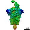
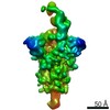
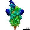
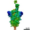
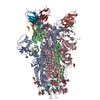
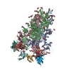
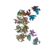
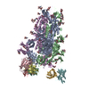
- PDB-7nd7: EM structure of SARS-CoV-2 Spike glycoprotein in complex with COV... -
+ Open data

- Basic information
| Entry | Database: PDB / ID: 7nd7 | |||||||||
|---|---|---|---|---|---|---|---|---|---|---|
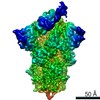
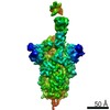
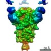
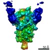
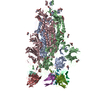
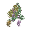
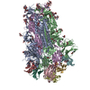
| Title | EM structure of SARS-CoV-2 Spike glycoprotein in complex with COVOX-316 Fab | |||||||||
 Components Components |
| |||||||||
 Keywords Keywords |  VIRAL PROTEIN/IMMUNE SYSTEM / SARS-CoV-2 / antibody / germline / V-gene / receptor-binding-domain / spike / neutralisation / protection / glycosylation / valency / VIRAL PROTEIN-IMMUNE SYSTEM complex VIRAL PROTEIN/IMMUNE SYSTEM / SARS-CoV-2 / antibody / germline / V-gene / receptor-binding-domain / spike / neutralisation / protection / glycosylation / valency / VIRAL PROTEIN-IMMUNE SYSTEM complex | |||||||||
| Function / homology |  Function and homology information Function and homology informationMaturation of spike protein / viral translation / Translation of Structural Proteins / Virion Assembly and Release / host cell surface / host extracellular space / suppression by virus of host tetherin activity / Induction of Cell-Cell Fusion / structural constituent of virion / host cell endoplasmic reticulum-Golgi intermediate compartment membrane ...Maturation of spike protein / viral translation / Translation of Structural Proteins / Virion Assembly and Release / host cell surface / host extracellular space / suppression by virus of host tetherin activity / Induction of Cell-Cell Fusion / structural constituent of virion / host cell endoplasmic reticulum-Golgi intermediate compartment membrane / entry receptor-mediated virion attachment to host cell / receptor-mediated endocytosis of virus by host cell / Attachment and Entry / membrane fusion / positive regulation of viral entry into host cell / receptor-mediated virion attachment to host cell / receptor ligand activity / host cell surface receptor binding / fusion of virus membrane with host plasma membrane / fusion of virus membrane with host endosome membrane / viral envelope / virion attachment to host cell / SARS-CoV-2 activates/modulates innate and adaptive immune responses / host cell plasma membrane / virion membrane / membrane / identical protein binding / plasma membraneSimilarity search - Function | |||||||||
| Biological species |   Severe acute respiratory syndrome coronavirus 2 Severe acute respiratory syndrome coronavirus 2 Homo sapiens (human) Homo sapiens (human) | |||||||||
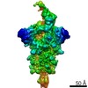
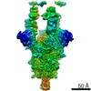
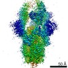
| Method | ELECTRON MICROSCOPY / single particle reconstruction / cryo EM / Resolution: 3.6 Å | |||||||||
 Authors Authors | Duyvesteyn, H.M.E. / Zhao, Y. / Ren, J. / Stuart, D. | |||||||||
| Funding support |  United Kingdom, United Kingdom,  China, 2items China, 2items
| |||||||||
 Citation Citation | Journal: Cell / Year: 2021 Title: The antigenic anatomy of SARS-CoV-2 receptor binding domain. Authors: Wanwisa Dejnirattisai / Daming Zhou / Helen M Ginn / Helen M E Duyvesteyn / Piyada Supasa / James Brett Case / Yuguang Zhao / Thomas S Walter / Alexander J Mentzer / Chang Liu / Beibei Wang ...Authors: Wanwisa Dejnirattisai / Daming Zhou / Helen M Ginn / Helen M E Duyvesteyn / Piyada Supasa / James Brett Case / Yuguang Zhao / Thomas S Walter / Alexander J Mentzer / Chang Liu / Beibei Wang / Guido C Paesen / Jose Slon-Campos / César López-Camacho / Natasha M Kafai / Adam L Bailey / Rita E Chen / Baoling Ying / Craig Thompson / Jai Bolton / Alex Fyfe / Sunetra Gupta / Tiong Kit Tan / Javier Gilbert-Jaramillo / William James / Michael Knight / Miles W Carroll / Donal Skelly / Christina Dold / Yanchun Peng / Robert Levin / Tao Dong / Andrew J Pollard / Julian C Knight / Paul Klenerman / Nigel Temperton / David R Hall / Mark A Williams / Neil G Paterson / Felicity K R Bertram / C Alistair Siebert / Daniel K Clare / Andrew Howe / Julika Radecke / Yun Song / Alain R Townsend / Kuan-Ying A Huang / Elizabeth E Fry / Juthathip Mongkolsapaya / Michael S Diamond / Jingshan Ren / David I Stuart / Gavin R Screaton /    Abstract: Antibodies are crucial to immune protection against SARS-CoV-2, with some in emergency use as therapeutics. Here, we identify 377 human monoclonal antibodies (mAbs) recognizing the virus spike and ...Antibodies are crucial to immune protection against SARS-CoV-2, with some in emergency use as therapeutics. Here, we identify 377 human monoclonal antibodies (mAbs) recognizing the virus spike and focus mainly on 80 that bind the receptor binding domain (RBD). We devise a competition data-driven method to map RBD binding sites. We find that although antibody binding sites are widely dispersed, neutralizing antibody binding is focused, with nearly all highly inhibitory mAbs (IC < 0.1 μg/mL) blocking receptor interaction, except for one that binds a unique epitope in the N-terminal domain. Many of these neutralizing mAbs use public V-genes and are close to germline. We dissect the structural basis of recognition for this large panel of antibodies through X-ray crystallography and cryoelectron microscopy of 19 Fab-antigen structures. We find novel binding modes for some potently inhibitory antibodies and demonstrate that strongly neutralizing mAbs protect, prophylactically or therapeutically, in animal models. | |||||||||
| History |
|
- Structure visualization
| Movie |
Movie viewer |
|---|---|
| Structure viewer | Molecule: MolmilJmol/JSmol |
- Downloads & links
-Download
| PDBx/mmCIF format | 7nd7.cif.gz | 707.8 KB | Display | PDBx/mmCIF format |
|---|---|---|---|---|
| PDB format | pdb7nd7.ent.gz | 588.8 KB | Display | PDB format |
| PDBx/mmJSON format | 7nd7.json.gz | Tree view | PDBx/mmJSON format | |
| Others |  Other downloads Other downloads |
-Validation report
| Arichive directory | https://data.pdbj.org/pub/pdb/validation_reports/nd/7nd7ftp://data.pdbj.org/pub/pdb/validation_reports/nd/7nd7 | HTTPS FTP |
|---|
-Related structure data
| Related structure data | 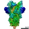 12278MC 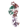 7behC 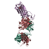 7beiC 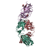 7bejC 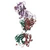 7bekC 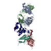 7belC 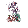 7bemC 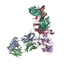 7benC 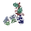 7beoC 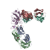 7bepC 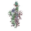 7nd3C 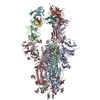 7nd4C  7nd5C  7nd6C  7nd8C  7nd9C  7ndaC 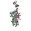 7ndbC 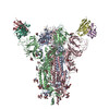 7ndcC  7nddC C: citing same article ( M: map data used to model this data |
|---|---|
| Similar structure data |
-Links
 PDBj
PDBj


- Assembly
| Deposited unit | 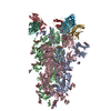
|
|---|---|
| 1 |
|
-Components
-Protein , 1 types, 3 molecules ABC
| #1: Protein | Spike protein / S glycoprotein / E2 / Peplomer protein Mass: 142399.375 Da / Num. of mol.: 3 Source method: isolated from a genetically manipulated source Source: (gene. exp.) Severe acute respiratory syndrome coronavirus 2Gene: S, 2 / Production host: Homo sapiens (human) / References: UniProt: P0DTC2 |
|---|
-Antibody , 2 types, 6 molecules HFJLGK
| #2: Antibody | Mass: 24329.373 Da / Num. of mol.: 3 Source method: isolated from a genetically manipulated source Source: (gene. exp.) Homo sapiens (human) / Production host: Homo sapiens (human)#3: Antibody | Mass: 22810.141 Da / Num. of mol.: 3 Source method: isolated from a genetically manipulated source Source: (gene. exp.) Homo sapiens (human) / Production host: Homo sapiens (human) |
|---|
-Sugars , 3 types, 43 molecules 
| #4: Polysaccharide | 2-acetamido-2-deoxy-beta-D-glucopyranose-(1-4)-2-acetamido-2-deoxy-beta-D-glucopyranose / Mass: 424.401 Da / Num. of mol.: 15Source method: isolated from a genetically manipulated source #5: Polysaccharide | / Mass: 570.542 Da / Num. of mol.: 3Source method: isolated from a genetically manipulated source #6: Sugar | ChemComp-NAG / N-Acetylglucosamine Type: D-saccharide, beta linking / Mass: 221.208 Da / Num. of mol.: 25 / Source method: obtained synthetically / Formula: C8H15NO6 Type: D-saccharide, beta linking / Mass: 221.208 Da / Num. of mol.: 25 / Source method: obtained synthetically / Formula: C8H15NO6 |
|---|
-Details
| Has ligand of interest | N |
|---|
-Experimental details
-Experiment
| Experiment | Method: ELECTRON MICROSCOPY |
|---|---|
| EM experiment | Aggregation state: PARTICLE / 3D reconstruction method: single particle reconstruction |
- Sample preparation
| Component |
| ||||||||||||||||||||||||
|---|---|---|---|---|---|---|---|---|---|---|---|---|---|---|---|---|---|---|---|---|---|---|---|---|---|
| Molecular weight | Value: 0.582 MDa / Experimental value: NO | ||||||||||||||||||||||||
| Source (natural) |
| ||||||||||||||||||||||||
| Source (recombinant) |
| ||||||||||||||||||||||||
| Buffer solution | pH: 4.6 | ||||||||||||||||||||||||
| Specimen | Embedding applied: NO / Shadowing applied: NO / Staining applied: NO / Vitrification applied: YES | ||||||||||||||||||||||||
| Specimen support | Grid material: COPPER / Grid mesh size: 200 divisions/in. / Grid type: C-flat-2/1 | ||||||||||||||||||||||||
| Vitrification | Cryogen name: ETHANE |
- Electron microscopy imaging
| Experimental equipment |  Model: Titan Krios / Image courtesy: FEI Company |
|---|---|
| Microscopy | Model: FEI TITAN KRIOS |
| Electron gun | Electron source: FIELD EMISSION GUN / Accelerating voltage: 300 kV / Illumination mode: OTHER |
| Electron lens | Mode: BRIGHT FIELDBright-field microscopy |
| Image recording | Electron dose: 46 e/Å2 / Film or detector model: GATAN K2 SUMMIT (4k x 4k) |
- Processing
| Software | Name: PHENIX / Version: 1.19_4092: / Classification: refinement | ||||||||||||||||||||||||
|---|---|---|---|---|---|---|---|---|---|---|---|---|---|---|---|---|---|---|---|---|---|---|---|---|---|
| EM software |
| ||||||||||||||||||||||||
| CTF correction | Type: PHASE FLIPPING AND AMPLITUDE CORRECTION | ||||||||||||||||||||||||
| Symmetry | Point symmetry: C3 (3 fold cyclic) | ||||||||||||||||||||||||
| 3D reconstruction | Resolution: 3.6 Å / Resolution method: FSC 0.143 CUT-OFF / Num. of particles: 162905 / Symmetry type: POINT | ||||||||||||||||||||||||
| Refine LS restraints |
|
