 Movie
Movie Controller

[English] 日本語
 Yorodumi
- PDB-7m6j: Human Septin Hexameric Complex SEPT2G/SEPT6/SEPT7 by Single Parti... -
+ Open data

- Basic information
| Entry | Database: PDB / ID: 7m6j | ||||||||||||
|---|---|---|---|---|---|---|---|---|---|---|---|---|---|
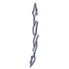
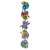
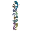
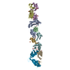
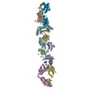
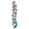
| Title | Human Septin Hexameric Complex SEPT2G/SEPT6/SEPT7 by Single Particle Cryo-EM | ||||||||||||
 Components Components |
| ||||||||||||
 Keywords Keywords |  CELL CYCLE / Complex / SPA / Cytoskeleton CELL CYCLE / Complex / SPA / Cytoskeleton | ||||||||||||
| Function / homology |  Function and homology information Function and homology informationseptin collar / regulation of embryonic cell shape / sperm annulus / septin complex / positive regulation of non-motile cilium assembly / photoreceptor connecting cilium / septin ring / cytoskeleton-dependent cytokinesis / regulation of exocytosis / non-motile cilium ...septin collar / regulation of embryonic cell shape / sperm annulus / septin complex / positive regulation of non-motile cilium assembly / photoreceptor connecting cilium / septin ring / cytoskeleton-dependent cytokinesis / regulation of exocytosis / non-motile cilium / ciliary membrane / smoothened signaling pathway / cell division site / intercellular bridge / axoneme / cleavage furrow / mitotic cytokinesis / cilium assembly / axon terminus / stress fiber / Anchoring of the basal body to the plasma membrane / MAPK6/MAPK4 signaling / kinetochore / spindle / microtubule cytoskeleton / synaptic vesicle / actin cytoskeleton / midbody / spermatogenesis / cell differentiation / molecular adaptor activity / cadherin binding / GTPase activity / GTP binding / structural molecule activity / extracellular exosome / nucleoplasm / identical protein binding / nucleus / plasma membrane / cytosol / cytoplasmSimilarity search - Function | ||||||||||||
| Biological species |  Homo sapiens (human) Homo sapiens (human) | ||||||||||||
| Method | ELECTRON MICROSCOPY / single particle reconstruction / cryo EM / Resolution: 3.6 Å | ||||||||||||
 Authors Authors | Mendonca, D.C. / Pereira, H.M. / van Heel, M. / Portugal, R.V. / Garratt, R.C. | ||||||||||||
| Funding support |  Brazil, 3items Brazil, 3items
| ||||||||||||
 Citation Citation | Journal: J Mol Biol / Year: 2021 Title: An atomic model for the human septin hexamer by cryo-EM. Authors: Deborah C Mendonça / Samuel L Guimarães / Humberto D'Muniz Pereira / Andressa A Pinto / Marcelo A de Farias / Andre S de Godoy / Ana P U Araujo / Marin van Heel / Rodrigo V Portugal / Richard C Garratt / Abstract: In order to form functional filaments, human septins must assemble into hetero-oligomeric rod-like particles which polymerize end-to-end. The rules governing the assembly of these particles and the ...In order to form functional filaments, human septins must assemble into hetero-oligomeric rod-like particles which polymerize end-to-end. The rules governing the assembly of these particles and the subsequent filaments are incompletely understood. Although crystallographic approaches have been successful in studying the separate components of the system, there has been difficulty in obtaining high resolution structures of the full particle. Here we report a first cryo-EM structure for a hexameric rod composed of human septins 2, 6 and 7 with a global resolution of ~3.6 Å and a local resolution of between ~3.0 Å and ~5.0 Å. By fitting the previously determined high-resolution crystal structures of the component subunits into the cryo-EM map, we are able to provide an essentially complete model for the particle. This exposes SEPT2 NC-interfaces at the termini of the hexamer and leaves internal cavities between the SEPT6-SEPT7 pairs. The floor of the cavity is formed by the two α helices including their polybasic regions. These are locked into place between the two subunits by interactions made with the α and α helices of the neighbouring monomer together with its polyacidic region. The cavity may serve to provide space allowing the subunits to move with respect to one another. The elongated particle shows a tendency to bend at its centre where two copies of SEPT7 form a homodimeric G-interface. Such bending is almost certainly related to the ability of septin filaments to recognize and even induce membrane curvature. | ||||||||||||
| History |
|
- Structure visualization
| Movie |
Movie viewer |
|---|---|
| Structure viewer | Molecule: MolmilJmol/JSmol |
- Downloads & links
-Download
| PDBx/mmCIF format | 7m6j.cif.gz | 302 KB | Display | PDBx/mmCIF format |
|---|---|---|---|---|
| PDB format | pdb7m6j.ent.gz | 244.5 KB | Display | PDB format |
| PDBx/mmJSON format | 7m6j.json.gz | Tree view | PDBx/mmJSON format | |
| Others |  Other downloads Other downloads |
-Validation report
| Arichive directory | https://data.pdbj.org/pub/pdb/validation_reports/m6/7m6jftp://data.pdbj.org/pub/pdb/validation_reports/m6/7m6j | HTTPS FTP |
|---|
-Related structure data
| Related structure data | 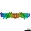 23698MC M: map data used to model this data C: citing same article ( |
|---|---|
| Similar structure data |

-Links
 PDBj
PDBj- Assembly
| Deposited unit | 
|
|---|---|
| 1 |
|
-Components
-Protein , 3 types, 6 molecules AFBECD
| #1: Protein | / Neural precursor cell expressed developmentally down-regulated protein 5 / NEDD-5 Mass: 31748.352 Da / Num. of mol.: 2 Source method: isolated from a genetically manipulated source Source: (gene. exp.) Homo sapiens (human) / Gene: SEPTIN2, DIFF6, KIAA0158, NEDD5, SEPT2 / Production host:  Escherichia coli (E. coli) / References: UniProt: Q15019 Escherichia coli (E. coli) / References: UniProt: Q15019#2: Protein | Mass: 48948.723 Da / Num. of mol.: 2 Source method: isolated from a genetically manipulated source Source: (gene. exp.) Homo sapiens (human) / Gene: SEPTIN6, KIAA0128, SEP2, SEPT6 / Production host: Escherichia coli (E. coli) / References: UniProt: Q14141#3: Protein | / CDC10 protein homologMass: 50456.645 Da / Num. of mol.: 2 Source method: isolated from a genetically manipulated source Source: (gene. exp.) Homo sapiens (human) / Gene: SEPTIN7, CDC10, SEPT7 / Production host: Escherichia coli (E. coli) / References: UniProt: Q16181 |
|---|
-Non-polymers , 3 types, 7 molecules 


| #4: Chemical | ChemComp-GDP / Guanosine diphosphate Type: RNA linking / Mass: 443.201 Da / Num. of mol.: 4 / Source method: obtained synthetically / Formula: C10H15N5O11P2 / Comment: GDP, energy-carrying molecule*YM Type: RNA linking / Mass: 443.201 Da / Num. of mol.: 4 / Source method: obtained synthetically / Formula: C10H15N5O11P2 / Comment: GDP, energy-carrying molecule*YM#5: Chemical | Guanosine triphosphate Mass: 523.180 Da / Num. of mol.: 2 / Source method: obtained synthetically / Formula: C10H16N5O14P3 / Comment: GTP, energy-carrying molecule*YM Mass: 523.180 Da / Num. of mol.: 2 / Source method: obtained synthetically / Formula: C10H16N5O14P3 / Comment: GTP, energy-carrying molecule*YM#6: Chemical | ChemComp-MG / |  Mass: 24.305 Da / Num. of mol.: 1 / Source method: obtained synthetically / Formula: Mg Mass: 24.305 Da / Num. of mol.: 1 / Source method: obtained synthetically / Formula: Mg |
|---|
-Details
| Has ligand of interest | N |
|---|
-Experimental details
-Experiment
| Experiment | Method: ELECTRON MICROSCOPY |
|---|---|
| EM experiment | Aggregation state: PARTICLE / 3D reconstruction method: single particle reconstruction |
- Sample preparation
| Component | Name: Human Septin Hexameric Complex SEPT2G/SEPT6/SEPT7 / Type: COMPLEX / Entity ID: #1-#3 / Source: RECOMBINANT |
|---|---|
| Source (natural) | Organism: Homo sapiens (human) |
| Source (recombinant) | Organism: Escherichia coli (E. coli) |
| Buffer solution | pH: 7.8 |
| Specimen | Embedding applied: NO / Shadowing applied: NO / Staining applied: NO / Vitrification applied: YES |
| Vitrification | Cryogen name: ETHANE |
- Electron microscopy imaging
| Experimental equipment |  Model: Titan Krios / Image courtesy: FEI Company |
|---|---|
| Microscopy | Model: FEI TITAN KRIOS |
| Electron gun | Electron source: FIELD EMISSION GUN / Accelerating voltage: 300 kV / Illumination mode: FLOOD BEAM |
| Electron lens | Mode: BRIGHT FIELDBright-field microscopy |
| Image recording | Electron dose: 30 e/Å2 / Film or detector model: FEI FALCON III (4k x 4k) |
- Processing
| Software | Name: PHENIX / Version: 1.18.2_3874: / Classification: refinement | ||||||||||||||||||||||||
|---|---|---|---|---|---|---|---|---|---|---|---|---|---|---|---|---|---|---|---|---|---|---|---|---|---|
| CTF correction | Type: PHASE FLIPPING AND AMPLITUDE CORRECTION | ||||||||||||||||||||||||
| 3D reconstruction | Resolution: 3.6 Å / Resolution method: FSC 1/2 BIT CUT-OFF / Num. of particles: 108262 / Symmetry type: POINT | ||||||||||||||||||||||||
| Refine LS restraints |
|
