Movie
Movie Controller


+ Open data

- Basic information
| Entry | Database: PDB / ID: 7l30 | ||||||
|---|---|---|---|---|---|---|---|
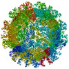
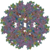
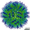
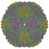
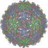
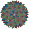
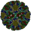
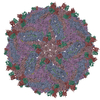
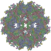
| Title | Binjari virus (BinJV) | ||||||
 Components Components |
| ||||||
 Keywords Keywords |  VIRUS / Flavivirus / glycoprotein / fusion VIRUS / Flavivirus / glycoprotein / fusion | ||||||
| Function / homology |  Function and homology informationflavivirin / symbiont-mediated suppression of host JAK-STAT cascade via inhibition of STAT2 activity / viral capsid / nucleoside-triphosphate phosphatase / double-stranded RNA binding / mRNA (nucleoside-2'-O-)-methyltransferase activity / mRNA 5'-cap (guanine-N7-)-methyltransferase activity / RNA helicase activity / membrane => GO:0016020 / host cell endoplasmic reticulum membrane ...flavivirin / symbiont-mediated suppression of host JAK-STAT cascade via inhibition of STAT2 activity / viral capsid / nucleoside-triphosphate phosphatase / double-stranded RNA binding / mRNA (nucleoside-2'-O-)-methyltransferase activity / mRNA 5'-cap (guanine-N7-)-methyltransferase activity / RNA helicase activity / membrane => GO:0016020 / host cell endoplasmic reticulum membrane / protein dimerization activity / RNA helicase / induction by virus of host autophagy / viral RNA genome replication / RNA-dependent RNA polymerase activity / serine-type endopeptidase activity / fusion of virus membrane with host endosome membrane / symbiont-mediated suppression of host type I interferon-mediated signaling pathway / host cell nucleus / virion attachment to host cell / virion membrane / structural molecule activity / extracellular region / ATP binding / metal ion binding Function and homology informationflavivirin / symbiont-mediated suppression of host JAK-STAT cascade via inhibition of STAT2 activity / viral capsid / nucleoside-triphosphate phosphatase / double-stranded RNA binding / mRNA (nucleoside-2'-O-)-methyltransferase activity / mRNA 5'-cap (guanine-N7-)-methyltransferase activity / RNA helicase activity / membrane => GO:0016020 / host cell endoplasmic reticulum membrane ...flavivirin / symbiont-mediated suppression of host JAK-STAT cascade via inhibition of STAT2 activity / viral capsid / nucleoside-triphosphate phosphatase / double-stranded RNA binding / mRNA (nucleoside-2'-O-)-methyltransferase activity / mRNA 5'-cap (guanine-N7-)-methyltransferase activity / RNA helicase activity / membrane => GO:0016020 / host cell endoplasmic reticulum membrane / protein dimerization activity / RNA helicase / induction by virus of host autophagy / viral RNA genome replication / RNA-dependent RNA polymerase activity / serine-type endopeptidase activity / fusion of virus membrane with host endosome membrane / symbiont-mediated suppression of host type I interferon-mediated signaling pathway / host cell nucleus / virion attachment to host cell / virion membrane / structural molecule activity / extracellular region / ATP binding / metal ion bindingSimilarity search - Function | ||||||
| Biological species |  Binjari virus Binjari virus | ||||||
| Method | ELECTRON MICROSCOPY / single particle reconstruction / cryo EM / Resolution: 4.4 Å | ||||||
 Authors Authors | Hardy, J.M. / Venugopal, H.V. / Newton, N.D. / Watterson, D. / Coulibaly, F.J. | ||||||
| Funding support |  Australia, 1items Australia, 1items
| ||||||
 Citation Citation | Journal: Sci Adv / Year: 2021 Title: The structure of an infectious immature flavivirus redefines viral architecture and maturation. Authors: Natalee D Newton / Joshua M Hardy / Naphak Modhiran / Leon E Hugo / Alberto A Amarilla / Summa Bibby / Hariprasad Venugopal / Jessica J Harrison / Renee J Traves / Roy A Hall / Jody Hobson- ...Authors: Natalee D Newton / Joshua M Hardy / Naphak Modhiran / Leon E Hugo / Alberto A Amarilla / Summa Bibby / Hariprasad Venugopal / Jessica J Harrison / Renee J Traves / Roy A Hall / Jody Hobson-Peters / Fasséli Coulibaly / Daniel Watterson / Abstract: Flaviviruses are the cause of severe human diseases transmitted by mosquitoes and ticks. These viruses use a potent fusion machinery to enter target cells that needs to be restrained during viral ...Flaviviruses are the cause of severe human diseases transmitted by mosquitoes and ticks. These viruses use a potent fusion machinery to enter target cells that needs to be restrained during viral assembly and egress. A molecular chaperone, premembrane (prM) maintains the virus particles in an immature, fusion-incompetent state until they exit the cell. Taking advantage of an insect virus that produces particles that are both immature and infectious, we determined the structure of the first immature flavivirus with a complete spike by cryo-electron microscopy. Unexpectedly, the prM chaperone forms a supporting pillar that maintains the immature spike in an asymmetric and upright state, primed for large rearrangements upon acidification. The collapse of the spike along a path defined by the prM chaperone is required, and its inhibition by a multivalent immunoglobulin M blocks infection. The revised architecture and collapse model are likely to be conserved across flaviviruses. | ||||||
| History |
|
- Structure visualization
| Movie |
Movie viewer |
|---|---|
| Structure viewer | Molecule: MolmilJmol/JSmol |
- Downloads & links
-Download
| PDBx/mmCIF format | 7l30.cif.gz | 310.8 KB | Display | PDBx/mmCIF format |
|---|---|---|---|---|
| PDB format | pdb7l30.ent.gz | 253.3 KB | Display | PDB format |
| PDBx/mmJSON format | 7l30.json.gz | Tree view | PDBx/mmJSON format | |
| Others |  Other downloads Other downloads |
-Validation report
| Arichive directory | https://data.pdbj.org/pub/pdb/validation_reports/l3/7l30ftp://data.pdbj.org/pub/pdb/validation_reports/l3/7l30 | HTTPS FTP |
|---|
-Related structure data
| Related structure data | 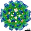 23147MC M: map data used to model this data C: citing same article ( |
|---|---|
| Similar structure data |

-Links
 PDBj
PDBj

- Assembly
| Deposited unit | 
|
|---|---|
| 1 | x 60
|
| 2 |
|
| 3 | x 5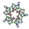
|
| 4 | x 6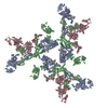
|
| 5 | 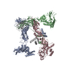
|
| Symmetry | Point symmetry: (Schoenflies symbol: I (icosahedral)) |
-Components
| #1: Protein | Mass: 53495.086 Da / Num. of mol.: 3 / Fragment: UNP residues 294-790 Source method: isolated from a genetically manipulated source Source: (gene. exp.) Binjari virus / Cell line (production host): C6/36 / Production host:  Aedes albopictus (Asian tiger mosquito) / References: UniProt: A0A5K6VMX2 Aedes albopictus (Asian tiger mosquito) / References: UniProt: A0A5K6VMX2#2: Protein | Mass: 18946.809 Da / Num. of mol.: 3 / Fragment: UNP residues 125-293 Source method: isolated from a genetically manipulated source Source: (gene. exp.) Binjari virus / Cell line (production host): C6/36 / Production host: Aedes albopictus (Asian tiger mosquito) / References: UniProt: A0A5K6VMX2 |
|---|
-Experimental details
-Experiment
| Experiment | Method: ELECTRON MICROSCOPY |
|---|---|
| EM experiment | Aggregation state: PARTICLE / 3D reconstruction method: single particle reconstruction |
- Sample preparation
| Component | Name: Binjari virus / Type: VIRUS Details: M and E from Binjari virus form a complex that assembles into anti-parallel dimers, (M-E)2, in the T=3 icosahedral particle. Entity ID: all / Source: MULTIPLE SOURCES | ||||||||||||||||||||
|---|---|---|---|---|---|---|---|---|---|---|---|---|---|---|---|---|---|---|---|---|---|
| Molecular weight | Value: 22 MDa / Experimental value: NO | ||||||||||||||||||||
| Source (natural) | Organism: Binjari virus / Strain: BFTA20 | ||||||||||||||||||||
| Source (recombinant) | Organism: Aedes albopictus (Asian tiger mosquito) / Cell: C6/36 | ||||||||||||||||||||
| Details of virus | Empty: NO / Enveloped: YES / Isolate: SPECIES / Type: VIRION | ||||||||||||||||||||
| Natural host | Organism: Ochlerotatus normanensis | ||||||||||||||||||||
| Virus shell | Diameter: 470 nm / Triangulation number (T number): 3 | ||||||||||||||||||||
| Buffer solution | pH: 8 | ||||||||||||||||||||
| Buffer component |
| ||||||||||||||||||||
| Specimen | Conc.: 4 mg/ml / Embedding applied: NO / Shadowing applied: NO / Staining applied: NO / Vitrification applied: YES | ||||||||||||||||||||
| Specimen support | Grid material: COPPER / Grid mesh size: 300 divisions/in. / Grid type: Quantifoil R1.2/1.3 | ||||||||||||||||||||
| Vitrification | Instrument: FEI VITROBOT MARK IV / Cryogen name: ETHANE / Humidity: 100 % / Chamber temperature: 277 K Details: 0 second wait time 2.5 second blot time -4 blot force 1 second drain time |
- Electron microscopy imaging
| Experimental equipment |  Model: Titan Krios / Image courtesy: FEI Company |
|---|---|
| Microscopy | Model: FEI TITAN KRIOS |
| Electron gun | Electron source: FIELD EMISSION GUN / Accelerating voltage: 300 kV / Illumination mode: FLOOD BEAM |
| Electron lens | Mode: BRIGHT FIELDBright-field microscopy / Calibrated magnification: 105000 X / Calibrated defocus min: 100 nm / Calibrated defocus max: 4300 nm / Cs: 2.7 mm |
| Specimen holder | Cryogen: NITROGEN / Specimen holder model: FEI TITAN KRIOS AUTOGRID HOLDER |
| Image recording | Average exposure time: 12.8 sec. / Electron dose: 43.2768 e/Å2 / Detector mode: SUPER-RESOLUTION / Film or detector model: GATAN K2 QUANTUM (4k x 4k) / Num. of grids imaged: 1 / Num. of real images: 6226 |
| Image scans | Movie frames/image: 32 |
- Processing
| EM software |
| ||||||||||||||||||||||||||||||||
|---|---|---|---|---|---|---|---|---|---|---|---|---|---|---|---|---|---|---|---|---|---|---|---|---|---|---|---|---|---|---|---|---|---|
| CTF correction | Type: PHASE FLIPPING AND AMPLITUDE CORRECTION | ||||||||||||||||||||||||||||||||
| Particle selection | Num. of particles selected: 55992 | ||||||||||||||||||||||||||||||||
| Symmetry | Point symmetry: I (icosahedral) | ||||||||||||||||||||||||||||||||
| 3D reconstruction | Resolution: 4.4 Å / Resolution method: FSC 0.143 CUT-OFF / Num. of particles: 26038 / Algorithm: BACK PROJECTION / Num. of class averages: 1 / Symmetry type: POINT | ||||||||||||||||||||||||||||||||
| Atomic model building | Protocol: RIGID BODY FIT / Space: REAL | ||||||||||||||||||||||||||||||||
| Atomic model building |
| ||||||||||||||||||||||||||||||||
| Refinement | Stereochemistry target values: GeoStd + Monomer Library + CDL v1.2 | ||||||||||||||||||||||||||||||||
| Displacement parameters | Biso mean: 82.99 Å2 | ||||||||||||||||||||||||||||||||
| Refine LS restraints |
|