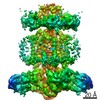
 Movie
Movie Controller


+ Open data

- Basic information
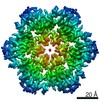
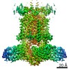
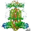
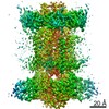
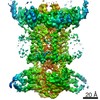
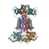
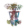
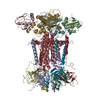
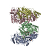
| Entry | Database: PDB / ID: 7cge | |||||||||
|---|---|---|---|---|---|---|---|---|---|---|
| Title | The overall structure of nucleotide free MlaFEDB complex | |||||||||
 Components Components |
| |||||||||
 Keywords Keywords |  MEMBRANE PROTEIN MEMBRANE PROTEIN | |||||||||
| Function / homology |  Function and homology information Function and homology informationphospholipid transfer activity / intermembrane phospholipid transfer / phospholipid transporter activity / phospholipid-translocating ATPase complex / phospholipid transport / Translocases; Catalysing the translocation of other compounds; Linked to the hydrolysis of a nucleoside triphosphate / ATPase-coupled transmembrane transporter activity / ATP-binding cassette (ABC) transporter complex / phospholipid binding / response to antibiotic ...phospholipid transfer activity / intermembrane phospholipid transfer / phospholipid transporter activity / phospholipid-translocating ATPase complex / phospholipid transport / Translocases; Catalysing the translocation of other compounds; Linked to the hydrolysis of a nucleoside triphosphate / ATPase-coupled transmembrane transporter activity / ATP-binding cassette (ABC) transporter complex / phospholipid binding / response to antibiotic / DNA damage response / ATP hydrolysis activity / extracellular region / ATP binding / membrane / plasma membrane / cytosolSimilarity search - Function | |||||||||
| Biological species |  Escherichia coli (E. coli) Escherichia coli (E. coli) | |||||||||
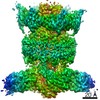
| Method | ELECTRON MICROSCOPY / single particle reconstruction / cryo EM / Resolution: 2.9 Å | |||||||||
 Authors Authors | Chi, X.M. / Fan, Q.X. / Zhang, Y.Y. / Liang, K. / Zhou, Q. / Li, Y.Y. | |||||||||
| Funding support |  China, 1items China, 1items
| |||||||||
 Citation Citation | Journal: Cell Res / Year: 2020 Title: Structural mechanism of phospholipids translocation by MlaFEDB complex. Authors: Ximin Chi / Qiongxuan Fan / Yuanyuan Zhang / Ke Liang / Li Wan / Qiang Zhou / Yanyan Li / Abstract: In Gram-negative bacteria, phospholipids are major components of the inner membrane and the inner leaflet of the outer membrane, playing an essential role in forming the unique dual-membrane barrier ...In Gram-negative bacteria, phospholipids are major components of the inner membrane and the inner leaflet of the outer membrane, playing an essential role in forming the unique dual-membrane barrier to exclude the entry of most antibiotics. Understanding the mechanisms of phospholipid translocation between the inner and outer membrane represents one of the major challenges surrounding bacterial phospholipid homeostasis. The conserved MlaFEDB complex in the inner membrane functions as an ABC transporter to drive the translocation of phospholipids between the inner membrane and the periplasmic protein MlaC. However, the mechanism of phospholipid translocation remains elusive. Here we determined three cryo-EM structures of MlaFEDB from Escherichia coli in its nucleotide-free and ATP-bound conformations, and performed extensive functional studies to verify and extend our findings from structural analyses. Our work reveals unique structural features of the entire MlaFEDB complex, six well-resolved phospholipids in three distinct cavities, and large-scale conformational changes upon ATP binding. Together, these findings define the cycle of structural rearrangement of MlaFEDB in action, and suggest that MlaFEDB uses an extrusion mechanism to extract and release phospholipids through the central translocation cavity. | |||||||||
| History |
|
- Structure visualization
| Movie |
Movie viewer |
|---|---|
| Structure viewer | Molecule: MolmilJmol/JSmol |
- Downloads & links
-Download
| PDBx/mmCIF format | 7cge.cif.gz | 367.3 KB | Display | PDBx/mmCIF format |
|---|---|---|---|---|
| PDB format | pdb7cge.ent.gz | 300.3 KB | Display | PDB format |
| PDBx/mmJSON format | 7cge.json.gz | Tree view | PDBx/mmJSON format | |
| Others |  Other downloads Other downloads |
-Validation report
| Arichive directory | https://data.pdbj.org/pub/pdb/validation_reports/cg/7cgeftp://data.pdbj.org/pub/pdb/validation_reports/cg/7cge | HTTPS FTP |
|---|
-Related structure data
| Related structure data | 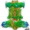 30355MC 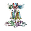 7cgnC 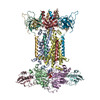 7ch0C M: map data used to model this data C: citing same article ( |
|---|---|
| Similar structure data |
-Links
 PDBj
PDBj


- Assembly
| Deposited unit | 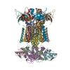
|
|---|---|
| 1 |
|
-Components
| #1: Protein | Mass: 27885.162 Da / Num. of mol.: 2 Source method: isolated from a genetically manipulated source Source: (gene. exp.) Escherichia coli (strain K12) (bacteria)Strain: K12 / Gene: mlaE, FAZ83_04790 / Production host: Escherichia coli K-12 (bacteria) / Strain (production host): K-12 / References: UniProt: A0A4S5B3V0, UniProt: P64606*PLUS#2: Protein | Mass: 29128.801 Da / Num. of mol.: 2 Source method: isolated from a genetically manipulated source Source: (gene. exp.) Escherichia coli (strain K12) (bacteria)Strain: K12 / Gene: mlaF, FAZ83_04795 / Production host: Escherichia coli K-12 (bacteria) / Strain (production host): K-12 / References: UniProt: A0A4V3YUQ9, UniProt: P63386*PLUS#3: Protein | Mass: 10690.313 Da / Num. of mol.: 2 Source method: isolated from a genetically manipulated source Source: (gene. exp.) Escherichia coli (strain K12) (bacteria)Strain: K12 / Gene: mlaB, FAZ83_04775 / Production host: Escherichia coli K-12 (bacteria) / Strain (production host): K-12 / References: UniProt: A0A4S5B5E3, UniProt: P64602*PLUS#4: Protein | Mass: 19593.133 Da / Num. of mol.: 6 Source method: isolated from a genetically manipulated source Source: (gene. exp.) Escherichia coli (strain K12) (bacteria)Strain: K12 / Gene: mlaD, FAZ83_04785 / Production host: Escherichia coli K-12 (bacteria) / Strain (production host): K-12 / References: UniProt: A0A6D2XU65, UniProt: P64604*PLUS#5: Chemical | ChemComp-PGW / ( Phosphatidylglycerol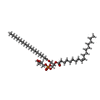  Mass: 749.007 Da / Num. of mol.: 12 / Source method: obtained synthetically / Formula: C40H77O10P / Comment: phospholipid*YM Mass: 749.007 Da / Num. of mol.: 12 / Source method: obtained synthetically / Formula: C40H77O10P / Comment: phospholipid*YMHas ligand of interest | N | |
|---|
-Experimental details
-Experiment
| Experiment | Method: ELECTRON MICROSCOPY |
|---|---|
| EM experiment | Aggregation state: PARTICLE / 3D reconstruction method: single particle reconstruction |
- Sample preparation
| Component | Name: cryo_EM map of nucleotide free MlaFEDB complex / Type: COMPLEX / Entity ID: #1-#4 / Source: RECOMBINANT |
|---|---|
| Source (natural) | Organism: Escherichia coli K-12 (bacteria) |
| Source (recombinant) | Organism: Escherichia coli K-12 (bacteria) |
| Buffer solution | pH: 8 |
| Specimen | Embedding applied: NO / Shadowing applied: NO / Staining applied: NO / Vitrification applied: YES |
| Vitrification | Cryogen name: ETHANE |
- Electron microscopy imaging
| Experimental equipment |  Model: Titan Krios / Image courtesy: FEI Company |
|---|---|
| Microscopy | Model: FEI TITAN KRIOS |
| Electron gun | Electron source: FIELD EMISSION GUN / Accelerating voltage: 300 kV / Illumination mode: FLOOD BEAM |
| Electron lens | Mode: BRIGHT FIELDBright-field microscopy / Alignment procedure: COMA FREE |
| Specimen holder | Cryogen: NITROGEN / Specimen holder model: FEI TITAN KRIOS AUTOGRID HOLDER |
| Image recording | Electron dose: 50 e/Å2 / Film or detector model: GATAN K3 BIOQUANTUM (6k x 4k) |
- Processing
| EM software | Name: RELION / Version: 3.0.6 / Category: 3D reconstruction |
|---|---|
| CTF correction | Type: PHASE FLIPPING AND AMPLITUDE CORRECTION |
| 3D reconstruction | Resolution: 2.9 Å / Resolution method: FSC 0.143 CUT-OFF / Num. of particles: 271252 / Symmetry type: POINT |
