 Movie
Movie Controller


+ Open data

- Basic information
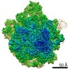
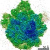
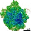
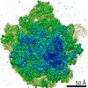
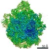
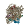
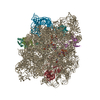
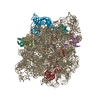
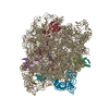
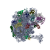
| Entry | Database: PDB / ID: 6pcr | |||||||||
|---|---|---|---|---|---|---|---|---|---|---|
| Title | E. coli 50S ribosome bound to compound 40o | |||||||||
 Components Components |
| |||||||||
 Keywords Keywords |  RIBOSOME / E. coli ribosome / streptogramin A analog / antibiotics RIBOSOME / E. coli ribosome / streptogramin A analog / antibiotics | |||||||||
| Function / homology |  Function and homology informationtranscriptional attenuation / endoribonuclease inhibitor activity / RNA-binding transcription regulator activity / negative regulation of cytoplasmic translation / DnaA-L2 complex / translation repressor activity / negative regulation of DNA-templated DNA replication initiation / ribosome assembly / DNA-templated transcription termination / mRNA 5'-UTR binding ...transcriptional attenuation / endoribonuclease inhibitor activity / RNA-binding transcription regulator activity / negative regulation of cytoplasmic translation / DnaA-L2 complex / translation repressor activity / negative regulation of DNA-templated DNA replication initiation / ribosome assembly / DNA-templated transcription termination / mRNA 5'-UTR binding / ribosomal large subunit assembly / large ribosomal subunit rRNA binding / large ribosomal subunit / cytoplasmic translation / cytosolic large ribosomal subunit / transferase activity / negative regulation of translation / rRNA binding / ribosome / structural constituent of ribosome / translation / response to antibiotic / mRNA binding / negative regulation of DNA-templated transcription / DNA binding / RNA binding / zinc ion binding / cytosol / cytoplasm Function and homology informationtranscriptional attenuation / endoribonuclease inhibitor activity / RNA-binding transcription regulator activity / negative regulation of cytoplasmic translation / DnaA-L2 complex / translation repressor activity / negative regulation of DNA-templated DNA replication initiation / ribosome assembly / DNA-templated transcription termination / mRNA 5'-UTR binding ...transcriptional attenuation / endoribonuclease inhibitor activity / RNA-binding transcription regulator activity / negative regulation of cytoplasmic translation / DnaA-L2 complex / translation repressor activity / negative regulation of DNA-templated DNA replication initiation / ribosome assembly / DNA-templated transcription termination / mRNA 5'-UTR binding / ribosomal large subunit assembly / large ribosomal subunit rRNA binding / large ribosomal subunit / cytoplasmic translation / cytosolic large ribosomal subunit / transferase activity / negative regulation of translation / rRNA binding / ribosome / structural constituent of ribosome / translation / response to antibiotic / mRNA binding / negative regulation of DNA-templated transcription / DNA binding / RNA binding / zinc ion binding / cytosol / cytoplasmSimilarity search - Function | |||||||||
| Biological species |  Escherichia coli (E. coli) Escherichia coli (E. coli) | |||||||||
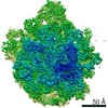
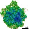
| Method | ELECTRON MICROSCOPY / single particle reconstruction / cryo EM / Resolution: 2.5 Å | |||||||||
 Authors Authors | Pellegrino, J. / Lee, D.J. / Fraser, J.S. / Seiple, I.B. | |||||||||
| Funding support |  United States, 2items United States, 2items
| |||||||||
 Citation Citation | Journal: Nature / Year: 2020 Title: Synthetic group A streptogramin antibiotics that overcome Vat resistance. Authors: Qi Li / Jenna Pellegrino / D John Lee / Arthur A Tran / Hector A Chaires / Ruoxi Wang / Jesslyn E Park / Kaijie Ji / David Chow / Na Zhang / Axel F Brilot / Justin T Biel / Gydo van Zundert ...Authors: Qi Li / Jenna Pellegrino / D John Lee / Arthur A Tran / Hector A Chaires / Ruoxi Wang / Jesslyn E Park / Kaijie Ji / David Chow / Na Zhang / Axel F Brilot / Justin T Biel / Gydo van Zundert / Kenneth Borrelli / Dean Shinabarger / Cindy Wolfe / Beverly Murray / Matthew P Jacobson / Estelle Mühle / Olivier Chesneau / James S Fraser / Ian B Seiple /   Abstract: Natural products serve as chemical blueprints for most antibiotics in clinical use. The evolutionary process by which these molecules arise is inherently accompanied by the co-evolution of resistance ...Natural products serve as chemical blueprints for most antibiotics in clinical use. The evolutionary process by which these molecules arise is inherently accompanied by the co-evolution of resistance mechanisms that shorten the clinical lifetime of any given class of antibiotics. Virginiamycin acetyltransferase (Vat) enzymes are resistance proteins that provide protection against streptogramins, potent antibiotics against Gram-positive bacteria that inhibit the bacterial ribosome. Owing to the challenge of selectively modifying the chemically complex, 23-membered macrocyclic scaffold of group A streptogramins, analogues that overcome the resistance conferred by Vat enzymes have not been previously developed. Here we report the design, synthesis, and antibacterial evaluation of group A streptogramin antibiotics with extensive structural variability. Using cryo-electron microscopy and forcefield-based refinement, we characterize the binding of eight analogues to the bacterial ribosome at high resolution, revealing binding interactions that extend into the peptidyl tRNA-binding site and towards synergistic binders that occupy the nascent peptide exit tunnel. One of these analogues has excellent activity against several streptogramin-resistant strains of Staphylococcus aureus, exhibits decreased rates of acetylation in vitro, and is effective at lowering bacterial load in a mouse model of infection. Our results demonstrate that the combination of rational design and modular chemical synthesis can revitalize classes of antibiotics that are limited by naturally arising resistance mechanisms. | |||||||||
| History |
|
- Structure visualization
| Movie |
Movie viewer |
|---|---|
| Structure viewer | Molecule: MolmilJmol/JSmol |
- Downloads & links
-Download
| PDBx/mmCIF format | 6pcr.cif.gz | 1.6 MB | Display | PDBx/mmCIF format |
|---|---|---|---|---|
| PDB format | pdb6pcr.ent.gz | 1.2 MB | Display | PDB format |
| PDBx/mmJSON format | 6pcr.json.gz | Tree view | PDBx/mmJSON format | |
| Others |  Other downloads Other downloads |
-Validation report
| Arichive directory | https://data.pdbj.org/pub/pdb/validation_reports/pc/6pcrftp://data.pdbj.org/pub/pdb/validation_reports/pc/6pcr | HTTPS FTP |
|---|
-Related structure data
| Related structure data | 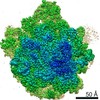 20305MC 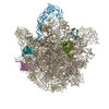 6pc5C 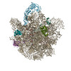 6pc6C  6pc7C  6pc8C  6pchC 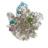 6pcqC 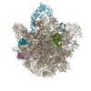 6pcsC  6pctC  6wyvC  6x3cC  6x3jC M: map data used to model this data C: citing same article ( |
|---|---|
| Similar structure data | |
| EM raw data | EMPIAR-10524 (Title: E. coli 50S ribosome bound to compound 40o / Data size: 446.9 Data #1: Unaligned movies of 50S ribosome complex bound to compound 40o [micrographs - multiframe]) |


-Links
 PDBj
PDBj


- Assembly
| Deposited unit | 
|
|---|---|
| 1 |
|
-Components
-RNA chain , 2 types, 2 molecules IJ
| #1: RNA chain | Mass: 941795.562 Da / Num. of mol.: 1 / Source method: isolated from a natural source / Source: (natural) Escherichia coli (E. coli) |
|---|---|
| #2: RNA chain | Mass: 38177.762 Da / Num. of mol.: 1 / Source method: isolated from a natural source / Source: (natural) Escherichia coli (E. coli) / References: GenBank: 1266940032 |
-50S ribosomal protein ... , 5 types, 5 molecules KLMNO
| #3: Protein | / Large ribosomal subunit protein uL2 Mass: 29663.244 Da / Num. of mol.: 1 / Source method: isolated from a natural source / Source: (natural) Escherichia coli (E. coli) / References: UniProt: P60422 |
|---|---|
| #4: Protein | Mass: 15008.471 Da / Num. of mol.: 1 / Source method: isolated from a natural source / Source: (natural) Escherichia coli (E. coli) / References: UniProt: A0A037Y8L6, UniProt: P02413*PLUS |
| #5: Protein | Mass: 22121.566 Da / Num. of mol.: 1 / Source method: isolated from a natural source / Source: (natural) Escherichia coli (E. coli) / References: UniProt: D7Z9F6, UniProt: P60723*PLUS |
| #6: Protein | / Large ribosomal subunit protein uL3 Mass: 22277.535 Da / Num. of mol.: 1 / Source method: isolated from a natural source / Source: (natural) Escherichia coli (E. coli) / References: UniProt: P60438 |
| #7: Protein | Mass: 16050.606 Da / Num. of mol.: 1 / Source method: isolated from a natural source / Source: (natural) Escherichia coli (E. coli) / References: UniProt: D7ZET0, UniProt: P0AA10*PLUS |
-Non-polymers , 1 types, 1 molecules 
| #8: Chemical | ChemComp-O8P / ( Mass: 742.613 Da / Num. of mol.: 1 / Source method: obtained synthetically / Formula: C34H40BrN5O9 / Feature type: SUBJECT OF INVESTIGATION Mass: 742.613 Da / Num. of mol.: 1 / Source method: obtained synthetically / Formula: C34H40BrN5O9 / Feature type: SUBJECT OF INVESTIGATION |
|---|
-Details
| Has ligand of interest | Y |
|---|
-Experimental details
-Experiment
| Experiment | Method: ELECTRON MICROSCOPY |
|---|---|
| EM experiment | Aggregation state: PARTICLE / 3D reconstruction method: single particle reconstruction |
- Sample preparation
| Component | Name: 50S E. coli ribosome / Type: RIBOSOME / Entity ID: #1-#7 / Source: NATURAL |
|---|---|
| Molecular weight | Experimental value: NO |
| Source (natural) | Organism: Escherichia coli (E. coli) |
| Buffer solution | pH: 7.5 |
| Specimen | Embedding applied: NO / Shadowing applied: NO / Staining applied: NO / Vitrification applied: YES |
| Vitrification | Cryogen name: ETHANE |
- Electron microscopy imaging
| Experimental equipment |  Model: Titan Krios / Image courtesy: FEI Company |
|---|---|
| Microscopy | Model: FEI TITAN KRIOS |
| Electron gun | Electron source: FIELD EMISSION GUN / Accelerating voltage: 300 kV / Illumination mode: FLOOD BEAM |
| Electron lens | Mode: BRIGHT FIELDBright-field microscopy |
| Image recording | Electron dose: 71.2 e/Å2 / Film or detector model: GATAN K2 SUMMIT (4k x 4k) |
- Processing
| CTF correction | Type: PHASE FLIPPING AND AMPLITUDE CORRECTION |
|---|---|
| Symmetry | Point symmetry: C1 (asymmetric) |
| 3D reconstruction | Resolution: 2.5 Å / Resolution method: FSC 0.143 CUT-OFF / Num. of particles: 44462 / Symmetry type: POINT |
