 Movie
Movie Controller

[English] 日本語
 Yorodumi
- PDB-6mwx: CryoEM structure of Chimeric Eastern Equine Encephalitis Virus wi... -
+ Open data

- Basic information
| Entry | Database: PDB / ID: 6mwx | |||||||||||||||
|---|---|---|---|---|---|---|---|---|---|---|---|---|---|---|---|---|
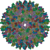
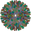
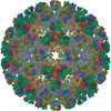
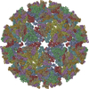
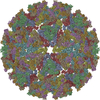
| Title | CryoEM structure of Chimeric Eastern Equine Encephalitis Virus with Fab of EEEV-69 Antibody | |||||||||||||||
 Components Components |
| |||||||||||||||
 Keywords Keywords | VIRUS/IMMUNE SYSTEM /  Alphavirus / EEEV / Eastern Equine Encephalitis Virus / Sindbis / Fab / VIRUS-IMMUNE SYSTEM complex Alphavirus / EEEV / Eastern Equine Encephalitis Virus / Sindbis / Fab / VIRUS-IMMUNE SYSTEM complex | |||||||||||||||
| Function / homology |  Function and homology informationtogavirin / T=4 icosahedral viral capsid / symbiont-mediated suppression of host gene expression / symbiont-mediated suppression of host toll-like receptor signaling pathway / host cell cytoplasm / symbiont entry into host cell / serine-type endopeptidase activity / fusion of virus membrane with host endosome membrane / host cell nucleus / virion attachment to host cell ...togavirin / T=4 icosahedral viral capsid / symbiont-mediated suppression of host gene expression / symbiont-mediated suppression of host toll-like receptor signaling pathway / host cell cytoplasm / symbiont entry into host cell / serine-type endopeptidase activity / fusion of virus membrane with host endosome membrane / host cell nucleus / virion attachment to host cell / host cell plasma membrane / virion membrane / structural molecule activity / proteolysis / RNA binding / membrane / plasma membrane / cytoplasm Function and homology informationtogavirin / T=4 icosahedral viral capsid / symbiont-mediated suppression of host gene expression / symbiont-mediated suppression of host toll-like receptor signaling pathway / host cell cytoplasm / symbiont entry into host cell / serine-type endopeptidase activity / fusion of virus membrane with host endosome membrane / host cell nucleus / virion attachment to host cell ...togavirin / T=4 icosahedral viral capsid / symbiont-mediated suppression of host gene expression / symbiont-mediated suppression of host toll-like receptor signaling pathway / host cell cytoplasm / symbiont entry into host cell / serine-type endopeptidase activity / fusion of virus membrane with host endosome membrane / host cell nucleus / virion attachment to host cell / host cell plasma membrane / virion membrane / structural molecule activity / proteolysis / RNA binding / membrane / plasma membrane / cytoplasmSimilarity search - Function | |||||||||||||||
| Biological species |  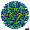 Eastern equine encephalitis virus Eastern equine encephalitis virus Mus musculus (house mouse) Mus musculus (house mouse) | |||||||||||||||
| Method | ELECTRON MICROSCOPY / single particle reconstruction / cryo EM / Resolution: 8.2 Å | |||||||||||||||
 Authors Authors | Hasan, S.S. / Sun, C. / Kim, A.S. / Watanabe, Y. / Chen, C.L. / Klose, T. / Buda, G. / Crispin, M. / Diamond, M.S. / Klimstra, W.B. / Rossmann, M.G. | |||||||||||||||
| Funding support |  United States, United States,  United Kingdom, 4items United Kingdom, 4items
| |||||||||||||||
 Citation Citation | Journal: Cell Rep / Year: 2018 Title: Cryo-EM Structures of Eastern Equine Encephalitis Virus Reveal Mechanisms of Virus Disassembly and Antibody Neutralization. Authors: S Saif Hasan / Chengqun Sun / Arthur S Kim / Yasunori Watanabe / Chun-Liang Chen / Thomas Klose / Geeta Buda / Max Crispin / Michael S Diamond / William B Klimstra / Michael G Rossmann / Abstract: Alphaviruses are enveloped pathogens that cause arthritis and encephalitis. Here, we report a 4.4-Å cryoelectron microscopy (cryo-EM) structure of eastern equine encephalitis virus (EEEV), an ...Alphaviruses are enveloped pathogens that cause arthritis and encephalitis. Here, we report a 4.4-Å cryoelectron microscopy (cryo-EM) structure of eastern equine encephalitis virus (EEEV), an alphavirus that causes fatal encephalitis in humans. Our analysis provides insights into viral entry into host cells. The envelope protein E2 showed a binding site for the cellular attachment factor heparan sulfate. The presence of a cryptic E2 glycan suggests how EEEV escapes surveillance by lectin-expressing myeloid lineage cells, which are sentinels of the immune system. A mechanism for nucleocapsid core release and disassembly upon viral entry was inferred based on pH changes and capsid dissociation from envelope proteins. The EEEV capsid structure showed a viral RNA genome binding site adjacent to a ribosome binding site for viral genome translation following genome release. Using five Fab-EEEV complexes derived from neutralizing antibodies, our investigation provides insights into EEEV host cell interactions and protective epitopes relevant to vaccine design. | |||||||||||||||
| History |
|
- Structure visualization
| Movie |
Movie viewer |
|---|---|
| Structure viewer | Molecule: MolmilJmol/JSmol |
- Downloads & links
-Download
| PDBx/mmCIF format | 6mwx.cif.gz | 896.9 KB | Display | PDBx/mmCIF format |
|---|---|---|---|---|
| PDB format | pdb6mwx.ent.gz | 738.3 KB | Display | PDB format |
| PDBx/mmJSON format | 6mwx.json.gz | Tree view | PDBx/mmJSON format | |
| Others |  Other downloads Other downloads |
-Validation report
| Arichive directory | https://data.pdbj.org/pub/pdb/validation_reports/mw/6mwxftp://data.pdbj.org/pub/pdb/validation_reports/mw/6mwx | HTTPS FTP |
|---|
-Related structure data
| Related structure data | 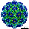 9279MC 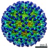 9249C  9274C 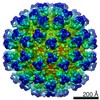 9275C 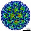 9278C 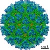 9280C  9281C  6muiC 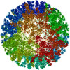 6mw9C 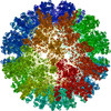 6mwcC  6mwvC 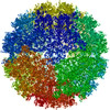 6mx4C  6mx7C C: citing same article ( M: map data used to model this data |
|---|---|
| Similar structure data |

-Links
 PDBj
PDBj


- Assembly
| Deposited unit | 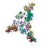
|
|---|---|
| 1 | x 60
|
| 2 |
|
| 3 | x 5
|
| 4 | x 6
|
| 5 | 
|
| Symmetry | Point symmetry: (Schoenflies symbol: I (icosahedral)) |
-Components
| #1: Protein | Mass: 47938.141 Da / Num. of mol.: 4 / Fragment: ectodomain (UNP residues 802-1242) Source method: isolated from a genetically manipulated source Source: (gene. exp.) Eastern equine encephalitis virus / Cell (production host): BHK-15 / Production host: Mesocricetus auratus (golden hamster) / References: UniProt: E9KXM2, UniProt: Q4QXJ7*PLUS#2: Protein | Mass: 47046.953 Da / Num. of mol.: 4 / Fragment: ectodomain (UNP residues 325-744) Source method: isolated from a genetically manipulated source Source: (gene. exp.) Eastern equine encephalitis virus / Cell (production host): BHK-15 / Production host: Mesocricetus auratus (golden hamster) / References: UniProt: E9KXL2, UniProt: Q4QXJ7*PLUS#3: Antibody | Mass: 23743.719 Da / Num. of mol.: 4 / Fragment: Fab / Source method: isolated from a natural source / Source: (natural) Mus musculus (house mouse)#4: Antibody | Mass: 23177.713 Da / Num. of mol.: 4 / Fragment: Fab / Source method: isolated from a natural source / Source: (natural) Mus musculus (house mouse) / References: UniProt: G0YP42 |
|---|
-Experimental details
-Experiment
| Experiment | Method: ELECTRON MICROSCOPY |
|---|---|
| EM experiment | Aggregation state: PARTICLE / 3D reconstruction method: single particle reconstruction |
- Sample preparation
| Component |
| ||||||||||||||||||||||||||||
|---|---|---|---|---|---|---|---|---|---|---|---|---|---|---|---|---|---|---|---|---|---|---|---|---|---|---|---|---|---|
| Source (natural) |
| ||||||||||||||||||||||||||||
| Details of virus | Empty: NO / Enveloped: YES / Isolate: OTHER / Type: VIRION | ||||||||||||||||||||||||||||
| Buffer solution | pH: 7.5 | ||||||||||||||||||||||||||||
| Specimen | Conc.: 1 mg/ml / Embedding applied: NO / Shadowing applied: NO / Staining applied: NO / Vitrification applied: YES | ||||||||||||||||||||||||||||
| Specimen support | Details: unspecified | ||||||||||||||||||||||||||||
| Vitrification | Instrument: GATAN CRYOPLUNGE 3 / Cryogen name: HELIUM / Humidity: 80 % / Chamber temperature: 295 K |
- Electron microscopy imaging
| Experimental equipment |  Model: Titan Krios / Image courtesy: FEI Company |
|---|---|
| Microscopy | Model: FEI TITAN KRIOS |
| Electron gun | Electron source: FIELD EMISSION GUN / Accelerating voltage: 300 kV / Illumination mode: FLOOD BEAM |
| Electron lens | Mode: BRIGHT FIELDBright-field microscopy |
| Image recording | Electron dose: 31 e/Å2 / Film or detector model: GATAN K2 SUMMIT (4k x 4k) |
- Processing
| CTF correction | Type: PHASE FLIPPING AND AMPLITUDE CORRECTION |
|---|---|
| 3D reconstruction | Resolution: 8.2 Å / Resolution method: FSC 0.143 CUT-OFF / Num. of particles: 5964 / Symmetry type: POINT |
| Atomic model building | Protocol: RIGID BODY FIT |
